Nervous system of the frog (1)
•Download as PPTX, PDF•
6 likes•7,465 views

Nahanap ko sa net. Baka makatulong sa expt 12 haha :D *All rights reserved sa kung sino man gumawa nito haha :P


Nervous system of the frog (1)
- 1. Nervous System of the FrogBy:Group 636 Marjorie Polintan37 Miguel Quiambao38 Inna Karla Ramos39 James Redulla40 Patricia Anne Reyes41 Zyril Mae Reyes42 Claudine Roxas
- 2. Nervous SystemServes as reception of stimuliConduction of impulsesCoordination and integration of the various functions of the organs
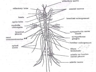
- 3. Three Divisions of the Frog’s Nervous System (according to morphology): 1. Central Nervous Systemalso known as the Cerebro-spinal Nervous Systemincludes the brain and the spinal cordserves as the great center of communication between the principal sense organs and the rest of the bodydivided into five major parts, namely:TelencephalonDiencephalonMesencephalonMetencephalonMyelencephalon
- 4. 2. Peripheral Nervous Systemincludes the ten pairs of cranial nerves (from the brain) and ten pairs of spinal nerves (from the spinal cord)connects the central nervous organs to the receptors and effectors of the body3. SymphatheticNervous Systemconsists of two slender nerve trunks or cord, each with a chain of ganglia on either side of the spinal columnhelps deliver information to the body about impending dangerresponsible for the fight-or-flight response
- 5. I. Central Nervous System (CNS)
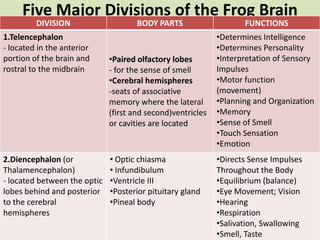
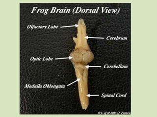
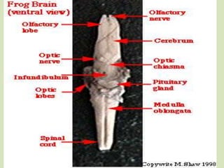
- 6. Five Major Divisions of the Frog BrainA
- 8. Other parts of the CNS:Choroid Plexuses- secretes the cerebro-spinal fluidForamen of Monro- a narrow passage that joins the lateral ventricles with the 3rd ventriclesAqueduct of Sylvius- joins the 3rd ventricle with the 4th ventricle Cerebro-spinal Fluid- serves as a protective liquid cushion and helps nourish the central nervous organs Spinal Cord- short and somewhat flattened; posterior prolongation of the brain; surrounded by meninges; extends from the medulla oblongata to the filumterminaleÂ
- 9. FilumTerminale- posterior tapering portion located in the urostyle; presents two enlargements, namely:Brachial Enlargement- associated with the nerve supply to the forelimbsLumbar Enlargement or Sciatic Enlargement- associated with the nerve supply to the hindlimbsMeninges- two connective tissue membranes which surrounds the spinal chord(Outer) Dura Mater- adhering to the bone(Inner Vascular) Pia Mater- adhering to the nervous tissueVascular Arachnoid- located beneath the dura mater; forms the middle layer of the meninges (for higher vertebrates)Â
- 12. II. Peripheral Nervous System (PNS)2 Divisions of the Peripheral Nervous SystemA.CranialNerves – consists of 10 pairs of nerves (left,right) -extending from the lateral surfaces of the brain to the parts of the body B.SpinalNerves- consists of 10pairs of nerves (left,right)Arise from the spinal cord Distributes to the limbs and trunk.emerge between vertebrae each spinal nerve is attached to the spinal cord by 2 roots (dorsal sensory root and ventral motor root)
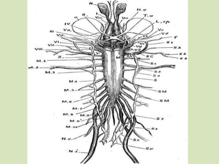
- 13. A. The 10 Paired Cranial Nerves Olfactory Function/Type: Sensory nerves for smellLocation: walls of nasal chambersOpticFunction/Type: Sensory nerves for visionLocation: retina of the eyesOculo-MotorFunction/Type: Motor nervesLocation: 4 muscles of the eye namely (superior rectus,inferiorrectus,medialrectus,inferior oblique muscles)TrochlearFunction/Type: Motor nervesLocation: superior oblique muscle of the eye
- 14. 5. TrigeminalFunction/Type: sensory and motor nervesLocation: muscles of the jaws, skin of the face, mouth and the tongue6. AbducensFunction/Type: motor nervesLocation: lateral or external rectus muscle of the eye7. FacialFunction/Type: motor and sensory (mostly motor)Location: muscles of the face and throat
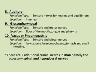
- 15. 8. AuditoryFunction/Type: Sensory nerves for hearing and equilibriumLocation: inner ear9. GlossopharyngealFunction/Type: Sensory and motor nervesLocation: floor of the mouth,tongue and pharynx10. Vagus or PneumogastricFunction/Type: Sensory and Motor nervesLocation: larynx,lungs,heart,esophagus,stomach and small intestine. *There are 2 additional cranial nerves in man namely the accessory spinal and hypoglossal nerves  Â
- 22. Some Mnemonics to help remember the names and order of the cranial nerves..On Old Olympus' Towering Top AFinn And German Viewed Oliver the optimistic octopus trots triumphantly about facing audiences glossily vaguely OOOTruly There Are Five Absolutely Gorgeous VixenSome mnemonics to remember the types of cranial nerves..Some Say Money Matters, But My Brother Says Big Brains Matter More.Sally Sells Mega Monkeys, But My Brother Sells Bigger Better Mega Monkeys.Some Stars Make Money, But My Brother Says Bugs Bunny Makes More.
- 23. B. The 10 Paired Spinal Nerves1st spinal nerve2nd spinal nerve3rd spinal nerve4th spinal nerve5th spinal nerve6th spinal nerve-Unite to form the Brachial Plexus to the shoulder and forelimb region-Supply the skin and muscles of the abdominal wall
- 24. -forms the Sciatic / lumbo-sacral plexus to the hindlimb*the 9th spinal nerve is the largest and is known as the sciatic nerve-Distributed to the urinary bladder, cloacaand oviducts7th spinal nerve8th spinal nerve9th spinal nerve*10th spinal nerve
- 26. III Sympathetic Nervous System (SNS)In man, Sympathetic and parasympathetic comprise Autonomic Division.It is branch of the autonomic nervous systemIt is always active at a basal level ( sympathetic tone) becomes more active during times of stress (fight-or-flight response)operates through a series of interconnected neurons. frequently considered part of the peripheral nervous system (PNS), although many lie within the central nervous system(CNS).
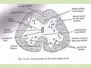
- 27. HISTOLOGY OF THE SPINAL CORD(mammalian) The spinal cords the reflex activities of the animals but which may be checked or modified by impulses from the brain.In the cross sections, the spinal cord shows 2 distinct areas:1. Gray matter – made up of nerve cell bodies, portion of the dendrites and axons, and the unmyelinated fibers.2.White matter- composed of myelinated fibers
- 28. Neurogliacells and their processes bind together and support the nervous elements in both areas.CanalisCentralis– located near the center of the gray matter -lined with a single layer of the EpithelialependymalcellsAt the sides of the gray matter, dorsal and ventral hornsor cornua are produced.Gray Commisuresconnects the gray matter on the two sides.Oblique crossings of the medullated fibers form the white commisures.Ventral fissure separates the right and the left columns of the white matter.
- 29. Dorsal Septum, composed of Pial tissues, extends from the base of this sulcus almost to the gray matterThe deep indentation at the bottom of the cord is the Ventral Fissure
- 33. Histology of the eyeeye- organ of visionDivided in to 2 segments:aqueous humor-anterior segment-contains watery fluidvitreous humor-posterior segment (adapted to the reception/transmission of images)-more viscuous/jelly like fluid
- 34. Outer coat-opaque,no light can enter-separated from the anterior segment by the crystalline lens and suspensory ligament.The three coats of the eye..1.Sclera2.Choroid3.retina
- 35. Sclera –protective ,outer,thick fibrous coat -tendons of eye are attached to it -continuous with the transparent cornea that permits light rays to enter2. Choroid-vascular layer between the sclera and retina -concerned with nutrition of ocular tissues -presence of numerous blood vessels -anterior portion:ciliary body and iris (mechanism for the accomodation of the refraction of the eye,projects over the anterior portion of the lens)
- 36. Pupil-central opening -Regulates the amt of light entering the eyeIris- colored porion of the eye3. Retina- innermost photosensitive coat. -contains receptors (rods,cones) -first link of the nervous pathways conveying impulses through the optic nerve of the brain.
- 38. END. :3