NMR Higher order structure (HOS) analysis of biologics for comparison studies of batch-to-batch consistency, biosimilars and formulation optimizations
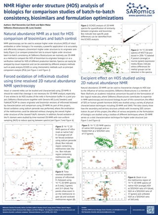
- 1. NMR Higher order structure (HOS) analysis of biologics for comparison studies of batch-to-batch consistency, biosimilars and formulation optimizations Authors: Olof Stenstr├Čm, Carl Diehl and Bj├Črn Walse SARomics Biostructures AB, Lund, Sweden www.saromics.com Natural abundance NMR as a tool for HOS comparison of biosimilars and batch control NMR spectroscopy can be used to analyze higher order structure (HOS) of intact antibodies or other biologics. For example, a powerful application is to accurately and efficiently compare a biosimilarŌĆÖs higher order structure to its originator anti- body (Figure 1) or compare production lots to ensure higher order structure batch-to-batch consistency. At SARomics Biostructures we routinely use 2D-NMR as a method to compare the HOS of biosimilars to originator biologics or as a verification method for HOS of different production batches. Spectra can easily be analysed by visual inspection and can be extended by different analysis methods such as peak analysis, ECHOS or using chemometric methods such as principal component analysis (PCA) (see Figure 1 and Figure 2). Contact: Carl Diehl, NMR Services Manager, Senior Scientist, carl.diehl@saromics.com Olof Stenstr├Čm, Scientist, olof.stenstrom@saromics.com Forced oxidation of infliximab studied using time resolved 2D natural abundance NMR spectroscopy Intact or cleaved mAbs can be studied and characterized using 2D-NMR. It should be noted that cleavage is not necessary for 2D-NMR analysis, especially if one wishes to do HOS studies of the mAb in formulation buffer. In a joint pro- ject, SARomics Biostructures together with Genovis AB (Lund, Sweden) used FabALACTICA┬« to cleave originator and biosimilar versions of infliximab followed by characterization and comparison using 2D-NMR. As part of this project, forced oxidation using sodium peroxide was performed, where the oxidization of the methionines could be detected without changing the overall higher order structure of the cleaved Fab/Fc domains. In addition, forced oxidation of the Fc domain were studied by time-resolved 2D-NMR with non-uniform sampling (NUS) to reduce spacing between spectra (see Figure 3 and Figure 4). Figure 3: 1 H-13 C 2D NMR spectra of inflix- imab as native Fab/ Fc (orange) and af- ter forced oxidation using sodium peroxide (blue). Box indicate region containing methionine side cha- ins. Box indicate region containing methionine side chains. Figure 5: 1 H-13 C 2D NMR spectra of native hGH (orange) and un- folded hGH at a SDS/hGH ratio of 37 (green). Figure 6: Zoom in on the methionine region of 1 H-13 C 2D NMR spectra of native hGH (orange), hGH at SDS/hGH ratio of 4 (blue), hGH at a SDS/hGH ratio of 37 (green) and unfolded hGH at a SDS/hGH ratio of 360 (red). Figure 4: Time- resolved oxidation process of infliximab Fc followed using 1 H-13 C 2D NMR spectra at 0 (red), 2 (green) and 16 h (blue). Top panels show zoom in on the methionine re- gion (A) and oxidized methionine region (B). Excipient effect on HOS studied using 2D natural abundance NMR Natural abundance 2D-NMR can be used to characterize changes to HOS due to the influence of various excipients. SARomics Biostructures is a member of Next- BioForm, an academic-industrial consortium focused on the formulation of bio- logical molecules, where SARomics Biostructures contributes its expertise in NMR spectroscopy and structural biology. As part of this consortium, the effect of SDS on human growth hormone (hGH) was studied using a variety of physical characterization techniques including 2D-NMR and SANS. The data clearly show how the secondary and tertiary structure unfolds with increasing SDS concen- tration. As part of this project, the effect of various excipients on the formulation of biologics are studied using a toolbox of different techniques, where 2D-NMR serves as a vital characterization technique for higher order structure (see Figure 5 and Figure 6). Reference: Sanchez-Fernandez,Adrian, Carl Diehl, Judith E. Houston,Anna E. Leung, James P.Tellam, Sarah E. Rogers, Sylvain Prevost, Stefan Ulvenlund, Helen Sj├Čgren, and Marie Wahlgren. ŌĆØAn integrative toolbox to unlock the structure and dynamics of proteinŌĆōsurfactant complexes.ŌĆØ Nanoscale Advances 2, no. 9 (2020): 4011-4023. Figure 2: 1 H-13 C 2D NMR spectra of NIST Fab pro- duced in 20%-13 C-labeled P. pastoris (orange) or murine (green) expression media. Boxes indicate where differences for methyl groups can be detected in the spectra. www.saromics.com Figure 1: ECHOS analysis of 2D-NMR spectra for quantification of similarity between originator and biosimilar. Box indicate how specific peak differences can be identified from the ECHOS analysis.

