Ophthalmic presentation on Gonioscopy - A Simple overview
- 1. GONIOSCOPY Presented by Karan kumar Satapathy (40) Srikanta kumar Panda (85) Guided by Prof. Dr. Sujata Priyambada Madam
- 2. WHAT IS GONIOSCOPY? ’ü▒ It is a biomicroscopic examination of angle of anterior chamber (irido- corneal angle) by using a device goniolens/ gonioscope/ gonioprism WHY THE ANGLE OF ANTERIOR CHAMBER CANNOT BE VISUALISE DIRECTLY? ’ü▒ Lack of transparency of corneoscleral junction. ’ü▒ Emergent light from angle structures undergo Total Internal Reflection at Corneal surface
- 3. critical angle of cornea-air surface: 46┬║
- 4. TYPES: Direct gonioscopy: ’üČ Done in supine position ’üČ Provides a direct view of the angle ’üČ Useful in bedridden patient, anaesthetized patient, children ’üČ E.g., Koeppe goniolens, Barkan goniolens Indirect gonioscopy: ’üČ Done in sitting position on slit lamp ’üČ Provides a mirror image of the opposite angle ’üČ E.g. Zeiss 4 mirror gonioscope, Posner 4 mirror gonioscope, Goldmann single mirror gonioscope
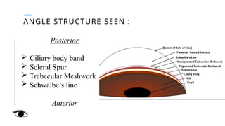
- 7. ANGLE STRUCTURE SEEN : Posterior ’āś Ciliary body band ’āś Scleral Spur ’āś Trabecular Meshwork ’āś SchwalbeŌĆÖs line Anterior
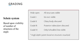
- 9. GRADING Scheie system Based upon visibility of number of structures of the angle.
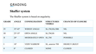
- 10. GRADING Shaffer system The Shaffer system is based on angularity. GRADE ANGLE CONFIGURATION STRUCTURES VISIBLE CHANCES OF CLOSURE IV 35┬║-45┬║ WIDEST ANGLE SL,TM,SS,CBB NIL III 25┬║-35┬║ OPEN ANGLE SL,TM,SS NIL II 20┬║ MODERATELY OPEN SL,TM POSSIBLE I 10┬║ VERY NARROW SL, anterior TM HIGHLY LIKELY 0 0┬║ CLOSED NONE CLOSED
- 11. APPLICATION OF GONIOSCOPY: ’ü▒Classification of Glaucoma ’ü▒Localization of foreign bodies, abnormal blood vessels, tumors in angle ’ü▒Demonstration of Peripheral Anterior Synechiae ’ü▒Surgical aid in goniotomy CONTRAINDICATION OF GONIOSCOPY: ’ü▒Hyphaema ’ü▒Compromised cornea (e.g., corneal ulcer) ’ü▒Lacerated or perforated globe
- 12. Thank you


