Definition of pulpitis, Factors causing injury to the pulp, aerodontalgia, classification of pulpitis, clinical features of various types of pulpitis, histopathology and its treatment are inlisted in this presentation.
The dental pulp is a soft tissue located within the center of a tooth that is surrounded by rigid dental structures. It contains blood vessels and nerves that make it highly sensitive. When bacteria enter through decay or cracks, they can cause inflammation of the pulp, known as pulpitis. Pulpitis can be reversible if caught early through mild symptoms like short pain from hot and cold. But over time, the inflammation can become irreversible as the bacteria spread, causing spontaneous, lingering pain that is worse when lying down and radiates elsewhere. This damages the pulp irreversibly.
This document discusses various pathologies that can affect the dental pulp and their sequels. It begins with definitions of pulp and pulpitis, describing pulpitis as an inflammatory response to noxious stimuli. Pulpitis is classified as reversible or irreversible. Causes and risk factors of pulpitis include mechanical, thermal, chemical, and bacterial factors. Sequels of untreated pulpitis include pulp necrosis, periapical abscesses, and periapical lesions such as granulomas or scars. Other topics covered include pulp degeneration, calcification, polyps, and dry socket. Throughout, the document provides details on clinical features, mechanisms, management approaches, and importance of prevention for these dental pulp conditions and their outcomes
The document discusses pulp and periapical diseases. It begins by describing the pathobiology of the periapex and classifying pulpoperipaical pathoses. It then discusses the classifications of Franklin Weine and the WHO for pulpoperipaical lesions. Key lesions discussed include acute and chronic apical periodontitis, periapical granuloma, periapical cyst, condensing osteitis, and Ludwig's angina. Differential diagnoses and clinical signs and symptoms are also covered.
1. Root canal treatment is needed when the pulp becomes inflamed or infected due to deep dental caries, leaky fillings, trauma, or excessive orthodontic forces.
2. The pulp can progress from a reversible inflammation to an irreversible one where recovery is not expected, and eventually become necrotic with complete breakdown of cellular organization and no reparative potential.
3. Treatment depends on the pulpal status and ranges from removal of irritants for reversible pulpitis to complete root canal instrumentation and filling for irreversible pulpitis or necrotic pulp to relieve symptoms and prevent or treat apical periodontitis.
This document discusses endodontic diagnosis and treatment planning. It begins by introducing endodontics and describing common causes of pulpitis like decay, trauma, and infection. Signs and symptoms of pulpitis include tooth pain from hot/cold, pressure, and swelling. Diagnosis involves subjective questions to the patient and objective examination of the tooth. Diagnostic tests include percussion, palpation, thermal sensitivity testing, electric pulp testing, and radiographs. Based on the diagnostic findings, the dentist determines if the pulp is normal, inflamed with reversible or irreversible pulpitis, or non-vital. The treatment plan is tailored to the diagnosis but commonly involves accessing the root canal, cleaning and shaping it, and filling
This document discusses endodontic diagnosis and treatment planning. It begins with an introduction to endodontics and causes of pulpitis. Signs and symptoms of pulpitis are then outlined. The diagnostic process involves subjective history, objective examination, and tests like percussion, palpation, thermal sensitivity, electric pulp testing, and radiographs. Based on the diagnosis, a treatment plan is formulated which may involve root canal treatment, referral, or extraction. The document provides details on diagnosing and treating different pulpal and periapical conditions like reversible/irreversible pulpitis, abscesses, cysts, and necrosis.
This document discusses diseases of the dental pulp, including pulpitis and its causes and classifications. Pulpitis, or inflammation of the dental pulp, is most commonly caused by microorganisms entering through caries or trauma. It can be acute or chronic and partial or total. Acute pulpitis causes severe pain from thermal changes. Chronic pulpitis may be asymptomatic or cause dull pain. Left untreated, pulpitis can lead to pulp necrosis and spread of infection to the periapical area, potentially causing a periapical abscess.
@Microbiology of pulp & periradicular pathosis part 1 2024.pptxismasajjad1
油
This document discusses endodontic microbiology and routes of microbial entry into the pulp and periradicular tissues, including caries, trauma, restorative procedures, periodontal disease, and more. It classifies endodontic infections as intraradicular or extraradicular and describes primary, secondary, and persistent intraradicular infections. Various microbial species involved in endodontic infections are mentioned. Pulpal diseases including reversible pulpitis, irreversible pulpitis, pulp necrosis, and previously treated teeth are defined. Periradicular lesions and non-endodontic lesions that can mimic periradicular pathosis are also outlined.
The document discusses various periapical diseases including their causes, symptoms, diagnosis and treatment. It states that pulpal inflammation can cause inflammatory changes in the periodontal ligament even before the pulp becomes necrotic due to their interrelationship. Microorganisms and their byproducts from infected or necrotic pulps can spread to the periapical area through various canals and cause inflammatory or immune reactions leading to both acute and chronic periapical diseases.
dental caries is the most common disease with which the tooth gets affected *ps if you treat the tooth like a human being and the pulp as the body parts so what happens to the body part of the tooth when the infection reaches to them ?
how would they react?
The document summarizes the response of the dental pulp to caries and other insults. It discusses how bacteria from caries produce toxins that trigger an inflammatory response in the pulp. Over time this can lead to pulpal disease. It also examines other sources of bacteria that may reach the pulp, such as periodontal disease or cracks. The document outlines the histologic changes that occur in the pulp at different stages of caries. It discusses factors like blood flow and interstitial fluid pressure that are altered during pulpal inflammation. The roles of the sympathetic and sensory nervous systems in modulating the inflammatory response are described. Mechanisms of pulpal repair and factors that encourage or limit repair are summarized. Potential iatrogenic effects
This document discusses dental emergencies and provides information on various conditions including dental caries, pulpitis, periapical abscesses, periodontitis, traumatic injuries like crown fractures and avulsions. It also covers topics like drainage techniques, alveolar osteitis, deep space infections, pediatric dental considerations. Nontraumatic emergencies involve conditions like tooth decay and resulting pulp inflammation or bone infection. Traumatic emergencies include tooth fractures, luxations, intrusions or avulsions from dental injury. The summary emphasizes that emergency management is temporary, communicating with consultants is important, and infections should be drained when possible while recognizing true dental versus medical emergencies.
Diagnosis Of Pulpal Pathology In PedodonticsDr. Shirin
油
This document provides an overview of dental pulp and classifications of pulpal diseases. It discusses the characteristics and clinical findings of normal pulp, reversible pulpitis, irreversible pulpitis, pulpal necrosis, and other conditions. A variety of diagnostic procedures and tests are also described, including reviewing history, clinical examination, percussion, radiographs, as well as newer pulp testing methods like laser Doppler flowmetry and pulse oximetry. The conclusion emphasizes the importance of gathering all available information to make an accurate diagnosis prior to providing endodontic treatment or other dental procedures.
The document discusses diseases of the dental pulp, including pulpitis and necrosis. It defines the pulp as the formative organ of the tooth that builds dentin. Pulpitis is the most common cause of dental pain and can be reversible or irreversible depending on the severity of inflammation. Untreated pulpitis can lead to necrosis or death of the pulp. Causes include mechanical, thermal, chemical, and bacterial factors. Management involves removal of irritants and root canal treatment if needed.
This document discusses diseases of the dental pulp, including causes, classifications, symptoms, diagnosis, and treatment. It covers inflammatory pulp diseases like reversible and irreversible pulpitis, as well as pulp degeneration conditions such as calcific degeneration and necrosis. Physical, chemical, and bacterial factors can cause pulp diseases. Classification includes inflammatory, degenerative, and necrotic categories. Symptoms, diagnosis, and treatment options vary depending on the specific pulp condition.
This document provides an overview of pulp therapy in deciduous and young permanent teeth. It discusses the histology and structure of the dental pulp, how the pulp reacts to dental caries and operative procedures, diagnosis and evaluation of pulp pathology, and techniques for indirect and direct pulp capping. The goal of pulp therapy is to maintain pulp vitality and integrity whenever possible by using appropriate medicaments and materials to encourage healing of the exposed pulp. Factors like the depth and rate of the carious lesion, use of proper cooling and gentle technique during procedures can impact the pulp's reaction and healing.
As heart is to the body, the pulp is to the tooth, providing a constant source of nutrition to maintain the vitality of a tooth. Every precaution should be taken to preserve vitality of the pulp. A simple dental infection if neglected , can proceed to life threatening complications. So early detection , early treatment and early prevention is very important.
This document discusses the management of tooth pulp. It describes the pulp as the formative organ that builds dentin during tooth development and after eruption. It also discusses patient history, clinical exam, categories of pulp pathology, types of pulpal pain, and techniques for managing deep carious lesions, including indirect pulp capping, direct pulp capping, and using corticosteroid-antibiotic pastes. The goal of pulp capping techniques is to protect the pulp from bacterial contamination if exposed and encourage reparative dentin formation.
The document discusses various pathologies that can affect the dental pulp, including pulpitis, pulp necrosis, and pulp degeneration. It defines reversible and irreversible pulpitis, and notes their causes, symptoms, diagnosis, and treatments. Pulp necrosis occurs when the pulp undergoes cell death due to noxious insults like bacteria or trauma. Various forms of pulp degeneration are also outlined. The document provides an overview of common pulp conditions and classifications.
Here discussing various cases of Obstructive jaundice namely Choledocholithiassis, Biliary atresia, Carcinoma Pancreas, Periampullary Carcinoma and Cholangiocarcinoma.
More Related Content
Similar to oral pathology 4-Periapical Pathology 4.ppt (20)
This document discusses diseases of the dental pulp, including pulpitis and its causes and classifications. Pulpitis, or inflammation of the dental pulp, is most commonly caused by microorganisms entering through caries or trauma. It can be acute or chronic and partial or total. Acute pulpitis causes severe pain from thermal changes. Chronic pulpitis may be asymptomatic or cause dull pain. Left untreated, pulpitis can lead to pulp necrosis and spread of infection to the periapical area, potentially causing a periapical abscess.
@Microbiology of pulp & periradicular pathosis part 1 2024.pptxismasajjad1
油
This document discusses endodontic microbiology and routes of microbial entry into the pulp and periradicular tissues, including caries, trauma, restorative procedures, periodontal disease, and more. It classifies endodontic infections as intraradicular or extraradicular and describes primary, secondary, and persistent intraradicular infections. Various microbial species involved in endodontic infections are mentioned. Pulpal diseases including reversible pulpitis, irreversible pulpitis, pulp necrosis, and previously treated teeth are defined. Periradicular lesions and non-endodontic lesions that can mimic periradicular pathosis are also outlined.
The document discusses various periapical diseases including their causes, symptoms, diagnosis and treatment. It states that pulpal inflammation can cause inflammatory changes in the periodontal ligament even before the pulp becomes necrotic due to their interrelationship. Microorganisms and their byproducts from infected or necrotic pulps can spread to the periapical area through various canals and cause inflammatory or immune reactions leading to both acute and chronic periapical diseases.
dental caries is the most common disease with which the tooth gets affected *ps if you treat the tooth like a human being and the pulp as the body parts so what happens to the body part of the tooth when the infection reaches to them ?
how would they react?
The document summarizes the response of the dental pulp to caries and other insults. It discusses how bacteria from caries produce toxins that trigger an inflammatory response in the pulp. Over time this can lead to pulpal disease. It also examines other sources of bacteria that may reach the pulp, such as periodontal disease or cracks. The document outlines the histologic changes that occur in the pulp at different stages of caries. It discusses factors like blood flow and interstitial fluid pressure that are altered during pulpal inflammation. The roles of the sympathetic and sensory nervous systems in modulating the inflammatory response are described. Mechanisms of pulpal repair and factors that encourage or limit repair are summarized. Potential iatrogenic effects
This document discusses dental emergencies and provides information on various conditions including dental caries, pulpitis, periapical abscesses, periodontitis, traumatic injuries like crown fractures and avulsions. It also covers topics like drainage techniques, alveolar osteitis, deep space infections, pediatric dental considerations. Nontraumatic emergencies involve conditions like tooth decay and resulting pulp inflammation or bone infection. Traumatic emergencies include tooth fractures, luxations, intrusions or avulsions from dental injury. The summary emphasizes that emergency management is temporary, communicating with consultants is important, and infections should be drained when possible while recognizing true dental versus medical emergencies.
Diagnosis Of Pulpal Pathology In PedodonticsDr. Shirin
油
This document provides an overview of dental pulp and classifications of pulpal diseases. It discusses the characteristics and clinical findings of normal pulp, reversible pulpitis, irreversible pulpitis, pulpal necrosis, and other conditions. A variety of diagnostic procedures and tests are also described, including reviewing history, clinical examination, percussion, radiographs, as well as newer pulp testing methods like laser Doppler flowmetry and pulse oximetry. The conclusion emphasizes the importance of gathering all available information to make an accurate diagnosis prior to providing endodontic treatment or other dental procedures.
The document discusses diseases of the dental pulp, including pulpitis and necrosis. It defines the pulp as the formative organ of the tooth that builds dentin. Pulpitis is the most common cause of dental pain and can be reversible or irreversible depending on the severity of inflammation. Untreated pulpitis can lead to necrosis or death of the pulp. Causes include mechanical, thermal, chemical, and bacterial factors. Management involves removal of irritants and root canal treatment if needed.
This document discusses diseases of the dental pulp, including causes, classifications, symptoms, diagnosis, and treatment. It covers inflammatory pulp diseases like reversible and irreversible pulpitis, as well as pulp degeneration conditions such as calcific degeneration and necrosis. Physical, chemical, and bacterial factors can cause pulp diseases. Classification includes inflammatory, degenerative, and necrotic categories. Symptoms, diagnosis, and treatment options vary depending on the specific pulp condition.
This document provides an overview of pulp therapy in deciduous and young permanent teeth. It discusses the histology and structure of the dental pulp, how the pulp reacts to dental caries and operative procedures, diagnosis and evaluation of pulp pathology, and techniques for indirect and direct pulp capping. The goal of pulp therapy is to maintain pulp vitality and integrity whenever possible by using appropriate medicaments and materials to encourage healing of the exposed pulp. Factors like the depth and rate of the carious lesion, use of proper cooling and gentle technique during procedures can impact the pulp's reaction and healing.
As heart is to the body, the pulp is to the tooth, providing a constant source of nutrition to maintain the vitality of a tooth. Every precaution should be taken to preserve vitality of the pulp. A simple dental infection if neglected , can proceed to life threatening complications. So early detection , early treatment and early prevention is very important.
This document discusses the management of tooth pulp. It describes the pulp as the formative organ that builds dentin during tooth development and after eruption. It also discusses patient history, clinical exam, categories of pulp pathology, types of pulpal pain, and techniques for managing deep carious lesions, including indirect pulp capping, direct pulp capping, and using corticosteroid-antibiotic pastes. The goal of pulp capping techniques is to protect the pulp from bacterial contamination if exposed and encourage reparative dentin formation.
The document discusses various pathologies that can affect the dental pulp, including pulpitis, pulp necrosis, and pulp degeneration. It defines reversible and irreversible pulpitis, and notes their causes, symptoms, diagnosis, and treatments. Pulp necrosis occurs when the pulp undergoes cell death due to noxious insults like bacteria or trauma. Various forms of pulp degeneration are also outlined. The document provides an overview of common pulp conditions and classifications.
Here discussing various cases of Obstructive jaundice namely Choledocholithiassis, Biliary atresia, Carcinoma Pancreas, Periampullary Carcinoma and Cholangiocarcinoma.
Local Anesthetic Use in the Vulnerable PatientsReza Aminnejad
油
Local anesthetics are a cornerstone of pain management, but their use requires special consideration in vulnerable groups such as pediatric, elderly, diabetic, or obese patients. In this presentation, well explore how factors like age and physiology influence local anesthetics' selection, dosing, and safety. By understanding these differences, we can optimize patient care and minimize risks.
Best Sampling Practices Webinar USP <797> Compliance & Environmental Monito...NuAire
油
Best Sampling Practices Webinar USP <797> Compliance & Environmental Monitoring
Are your cleanroom sampling practices USP <797> compliant? This webinar, hosted by Pharmacy Purchasing & Products (PP&P Magazine) and sponsored by NuAire, features microbiology expert Abby Roth discussing best practices for surface & air sampling, data analysis, and compliance.
Key Topics Covered:
鏝 Viable air & surface sampling best practices
鏝 USP <797> requirements & compliance strategies
鏝 How to analyze & trend viable sample data
鏝 Improving environmental monitoring in cleanrooms
・ Watch Now: https://www.nuaire.com/resources/best-sampling-practices-cleanroom-usp-797
Stay informedfollow Abby Roth on LinkedIn for more cleanroom insights!
Stability of Dosage Forms as per ICH GuidelinesKHUSHAL CHAVAN
油
This presentation covers the stability testing of pharmaceutical dosage forms according to ICH guidelines (Q1A-Q1F). It explains the definition of stability, various testing protocols, storage conditions, and evaluation criteria required for regulatory submissions. Key topics include stress testing, container closure systems, stability commitment, and photostability testing. The guidelines ensure that pharmaceutical products maintain their identity, purity, strength, and efficacy throughout their shelf life. This resource is valuable for pharmaceutical professionals, researchers, and regulatory experts.
An X-ray generator is a crucial device used in medical imaging, industry, and research to produce X-rays. It operates by accelerating electrons toward a metal target, generating X-ray radiation. Key components include the X-ray tube, transformer assembly, rectifier system, and high-tension circuits. Various types, such as single-phase, three-phase, constant potential, and high-frequency generators, offer different efficiency levels. High-frequency generators are the most advanced, providing stable, high-quality imaging with minimal radiation exposure. X-ray generators play a vital role in diagnostics, security screening, and industrial testing while requiring strict radiation safety measures.
1. Explain the physiological control of glomerular filtration and renal blood flow
2. Describe the humoral and autoregulatory feedback mechanisms that mediate the autoregulation of renal plasma flow and glomerular filtration rate
This presentation provides a detailed exploration of the morphological and microscopic features of pneumonia, covering its histopathology, classification, and clinical significance. Designed for medical students, pathologists, and healthcare professionals, this lecture differentiates bacterial vs. viral pneumonia, explains lobar, bronchopneumonia, and interstitial pneumonia, and discusses diagnostic imaging patterns.
Key Topics Covered:
Normal lung histology vs. pneumonia-affected lung
Morphological changes in lobar, bronchopneumonia, and interstitial pneumonia
Microscopic features: Fibroblastic plugs, alveolar septal thickening, inflammatory cell infiltration
Stages of lobar pneumonia: Congestion, Red hepatization, Gray hepatization, Resolution
Common causative pathogens (Streptococcus pneumoniae, Klebsiella pneumoniae, Mycoplasma, etc.)
Clinical case study with diagnostic approach and differentials
Who Should Watch?
This is an essential resource for medical students, pathology trainees, and respiratory health professionals looking to enhance their understanding of pneumonias morphological aspects.
Pharm test bank- 12th lehne pharmacology nursing classkoxoyav221
油
A pediatric nursing course is designed to prepare nursing students to provide specialized care for infants, children, and adolescents. The course integrates developmental, physiological, and psychological aspects of pediatric health and illness, emphasizing family-centered care. Below is a detailed breakdown of what you can expect in a pediatric nursing course:
1. Course Overview
Focuses on growth and development, health promotion, and disease prevention.
Covers common pediatric illnesses and conditions.
Emphasizes family dynamics, cultural competence, and ethical considerations in pediatric care.
Integrates clinical skills, including medication administration, assessment, and communication with children and families.
2. Key Topics Covered
A. Growth and Development
Neonates (0-28 days): Reflexes, feeding patterns, thermoregulation.
Infants (1 month - 1 year): Milestones, immunization schedule, nutrition.
Toddlers (1-3 years): Language development, toilet training, injury prevention.
Preschoolers (3-5 years): Cognitive and social development, school readiness.
School-age children (6-12 years): Psychosocial development, peer relationships.
Adolescents (13-18 years): Puberty, identity formation, risk-taking behaviors.
B. Pediatric Assessment
Head-to-toe assessment in children (differences from adults).
Vital signs (normal ranges vary by age).
Pain assessment using age-appropriate scales (FLACC, Wong-Baker, Numeric).
C. Pediatric Disease Conditions
Respiratory disorders: Asthma, bronchiolitis, pneumonia, cystic fibrosis.
Cardiac conditions: Congenital heart defects, Kawasaki disease.
Neurological disorders: Seizures, meningitis, cerebral palsy.
Gastrointestinal disorders: GERD, pyloric stenosis, intussusception.
Endocrine conditions: Diabetes mellitus type 1, congenital hypothyroidism.
Hematologic disorders: Sickle cell anemia, hemophilia, leukemia.
Infectious diseases: Measles, mumps, rubella, chickenpox.
Mental health concerns: Autism spectrum disorder, ADHD, eating disorders.
D. Pediatric Pharmacology
Medication administration (oral, IV, IM, subcutaneous).
Weight-based dosing calculations (mg/kg).
Common pediatric medications (antibiotics, analgesics, vaccines).
Parenteral nutrition and fluid management.
E. Pediatric Emergency & Critical Care
Pediatric Advanced Life Support (PALS) basics.
Recognizing signs of deterioration (early vs. late signs).
Shock, dehydration, respiratory distress management.
F. Family-Centered Care & Communication
Parental involvement in care decisions.
Therapeutic communication with children at different developmental stages.
Cultural considerations in pediatric care.
G. Ethical and Legal Issues in Pediatric Nursing
Informed consent for minors.
Mandatory reporting of abuse and neglect.
Palliative care and end-of-life considerations in pediatrics.
3. Clinical Component
Hands-on experience in pediatric hospital units, clinics, or community settings.
Performing assessments and interventions under supervision.
Case study disc
3. Introduction
Introduction
Calcified structure of the tooth protects the
vital soft tissue ( pulp) but Caries and
trauma and other things may be breached
the protective calcified of the tooth.
Dental caries can facilitate and leads to
opening of pathways to various microbes
The pulp tissue resist as inflammation
response which leads pulpitis
4. Conti------
Conti------
Once pulp is exposed the microbes invade
the pulp then breakdown of pulp tissue
The pulp loses its vitality
An inflammatory is mounted while the
microbes multiply .
microbes multiply .
The inflammatory of the pulp is followed
by death or nicrosis
5. contii
contii
The presence of microbes and their toxins
in the root canal can evokes an
inflammatory response in the periapical
area and thus inflammation extends to the
periapical area.
periapical area.
6. Types of disorders of pulp and
Types of disorders of pulp and
periapical
periapical
Pulpits
Pulpits
Necrotic of pulp
Necrotic of pulp
Pulp degeneration /pulp denticles /pulp stones
and pulp calcifications
Acute apical periodontitis of pulpal origin
Periapical abscess
Periapical abscess
Chronic apical periodontitis
Periapical abscess with sinus
Radicular cyst
Radicular cyst
others
others
7. Causes
Causes
The causes of pulpal disorder can be
physical, chemical and bacteria.
The dental caries is the commonest cause
of pulpal disease
of pulpal disease
8. Pulpitis =
Pulpitis = Inflammation of the Pulp
Inflammation of the Pulp
Irritant
Irritant
Chemical irritation
Chemical irritation
Fillings
Fillings
Erosion
Erosion
Bleaching
Bleaching
Thermal changes
Thermal changes
Uninsulated large fillings
Drilling
Drilling
Mechanical damage
Mechanical damage
Trauma
Trauma
Bruxism
Bruxism
Attrition
Attrition
Abrasion
Abrasion
Direct irritation
Direct irritation
Bacterial irritation from
caries
caries
Cracked tooth
Cracked tooth
Root fractures
Root fractures
Immune response
Immune response
Chemical mediators that
initiate inflammation
9. Microbial Irritant
Microbial Irritant
Microbes produce toxins
Initially pulp is infiltrated by chronic
inflammatory cells
inflammatory cells
Macrophages, lymphocytes & plasma cells
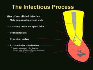
10. The Infectious Process
The Infectious Process
Sites of established infection
Main pulp canal space and walls
Accessory canals and apical delta
Dentinal tubules
Dentinal tubules
Cementum surface
Cementum surface
Extraradicular colonizations
Relative importance? few data, but
the root canal infection is of course paramount
Brynolf 1966, Langeland et al. 1977
11. The Infectious Process
The Infectious Process
Pulpitis Necrosis
Canal
infection
Apical
periodontitis
Time
Spread to
apex
Increasing infectious load;
increasingly difficult to treat
12. Inflammation =>
Inflammation => Necrosis
Necrosis
Pulp can impede spread of infection
Factors
Factors
Virulence of bacteria
Ability of pulp to release inflammatory factors to
prevent increase in intrapulpal pressure
Host resistance
Host resistance
Lymph drainage
Lymph drainage
Necrosis: coronal => apical
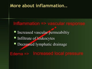
13. Inflammation =>
Edema =>
vascular response
Increased local pressure
More about inflammation..
Increased vascular permeabiltiy
Infiltrate of leukocytes
Decreased lymphatic drainage
14. So whats really going on?
So whats really going on?
Pulp is enclosed within calcified walls
Low compliance system
Circulation slows due to compression of venous return
Odontoblasts are altered or destroyed
Increase in tissue pressure
Compression of venules in area of injury
Progresses coronal => Apex
15. The sum total of the
The sum total of the
inflammatory response may
cause more damage that the
initial irritants alone!
16. PULPITIS
PULPITIS
Inflammation of dental pulp
Main source for dental pain
Causes
Causes
Dental caries- the
Dental caries- the most common
most common cause
cause
Traumatic exposure to pulp
Repeated dental procedure
17. Pathogenesis
Pathogenesis
Any causes above
Any causes above
Exposure to pulp
Exposure to pulp
Invasion by bacteria- Streptococus
Inflammation of pulp
18. Types
Types
Acute (rapid, severe onset, short duration)
Open communication between pulp cavity & oral cavity)
closed
closed
Chronic (slow , long duration, mild pain)
Open
Open
Closed
Closed
19. Acute-Closed
Acute-Closed
Micro organism- virulent & large no.
Clinical features
Clinical features
Early stage - Hypersensitivity to hot & cold
Later more persistent
Pain- sharp, severe & stabbing
Sometimes not localized
Tender
Tender
Tooth discoloration
Tooth discoloration
Swelling of gum
Swelling of gum
20. Acute-Open
Acute-Open
Common
Common
Acute exposure with micro organisms
Occurs at late stage of caries
Abscess formed drain out of cavity
21. Clinical features
Clinical features
Hypersensitivity hot & cold in early stage
Less pain
Less pain
Slight tender
Slight tender
22. Chronic-closed
Chronic-closed
No communication B/W pulp & oral cavity
Pulp tissue destruction at the site of micro organism entry
Infection remains localized for long time with remaing
pulp tissue intact or destruction occurs slowly
Clinical features
Clinical features
Hypersensitivity
Hypersensitivity
Early stage- to hot and cold
Late stage- only to hot but relieved by cold
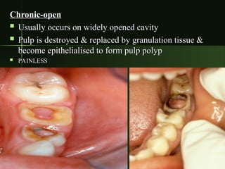
23. Chronic-open
Chronic-open
Usually occurs on widely opened cavity
Pulp is destroyed & replaced by granulation tissue &
become epithelialised to form pulp polyp
PAINLESS
PAINLESS
24. Diagnosis
Diagnosis
Test of healthy pulp
Tapping of tooth directly
Sensitivity if present indicates the spread of inflammation
to surrounding tissue
hot & cold sensitivity
If pain persists even after stimulus removal or
Pain persists spontaneously
Pulp may not be healthy to save
25. Test of pulp- dead or alive
Test of pulp- dead or alive
Electric pulp tester
It helps to recognize the pulp whether its alive or
dead but
dead but
If person feels the electric charge delivered to the
tooth the pulp is alive
26. Treatment
Treatment
If pulp viable just remove irritant n healed itself
Removal of caries n restoration by filling
If pulp dead
If pulp dead
RCT
RCT
Tooth extraction
Tooth extraction
antibiotic is given- penicillin in acute cases
27. Time-Course of Apical Peridontitis
Time-Course of Apical Peridontitis
Dynamics of pulpal infection
Bacterial succession and variations in
virulence and pathogenicity
Host factors modulating inflammation
and spread of the infection
Ultimate consequences of root canal
infection
infection
28. Microbes
Microbes
Type:
Type:
Dependent on the environment, nutrients, and
competition
competition
Primary infection:
Primary infection:
Obligate anaerobes and Gram Negative bacteria.
Secondary infection:
Facultative and Gram Positive bacteria. Including E.
Faecalis and candida.
Baumgartner
29. Inflammation of the periapical region
Inflammation of the periapical region
Relationship between pulpal and periapical
pathosis
pathosis
Periapical pathology follows pulp pathology
Periapical disease meets a more effective
resistance that pulpal disease
Repair is more often achieved
30. From Pulpal to Periapical Pathosis
From Pulpal to Periapical Pathosis
31. Periapical Pathosis
Periapical Pathosis
Bacterial endotoxins & inflammatory
mediators trigger surrounding immune cells
Defense cells
Defense cells
Prevent spread of infection into bone
32. Periapical Pathosis
Periapical Pathosis
Bone is replaced by highly vascularized
inflammatory tissue which can much better
eliminate invading microbes than the original
bone tissue could have.
35. Symptomatic Apical Periodontitis
Symptomatic Apical Periodontitis
Clinical features
Clinical features
Localized
Localized
Frequently spontaneous
Intense throbbing pain
Painful to touch
Painful to touch
None to minimal swelling
36. Symptomatic Apical Periodontitis
Symptomatic Apical Periodontitis
Histology
Histology
Inflammation of the PDL with acute and
chronic inflammatory cells
X-ray exam
X-ray exam
no change to slight thickening of
periodontal membrane
Treatment
Treatment
RCT or extraction
RCT or extraction
37. Asymptomatic Apical Periodontitis
Asymptomatic Apical Periodontitis
Clinical features
Clinical features
Represents a
Represents a
stand-off
stand-off
between local
between local
resistance and noxious stimuli
Indicative of pulpal necrosis
Common
Common
Painless
Painless
Slow growing
Slow growing
May transform into a cyst or granuloma
38. Asymptomatic Apical Periodontitis
Asymptomatic Apical Periodontitis
Histology
Histology
Proliferation of fibroblasts and endothelial
cells
cells
Lymphocytes, plasma cells and phagocytes
Foam cells and cholesterol clefts
Epithelial rest of Malassez
X-ray
X-ray
Large radiolucency up to 1cm
Treatment => RCTx or extraction
40. Periapical Abscess
Periapical Abscess
A localized collection of pus in a cavity
formed by the disintegration of tissues.
Indicative of pupal death
Type is based on the degree of exudate
formation, severity of pain and the presence
of symptoms
of symptoms
Symptomatic apical abscess
Asymptomatic apical abscess
41. Periapical Abscess
Periapical Abscess
Clinical features
Clinical features
Rapid onset of extreme pain
Painful to percussion
Not localized adjacent teeth can be painful
SWELLING present
SWELLING present
Sinus tract can form
Potentially life threatening
42. Periapical Abscess
Periapical Abscess
Histology
Histology
Resembles and acute apical periodontitis
Involvement of the adjacent bone and soft tissue
Pus and tissue necrosis
X-ray
X-ray
Widened PDL to large alveolar radiolucency
Treatment
Treatment
Rx for antibiotics
Rx for antibiotics
Establish drainage
Establish drainage
43. Untreated Apical Abscess
Untreated Apical Abscess
Cellulitis
Cellulitis
Infection travels through the facial planes of least resistance
Fever
Fever
Osteomyelitis
Osteomyelitis
Infection within bone through the medullary spaces
Parulis =
Parulis =
gum boil
gum boil
Ludwig
Ludwig
s angina
s angina
Swelling in floor of mouth elevates tongue and blocks
airway
airway
Cavernous sinus thrombosis
Infection from MX premolars and molars extends into the
cranial vault
cranial vault
45. Spread of infection
Spread of infection
The path of least resistance
Buccal plate is the most common route due to
the thin buccal bone
Outside on face
Outside on face
Palate
Palate
Neck below mylohyoid
PDL
PDL
Pulp canal
Pulp canal
Maxillary sinus
Maxillary sinus
Mandibular canal
Mandibular canal
51. Apical Periodontal Cyst / Granuloma
Apical Periodontal Cyst / Granuloma
Clinical features
Clinical features
The most common cyst of the jaws
May be asymptomatic of become
symptomatic
symptomatic
Slow continuous enlargement
X-ray
X-ray
Well-circumscribed radiolucency
Associated with apices of teeth
May cause resorption of teeth and bone
52. Apical Periodontal Cyst
Apical Periodontal Cyst
Histology
Histology
Inflammatory cells
Inflammatory cells
Prominent epithelial lining without keratin
Body of cyst filled with semifluid material
Treatment => Usually require apical
surgery if persistant
54. Apical Cyst vs. Granuloma
Apical Cyst vs. Granuloma
A cyst is
A cyst is lined by squamous epithelium
lined by squamous epithelium and containing
and containing
necrotic material in the lumen. The cyst wall or capsule
contains dense fibrous connective tissue with slight chronic
inflammation and cholesterin slits surrounded by foreign
body-type giant cells. There are "foam" cells in the epithelial
lining.
lining.
A lesion with
A lesion with highly vascular tissue
highly vascular tissue containing macrophages,
containing macrophages,
fibroblasts, collagen, and immune cells (neutrophils, plasma
cells, T and B cells, lymphocytes, eosinophils
56. condensing osteitis
condensing osteitis
Bone sclerosis around apices of tooth with pulpitis
Occurs when there is high tissue resistance to low
grade infection
grade infection
Clinical features
Clinical features
Adolescents and young adults
Most common in mandibular first molars
Tooth usually has large carious lesion
No symptoms
No symptoms
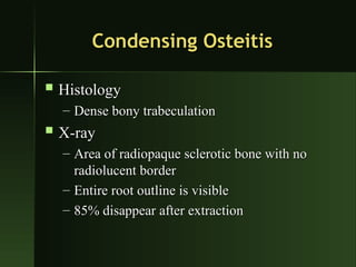
57. Condensing Osteitis
Condensing Osteitis
Histology
Histology
Dense bony trabeculation
X-ray
X-ray
Area of radiopaque sclerotic bone with no
radiolucent border
radiolucent border
Entire root outline is visible
85% disappear after extraction
58. Condensing Osteitis
Condensing Osteitis
Treatment
Treatment
None
None
RCTx
RCTx
Bone Scar
Bone Scar
The residual area of condensing osteitis that remains
after resolution of inflammation
Differential diagnosis
Idiopathic osteosclerosis
Periapical cemental dysplasia
60. Osteomyelitis
Osteomyelitis
If the periapical infection and inflammation extend
through the marrow spaces of the jaw, the result is
osteomyelitis. In this case, you can identify the offending
tooth causing the diffuse and irregular bone destruction.