ºÝºÝߣ

ºÝºÝߣShare a Scribd company logo
PATHOLOGY:
 Arteriosclerosis


nianderthalNOTES
ARTERIOSCLEROSIS
DEF:
-hardening of the arteries
-generic term reflecting arterial wall thickening and
  loss of elasticity

THREE GENERAL PATTERNS:
1. Arteriolosclerosis
2. Monckeberg medial sclerosis
3. Atherosclerosis
1. Arteriolosclerosis
-affects small arteries and arterioles
-anatomic variants:
a. hyaline
b. hyperplastic
2. Monckeberg medial sclerosis
-has calcific deposits in muscular arteries that
   may undergo metaplasia to bone
-lesions do not encroach vessel lumen thus NOT
   CLINICALLY SIGNIFICANT
3. Atherosclerosis
-most frequent and clinically important pattern
-characterized by intimal lesions called
  ATHEROMAS that protrude into the vessel
  lumen
-consists of a raised lesion:
a. Grumuous Core – made of lipid, (cholesterol
   or cholesterol ester) yellow, soft
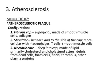
b. Fibrous Cap – white
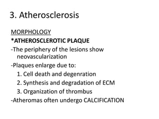
3. Atherosclerosis
EPIDEMIOLOGY
-Risk factors have a multiplicative effect
-Constitutional Risk Factors in IHD
a. Age – clinically manifests middle age or later
b. Gender – premenopausal women are relatively
    protected due to favorable influence of estrogen
              -but clinical trials have failed to demonstrate
    any utility of hormonal therapy for vascular diseases
    prevention
a. Genetics – MOST SIGNIFICANT INDEPENDENT RISK
    FACTOR
3. Atherosclerosis
EPIDEMIOLOGY
-Modifiable Risk Factors in IHD
a. Hyperlipidemia – esp. hypercholesterolemia,
   increased LDL, decreased HDL, increased
   lipoprotein a
*STATINS – a class of drugs that lower circulating
   cholesterol levels by inhibiting HMG-Coa
   reductase, the rate-limiting enzyme in
   hepatic cholesterol biosynthesis
3. Atherosclerosis
EPIDEMIOLOGY
-Modifiable Risk Factors in IHD
b. Hypertension – both systolic and diastolic
   components are important
   - MOST IMPORTANT CAUSE OF LEFT
   VENTRICULAR HYPERTROPHY
c. Cigarette smoking – prolonged smoking of
   one pack of cigarettes or more daily doubles
   the death rate from IHD
3. Atherosclerosis
EPIDEMIOLOGY
-Modifiable Risk Factors in IHD
d. Diabetes mellitus – induces
   hypercholesterolemia
3. Atherosclerosis
EPIDEMIOLOGY
-Additional Risk Factors
a. Inflammation – present during all stages of
   atherogenesis and is intimately linked with
   atherosclerotic plaque formation and rupture
*C-reactive Protein (CRP) has emerged as one of
   the simplest and most sensitive inflammatory
   markers; decreased by STATINS
3. Atherosclerosis
EPIDEMIOLOGY
-Additional Risk Factors
b. Hyperhomocysteinemia – seen in CAD, PAD,
   stroke and venous thrombosis
   -elevated homocysteine levels can be caused
   by low folate and Vitamin B12 intake
c. Metabolic syndrome
3. Atherosclerosis
EPIDEMIOLOGY
-Additional Risk Factors
d. Lipoprotein (a) – an altered form of LDL,
   associated with CAD and CVD
e. Factors affecting Hemostasis
f. Other Factors – lack of exercise; competitive
   stressful lifestyle (type A personality); obesity
3. Atherosclerosis
PATHOGENESIS
Hypotheses:
a. Intimal Cellular Proliferation
b. Repetitive Formation and Organization of Thrombi
c. Response-To-Injury
   -views atherosclerosis as a chronic inflammatory and
   healing response of the arterial wall to injury
   -lesion progression occurs through the interaction of
   modified lipoproteins, monocyte-derived
   macrophages, and T-lymphocytes with the normal
   cellular constituents of the arterial wall
3. Atherosclerosis
PATHOGENESIS
c. Response-To-Injury
    -PATHOGENIC EVENTS INVOLVED:
       i. Endothelial Injury
               -increased vascular permeability
               -leukocyte adhesion
               -thrombosis
       ii. Accumulation of Lipoproteins in vessel wall
               -usually LDL and its oxidized forms
       iii. Monocyte adhesion to the endothelium, followed
    by migration into the intima and transformation into
    macrophages and foam cells
3. Atherosclerosis
PATHOGENESIS
c. Response-To-Injury
    -PATHOGENIC EVENTS INVOLVED:
       iv. Platelet adhesion
       v. Factor release from activated platelets, macrophages
    and vascular wall cells, inducing smooth muscle cell
    recruitment
       vi. Smooth muscle cell proliferation and ECM
    production
       vii. Lipid accumulation
                -extracellular and within cells (macropahes and
    smooth muscle cells)
3. Atherosclerosis
PATHOGENESIS
MAJOR MECHANISMS
+Endothelial Cell Injury
   -results in intimal thickening
   -early human lesions begin at sites of morphologically
   intact endothelium, THUS, endothelial dysfunction
   underlies human atherosclerosis
   -can be caused by:
      -hypertension                     -hyperlipidemia
      -toxins from cigarette smoke      -infectious agents
      -inflammatory cytokines           -homocysteine
3. Atherosclerosis
PATHOGENESIS
MAJOR MECHANISMS
+Endothelial Cell Injury
   2 MOST IMPORTANT CAUSES OF
   ENDOTHELIAL DYSFUNCTION
     -hemodynamic disturbances
     -hypercholesterolemia
3. Atherosclerosis
PATHOGENESIS
+Endothelial Cell Injury
*Hemodynamic Disturbances
-plaques tend to occur at areas of disturbed flow
    patterns:
a. Ostia of exiting vessels
b. Branch points
c. Along the Posterior wall of the abdominal aorta
3. Atherosclerosis
PATHOGENESIS
+Endothelial Cell Injury
*Hemodynamic Disturbances
-nonturbulent laminar flow in normal
   vasculature leads to the induction of
   endothelial genes whose products protect
   against atherosclerosis e.g. SUPEROXIDE
   DISMUTASE
3. Atherosclerosis
PATHOGENESIS
+Endothelial Cell Injury
*Lipids
-Lipoprotein abnormalities present in many MI survivors:
    -Increased LDL
    -Decreased HDL
    - Increased Lipoprotein (a)
-DOMINANT LIPIDS IN ATHEROMATOUS PLAQUES ARE:
    -Cholesterol
    -Cholesterol Esters
3. Atherosclerosis
PATHOGENESIS
+Endothelial Cell Injury
*Lipids
-Lowering serum cholesterol by diet or drugs
   slows the rate of progression of
   atherosclerosis, causes regression of some
   plaques, and reduces the risk of
   cardiovascular events
3. Atherosclerosis
PATHOGENESIS
+Endothelial Cell Injury
*Lipids
-Mechanisms by which HYPERLIPIDEMIA
   contributes to ATHEROGENESIS:
   -impair endothelial cell function by increasing
   local oxygen free radical production
   -accumulate lipoproteins within the intima,
   which are then oxidized by oxygen free radicals
3. Atherosclerosis
PATHOGENESIS
+Endothelial Cell Injury
*Lipids
   -Oxidized LDL is ingested by macrophages
   through a SCAVENGER RECEPTOR, distinct from
   LDL receptor
   -MACROPHAGES become FOAM CELLS after
   accumulation of Oxidized LDL within the
   phagocyte
3. Atherosclerosis
PATHOGENESIS
+Endothelial Cell Injury
*Inflammation
-dysfunctional arterial endothelial cells express adhesion
    molecules that encourage leukocyte adhesion
-VCAM1 – binds monocytes and T cells
-Monocyte recruitment and differentiation is theoretically
    protective because they remove harmful lipid
    particles, but the presence of OXIDIZED LDL
    AUGMENTS Macrophage activation and cytokine
    production
3. Atherosclerosis
PATHOGENESIS
+Endothelial Cell Injury
*Infection
-Microorganisms detected in atherosclerotic
   plaques:
   -Herpesvirus
   -Cytomegalovirus
   -Chlamydiae pneumoniae
3. Atherosclerosis
PATHOGENESIS
+Smooth Muscle Proliferation
-intimal smooth muscle cell proliferation and ECM
    deposition convert a fatty streak into a mature
    atheroma
    *FATTY STREAK = EARLIEST LESION
-Growth Factors involved:
    -PDGF
    -FGF
    -TGF-a
3. Atherosclerosis
PATHOGENESIS
+Smooth Muscle Proliferation
-Smooth muscle cells synthesize ECM that
   stabilizes atherosclerotic plaques
- Active Inflammatory cells in atheromas can
   cause intimal smooth muscle cell apoptosis,
   and increase ECM catabolism leading to
   unstable plaques
3. Atherosclerosis
MORPHOLOGY
*FATTY STREAKS
-EARLIEST LESIONS IN ATHEROSCLEROSIS
-begin as minute, flat yellow spots and then colaesce
-not significantly raised and do not cause flow
    disturbance
-virtually in all children older than 10 years
-coronary fat streaks begin to form in adolescence, at the
    same anatomic sites that later tend to develop
    plaques
3. Atherosclerosis
MORPHOLOGY
*ATHEROSCLEROTIC PLAQUE
-KEY PROCESSES: Intimal Thickening & Lipid
    Accumulation
-Gross:
    -Color: White or Yellow (Ulcerated Plaques: Red-
    brown)
    -Size: 0.3-1.5 cm in diameter (can coalesce)
-In humans, the ABDOMINAL AORTA affected MORE
    than THORACIC AORTA
3. Atherosclerosis
MORPHOLOGY
*ATHEROSCLEROTIC PLAQUE
-Commonly involved Vessels (Descending Order):
   1. Lower Abdominal Aorta
   2. Coronary Arteries
   3. Popliteal Arteries
   4. Internal Carotid Arteries
   5. Circle of Willis
3. Atherosclerosis
MORPHOLOGY
*ATHEROSCLEROTIC PLAQUE
-Principal Components (CEL):
1. Cells
  -smooth muscle cells, macrophages, T cells
2. ECM
  -collagen, elastic fibers, proteoglycans
3. Lipids
  -intracellular and extracellular
3. Atherosclerosis
MORPHOLOGY
*ATHEROSCLEROTIC PLAQUE
-Configuration:
  1. Fibrous cap – superficial; made of smooth muscle
  cells, collagen
  2. Shoulder – beneath and to the side of the cap; more
  cellular with macrophages, T cells, smooth muscle cells
  3. Necrotic core – deep into cap, made of lipid
  primarily cholesterol and cholesterol esters, debris
  from dead cells, foam cells, fibrin, thrombus, other
  plasma proteins
3. Atherosclerosis
MORPHOLOGY
*ATHEROSCLEROTIC PLAQUE
-The periphery of the lesions show
  neovascularization
-Plaques enlarge due to:
  1. Cell death and degenration
  2. Synthesis and degradation of ECM
  3. Organization of thrombus
-Atheromas often undergo CALCIFICATION
3. Atherosclerosis
MORPHOLOGY
*ATHEROSCLEROTIC PLAQUE
-Plaques are susceptible to the following
  CLINICAL CHANGES:
1. Rupture, ulceration, erosion
2. Hemorrhage into a plaque
3. Atheroembolism
4. Aneurysm formation
3. Atherosclerosis
CONSEQUENCES OF ATHEROSCLEROTIC DISEASE
-Large Elastic and Large and Medium Muscular arteries
   are the MAJOR TARGETS of ATHEROSCLEROSIS
-Smaller vessels can become occluded, compromising
   distal tissue perfusion
-Ruptured plaque can embolize atherosclerotic debris and
   cause distal vessel obstruction, or can lead to acute
   thrombosis
-Destruction of the underlying vessel wall can lead to
   aneurysm formation, which can rupture and/or be a
   thrombi
3. Atherosclerosis
CONSEQUENCES OF ATHEROSCLEROTIC DISEASE
*ATHEROSCLEROTIC STENOSIS
-plaques can gradually occlude vessel lumens,
  compromising blood flow causing ischemic
  injury
-outward remodeling preserves lumen diameter
-effects of vascular occlusion depend on arterial
  supply and metabolic demand of affected
  tissue
3. Atherosclerosis
CONSEQUENCES OF ATHEROSCLEROTIC DISEASE
*ACUTE PLAQUE CHANGE
-plaque erosion or rupture is typically promptly followed
   by partial or complete vascular thrombosis resulting in
   acute tissue infarction
-THREE CATEGORIES OF PLAQUE CHANGES:
1. Rupture/Fissuring – exposing thrombogenic
    constituents
2. Erosion/Ulceration – exposing subendothelial
    basement membrane to blood
3. Hemorrhage into the atheroma
3. Atherosclerosis
CONSEQUENCES OF ATHEROSCLEROTIC DISEASE
*ACUTE PLAQUE CHANGE
-the precipitating lesion in patients who develop
  MI or other coronary syndromes is NOT
  NECESSARILY a severely stenotic and
  hemodynamically significant lesion BEFORE
  ITS ACUTE CHANGE
3. Atherosclerosis
CONSEQUENCES OF ATHEROSCLEROTIC DISEASE
*ACUTE PLAQUE CHANGE
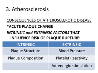
INTRINSIC and EXTRINSIC FACTORS THAT
  INFLUENCE RISK OF PLAQUE RUPTURE:
     INTRINSIC            EXTRINSIC
  Plaque Structure      Blood Pressure
Plaque Composition     Platelet Reactivity
                     Adrenergic stimulation
3. Atherosclerosis
CONSEQUENCES OF ATHEROSCLEROTIC DISEASE
*ACUTE PLAQUE CHANGE
VULNERABLE Plaques:
-contain large areas of foam cells and extracellular lipids
-with thin fibrous caps
   *collagen represents the major structural component of
   the fibrous cap and accounts for its mechanical strength
   and stability
-contain few smooth muscle cells
-have clusters of inflammatory cells
   *STATINS stabilize plaques by reducing plaque
   inflammation
3. Atherosclerosis
CONSEQUENCES OF ATHEROSCLEROTIC DISEASE
*ACUTE PLAQUE CHANGE
-Peak time of onset of acute myocardial
  infarction is between 6 AM and 12 NOON
3. Atherosclerosis
CONSEQUENCES OF ATHEROSCLEROTIC DISEASE
*THROMBOSIS
*VASOCONSTRICTION
-compromises lumen size and by increasing local
   mechanical forces can potentiate plaque disruption
-stimulated by:
1. Adrenergic agonists
2. Local platelet contents
3. impaired secretion of cell relaxing factors
4. mediators released from perivascular inflammtory
    cells

More Related Content

PATHOLOGY - Arteriosclerosis

  • 2. ARTERIOSCLEROSIS DEF: -hardening of the arteries -generic term reflecting arterial wall thickening and loss of elasticity THREE GENERAL PATTERNS: 1. Arteriolosclerosis 2. Monckeberg medial sclerosis 3. Atherosclerosis
  • 3. 1. Arteriolosclerosis -affects small arteries and arterioles -anatomic variants: a. hyaline b. hyperplastic
  • 4. 2. Monckeberg medial sclerosis -has calcific deposits in muscular arteries that may undergo metaplasia to bone -lesions do not encroach vessel lumen thus NOT CLINICALLY SIGNIFICANT
  • 5. 3. Atherosclerosis -most frequent and clinically important pattern -characterized by intimal lesions called ATHEROMAS that protrude into the vessel lumen -consists of a raised lesion: a. Grumuous Core – made of lipid, (cholesterol or cholesterol ester) yellow, soft b. Fibrous Cap – white
  • 6. 3. Atherosclerosis EPIDEMIOLOGY -Risk factors have a multiplicative effect -Constitutional Risk Factors in IHD a. Age – clinically manifests middle age or later b. Gender – premenopausal women are relatively protected due to favorable influence of estrogen -but clinical trials have failed to demonstrate any utility of hormonal therapy for vascular diseases prevention a. Genetics – MOST SIGNIFICANT INDEPENDENT RISK FACTOR
  • 7. 3. Atherosclerosis EPIDEMIOLOGY -Modifiable Risk Factors in IHD a. Hyperlipidemia – esp. hypercholesterolemia, increased LDL, decreased HDL, increased lipoprotein a *STATINS – a class of drugs that lower circulating cholesterol levels by inhibiting HMG-Coa reductase, the rate-limiting enzyme in hepatic cholesterol biosynthesis
  • 8. 3. Atherosclerosis EPIDEMIOLOGY -Modifiable Risk Factors in IHD b. Hypertension – both systolic and diastolic components are important - MOST IMPORTANT CAUSE OF LEFT VENTRICULAR HYPERTROPHY c. Cigarette smoking – prolonged smoking of one pack of cigarettes or more daily doubles the death rate from IHD
  • 9. 3. Atherosclerosis EPIDEMIOLOGY -Modifiable Risk Factors in IHD d. Diabetes mellitus – induces hypercholesterolemia
  • 10. 3. Atherosclerosis EPIDEMIOLOGY -Additional Risk Factors a. Inflammation – present during all stages of atherogenesis and is intimately linked with atherosclerotic plaque formation and rupture *C-reactive Protein (CRP) has emerged as one of the simplest and most sensitive inflammatory markers; decreased by STATINS
  • 11. 3. Atherosclerosis EPIDEMIOLOGY -Additional Risk Factors b. Hyperhomocysteinemia – seen in CAD, PAD, stroke and venous thrombosis -elevated homocysteine levels can be caused by low folate and Vitamin B12 intake c. Metabolic syndrome
  • 12. 3. Atherosclerosis EPIDEMIOLOGY -Additional Risk Factors d. Lipoprotein (a) – an altered form of LDL, associated with CAD and CVD e. Factors affecting Hemostasis f. Other Factors – lack of exercise; competitive stressful lifestyle (type A personality); obesity
  • 13. 3. Atherosclerosis PATHOGENESIS Hypotheses: a. Intimal Cellular Proliferation b. Repetitive Formation and Organization of Thrombi c. Response-To-Injury -views atherosclerosis as a chronic inflammatory and healing response of the arterial wall to injury -lesion progression occurs through the interaction of modified lipoproteins, monocyte-derived macrophages, and T-lymphocytes with the normal cellular constituents of the arterial wall
  • 14. 3. Atherosclerosis PATHOGENESIS c. Response-To-Injury -PATHOGENIC EVENTS INVOLVED: i. Endothelial Injury -increased vascular permeability -leukocyte adhesion -thrombosis ii. Accumulation of Lipoproteins in vessel wall -usually LDL and its oxidized forms iii. Monocyte adhesion to the endothelium, followed by migration into the intima and transformation into macrophages and foam cells
  • 15. 3. Atherosclerosis PATHOGENESIS c. Response-To-Injury -PATHOGENIC EVENTS INVOLVED: iv. Platelet adhesion v. Factor release from activated platelets, macrophages and vascular wall cells, inducing smooth muscle cell recruitment vi. Smooth muscle cell proliferation and ECM production vii. Lipid accumulation -extracellular and within cells (macropahes and smooth muscle cells)
  • 16. 3. Atherosclerosis PATHOGENESIS MAJOR MECHANISMS +Endothelial Cell Injury -results in intimal thickening -early human lesions begin at sites of morphologically intact endothelium, THUS, endothelial dysfunction underlies human atherosclerosis -can be caused by: -hypertension -hyperlipidemia -toxins from cigarette smoke -infectious agents -inflammatory cytokines -homocysteine
  • 17. 3. Atherosclerosis PATHOGENESIS MAJOR MECHANISMS +Endothelial Cell Injury 2 MOST IMPORTANT CAUSES OF ENDOTHELIAL DYSFUNCTION -hemodynamic disturbances -hypercholesterolemia
  • 18. 3. Atherosclerosis PATHOGENESIS +Endothelial Cell Injury *Hemodynamic Disturbances -plaques tend to occur at areas of disturbed flow patterns: a. Ostia of exiting vessels b. Branch points c. Along the Posterior wall of the abdominal aorta
  • 19. 3. Atherosclerosis PATHOGENESIS +Endothelial Cell Injury *Hemodynamic Disturbances -nonturbulent laminar flow in normal vasculature leads to the induction of endothelial genes whose products protect against atherosclerosis e.g. SUPEROXIDE DISMUTASE
  • 20. 3. Atherosclerosis PATHOGENESIS +Endothelial Cell Injury *Lipids -Lipoprotein abnormalities present in many MI survivors: -Increased LDL -Decreased HDL - Increased Lipoprotein (a) -DOMINANT LIPIDS IN ATHEROMATOUS PLAQUES ARE: -Cholesterol -Cholesterol Esters
  • 21. 3. Atherosclerosis PATHOGENESIS +Endothelial Cell Injury *Lipids -Lowering serum cholesterol by diet or drugs slows the rate of progression of atherosclerosis, causes regression of some plaques, and reduces the risk of cardiovascular events
  • 22. 3. Atherosclerosis PATHOGENESIS +Endothelial Cell Injury *Lipids -Mechanisms by which HYPERLIPIDEMIA contributes to ATHEROGENESIS: -impair endothelial cell function by increasing local oxygen free radical production -accumulate lipoproteins within the intima, which are then oxidized by oxygen free radicals
  • 23. 3. Atherosclerosis PATHOGENESIS +Endothelial Cell Injury *Lipids -Oxidized LDL is ingested by macrophages through a SCAVENGER RECEPTOR, distinct from LDL receptor -MACROPHAGES become FOAM CELLS after accumulation of Oxidized LDL within the phagocyte
  • 24. 3. Atherosclerosis PATHOGENESIS +Endothelial Cell Injury *Inflammation -dysfunctional arterial endothelial cells express adhesion molecules that encourage leukocyte adhesion -VCAM1 – binds monocytes and T cells -Monocyte recruitment and differentiation is theoretically protective because they remove harmful lipid particles, but the presence of OXIDIZED LDL AUGMENTS Macrophage activation and cytokine production
  • 25. 3. Atherosclerosis PATHOGENESIS +Endothelial Cell Injury *Infection -Microorganisms detected in atherosclerotic plaques: -Herpesvirus -Cytomegalovirus -Chlamydiae pneumoniae
  • 26. 3. Atherosclerosis PATHOGENESIS +Smooth Muscle Proliferation -intimal smooth muscle cell proliferation and ECM deposition convert a fatty streak into a mature atheroma *FATTY STREAK = EARLIEST LESION -Growth Factors involved: -PDGF -FGF -TGF-a
  • 27. 3. Atherosclerosis PATHOGENESIS +Smooth Muscle Proliferation -Smooth muscle cells synthesize ECM that stabilizes atherosclerotic plaques - Active Inflammatory cells in atheromas can cause intimal smooth muscle cell apoptosis, and increase ECM catabolism leading to unstable plaques
  • 28. 3. Atherosclerosis MORPHOLOGY *FATTY STREAKS -EARLIEST LESIONS IN ATHEROSCLEROSIS -begin as minute, flat yellow spots and then colaesce -not significantly raised and do not cause flow disturbance -virtually in all children older than 10 years -coronary fat streaks begin to form in adolescence, at the same anatomic sites that later tend to develop plaques
  • 29. 3. Atherosclerosis MORPHOLOGY *ATHEROSCLEROTIC PLAQUE -KEY PROCESSES: Intimal Thickening & Lipid Accumulation -Gross: -Color: White or Yellow (Ulcerated Plaques: Red- brown) -Size: 0.3-1.5 cm in diameter (can coalesce) -In humans, the ABDOMINAL AORTA affected MORE than THORACIC AORTA
  • 30. 3. Atherosclerosis MORPHOLOGY *ATHEROSCLEROTIC PLAQUE -Commonly involved Vessels (Descending Order): 1. Lower Abdominal Aorta 2. Coronary Arteries 3. Popliteal Arteries 4. Internal Carotid Arteries 5. Circle of Willis
  • 31. 3. Atherosclerosis MORPHOLOGY *ATHEROSCLEROTIC PLAQUE -Principal Components (CEL): 1. Cells -smooth muscle cells, macrophages, T cells 2. ECM -collagen, elastic fibers, proteoglycans 3. Lipids -intracellular and extracellular
  • 32. 3. Atherosclerosis MORPHOLOGY *ATHEROSCLEROTIC PLAQUE -Configuration: 1. Fibrous cap – superficial; made of smooth muscle cells, collagen 2. Shoulder – beneath and to the side of the cap; more cellular with macrophages, T cells, smooth muscle cells 3. Necrotic core – deep into cap, made of lipid primarily cholesterol and cholesterol esters, debris from dead cells, foam cells, fibrin, thrombus, other plasma proteins
  • 33. 3. Atherosclerosis MORPHOLOGY *ATHEROSCLEROTIC PLAQUE -The periphery of the lesions show neovascularization -Plaques enlarge due to: 1. Cell death and degenration 2. Synthesis and degradation of ECM 3. Organization of thrombus -Atheromas often undergo CALCIFICATION
  • 34. 3. Atherosclerosis MORPHOLOGY *ATHEROSCLEROTIC PLAQUE -Plaques are susceptible to the following CLINICAL CHANGES: 1. Rupture, ulceration, erosion 2. Hemorrhage into a plaque 3. Atheroembolism 4. Aneurysm formation
  • 35. 3. Atherosclerosis CONSEQUENCES OF ATHEROSCLEROTIC DISEASE -Large Elastic and Large and Medium Muscular arteries are the MAJOR TARGETS of ATHEROSCLEROSIS -Smaller vessels can become occluded, compromising distal tissue perfusion -Ruptured plaque can embolize atherosclerotic debris and cause distal vessel obstruction, or can lead to acute thrombosis -Destruction of the underlying vessel wall can lead to aneurysm formation, which can rupture and/or be a thrombi
  • 36. 3. Atherosclerosis CONSEQUENCES OF ATHEROSCLEROTIC DISEASE *ATHEROSCLEROTIC STENOSIS -plaques can gradually occlude vessel lumens, compromising blood flow causing ischemic injury -outward remodeling preserves lumen diameter -effects of vascular occlusion depend on arterial supply and metabolic demand of affected tissue
  • 37. 3. Atherosclerosis CONSEQUENCES OF ATHEROSCLEROTIC DISEASE *ACUTE PLAQUE CHANGE -plaque erosion or rupture is typically promptly followed by partial or complete vascular thrombosis resulting in acute tissue infarction -THREE CATEGORIES OF PLAQUE CHANGES: 1. Rupture/Fissuring – exposing thrombogenic constituents 2. Erosion/Ulceration – exposing subendothelial basement membrane to blood 3. Hemorrhage into the atheroma
  • 38. 3. Atherosclerosis CONSEQUENCES OF ATHEROSCLEROTIC DISEASE *ACUTE PLAQUE CHANGE -the precipitating lesion in patients who develop MI or other coronary syndromes is NOT NECESSARILY a severely stenotic and hemodynamically significant lesion BEFORE ITS ACUTE CHANGE
  • 39. 3. Atherosclerosis CONSEQUENCES OF ATHEROSCLEROTIC DISEASE *ACUTE PLAQUE CHANGE INTRINSIC and EXTRINSIC FACTORS THAT INFLUENCE RISK OF PLAQUE RUPTURE: INTRINSIC EXTRINSIC Plaque Structure Blood Pressure Plaque Composition Platelet Reactivity Adrenergic stimulation
  • 40. 3. Atherosclerosis CONSEQUENCES OF ATHEROSCLEROTIC DISEASE *ACUTE PLAQUE CHANGE VULNERABLE Plaques: -contain large areas of foam cells and extracellular lipids -with thin fibrous caps *collagen represents the major structural component of the fibrous cap and accounts for its mechanical strength and stability -contain few smooth muscle cells -have clusters of inflammatory cells *STATINS stabilize plaques by reducing plaque inflammation
  • 41. 3. Atherosclerosis CONSEQUENCES OF ATHEROSCLEROTIC DISEASE *ACUTE PLAQUE CHANGE -Peak time of onset of acute myocardial infarction is between 6 AM and 12 NOON
  • 42. 3. Atherosclerosis CONSEQUENCES OF ATHEROSCLEROTIC DISEASE *THROMBOSIS *VASOCONSTRICTION -compromises lumen size and by increasing local mechanical forces can potentiate plaque disruption -stimulated by: 1. Adrenergic agonists 2. Local platelet contents 3. impaired secretion of cell relaxing factors 4. mediators released from perivascular inflammtory cells