Peranan USG pada kehamilan kembar - Prof SRK.pptx
?Download as PPTX, PDF?
0 likes?46 views

Presentasi kehamilan kembar terbaru dengan bukti evidence terbaru


Peranan USG pada kehamilan kembar - Prof SRK.pptx
- 1. Peranan USG pada kehamilan kembar Sofie Rifayani Krisnadi Departemen Obstetri dan Ginekologi Fakultas Kedokteran Universitas Padjadjaran RSUP Dr. Hasan Sadikin BANDUNG
- 2. ?Kehamilan kembar berhubungan dengan risiko tinggi Mortalitas dan Morbiditas Perinatal. ?Tahun 2009, Tingkat kelahiran mati (stillbirth): 12/1000 kelahiran kembar dan 31/1000 kelahiran triplet dibandingkan 5 / 1000 kelahiran tunggal. ?65% kematian neonatal pada kehamilan multipel adalah preterm, dibandingkan 43% kematian neonatal pada kehamilan tunggal. Insidensi
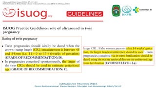
- 3. Penilaian ultrasound terhadap : ? Biometri janin ? Anatomi ? Doppler velosimetri ? Jumlah cairan amnion Gangguan pertumbuhan janin kembar dapat dinilai dengan parameter yang sama dengan kehamilan Tunggal. Identifikasi dan monitor : ? Uncomplicated twin pregnancy. ? Kehamilan kembar terhadap outcome yang buruk seperti pada kondisi TTTS, selective FGR (sFGR), twin anemia¨C polycythemia sequence (TAPS), twin reversed arterial perfusion (TRAP) sequence, conjoined twins.
- 5. CRL
- 7. Khalil A, Rodgers M, Baschat A, Bhide A, Gratacos E, et.al. ISUOG Practice Guidelines: role of ultrasound in twin pregnancy. Ultrasound Obstet Gynecol 2016; 47: 247¨C263.
- 10. Khalil A, Rodgers M, Baschat A, Bhide A, Gratacos E, et.al. ISUOG Practice Guidelines: role of ultrasound in twin pregnancy. Ultrasound Obstet Gynecol 2016; 47: 247¨C263. Routine monitoring of twin pregnancy with ultrasound
- 12. NT
- 13. NB DV
- 15. Khalil A, Archer R, Hutchinson V, et al. Noninvasive prenatal screening in twin pregnancies with cell-free DNA using the IONA test: a prospective multicenter study. Am J Obstet Gynecol 2021;225:79.e1-13
- 16. Dugoff L, Koelper NC, Chasen ST, Russo ML, Roman AS, et.al. Cell-free DNA screening for trisomy 21 in twin pregnancy: a large multicenter cohort study. Am J Obstet Gynecol. 2023 Oct;229(4):435.e1-435.e7.
- 19. Screening with cervical length at 18¨C22 weeks in multiple pregnancies is recommended by ISUOG, where the cut off used is <25 mm and SOGC. BUT routine cervical length screening is NOT currently recommended by NICEor the ACOG
- 20. Society for Maternal-Fetal Medicine (SMFM). McIntosh J, Feltovich H, Berghella V, Manuck T. The role of routine cervical length screening in selected high- and low-risk women for preterm birth prevention. Am J Obstet Gynecol. 2016;215(3):B2-7. Langkah-langkah untuk pengukuran panjang serviks yang tepat (1) Pastikan pasien mengosongkan kandung kemihnya. (2) Siapkan probe yang telah dibersihkan. (3) Masukkan probe dengan lembut ke dalam vagina pasien. (4) Arahkan probe ke forniks anterior. (5) Dapatkan gambar sagital, sumbu panjang dari seluruh serviks. (6) Tarik probe sampai gambar kabur dan kemudian masukkan kembali dengan lembut sampai gambar jelas (ini memastikan Anda tidak menggunakan tekanan berlebihan). (7) Perbesar gambar sehingga serviks menempati dua pertiga bagian layar. (8) Pastikan os internal dan eksternal terlihat jelas. (9) Ukur panjang serviks di sepanjang kanal endoserviks antara os internal dan eksternal. (10) Ulangi proses ini dua kali untuk mendapatkan 3 set gambar / pengukuran. (11) Gunakan pengukuran terbaik yang terpendek
- 21. Transvaginal ultrasound images of a normal cervix with slightly hyperechoic cervical mucosa Transvaginal ultrasound measurement of cervical length in the same patient, with a full bladder (a) and with an empty bladder (b) Bladder filled
- 22. Transvaginal ultrasound measurements of cervical length in the same patient, illustrating the effect on the measurement of pressure exerted by the probe: (a) avoiding pressure (correct); (b) with excessive probe pressure (incorrect), which elongates the cervix and creates a difference in width between the cervical lips. Coutinho CM, Sotiriadis A, Odibo A, Khalil A, DˇŻAntonio F, et.al, on behalf of the ISUOG Clinical Standards Committee. ISUOG Practice Guidelines: role of ultrasound in the prediction of spontaneous preterm birth. Ultrasound Obstet Gynecol 2022; 60: 435¨C456
- 23. Transvaginal ultrasound measurements of cervical length in the same patient, illustrating the potential effect on the identification of dynamic changes of the cervix of different degrees of pressure exerted by the probe. (a) Excessive pressure (incorrect) elongates the cervix, producing a difference in the widths of the cervical lips. (b,c) Gentle probe pressure (correct) allows identification of funneling and the short cervical length, with dynamic changes during the course of the examination Coutinho CM, Sotiriadis A, Odibo A, Khalil A, DˇŻAntonio F, et.al, on behalf of the ISUOG Clinical Standards Committee. ISUOG Practice Guidelines: role of ultrasound in the prediction of spontaneous preterm birth. Ultrasound Obstet Gynecol 2022; 60: 435¨C456
- 25. Townsend R, Khalil A. Ultrasound surveillance in twin pregnancy: An update for practitioners. Ultrasound. 2018;26(4):193-205.
- 26. Townsend R, Khalil A. Ultrasound surveillance in twin pregnancy: An update for practitioners. Ultrasound. 2018;26(4):193-205. In monochorionic twin pregnancy complicated by sFGR, fetal growth should be assessed at least every 2 weeks, and fetal Doppler (umbilical artery and MCA) at least weekly. If the umbilical artery Doppler is abnormal, assessment of the DV blood flow should be undertaken. The aim in managing these pregnancies is to prolong the pregnancy at least until viability is achieved, while at the same time avoiding single IUD with its associated serious consequences for the surviving cotwin.
- 27. Mustafa HJ, Javinani A, Heydari MH, et al. Selective intrauterine growth restriction without concomitant twin-to-twin transfusion syndrome, natural history, and risk factors for fetal death: A systematic review and meta-analysis. Am J Obstet Gynecol MFM 2023;5:101105.
- 28. ? As these twins have separate circulations, the pregnancy can be followed as in growth- restricted singleton pregnancy, monitoring for progressive deterioration of umbilical artery, MCA and DV Doppler, and of biophysical profile scores.
- 29. D'antonio F, Prasad S, Masciullo L, Eltaweel N, Khalil A. Selective fetal growth restriction in dichorionic diamniotic twin pregnancy: systematic review and meta-analysis of pregnancy and perinatal outcomes. Ultrasound Obstet Gynecol. 2024 Feb;63(2):164-172.
- 30. In dichorionic twin pregnancy complicated by sFGR, follow-up visits could be less frequent, as delivery is usually not recommended before 32¨C34 weeks gestation.
- 31. Dampak kehamilan kembar Monokorioik meningkatkan risiko terhadap kejadian : a)Twin-twin transfusion syndrome (TTTS) b)Twin anemia¨Cpolycythemia sequence (TAPS) c)Twin reversed arterial perfusion (TRAP) sequence d)Monochorionic monoamniotic (MCMA) twins e)Conjoined twins
- 32. Screening, diagnosis, staging and management of TTTS a) Up to one third of twin pregnancies are monochorionic. In nearly all monochorionic twins, the placenta contains vascular anastomoses connecting the two fetal circulations. b) It is the angioarchitecture of these vascular anastomoses that determines the risk profile. c) Monochorionic twins are at risk of developing TTTS when there is unequal hemodynamic and amniotic fluid balance. d) The diagnosis of TTTS requires the presence of significant amniotic fluid amount. e) TTTS affects 10¨C15% of monochorionic twin pregnancies and is associated with increased perinatal mortality and morbidity; if untreated, it leads to fetal demise in up to 90% of cases, with morbidity rates in survivors of over 50%. f) Early diagnosis, however, may allow intervention with fetoscopic laser ablation, which significantly improves the prognosis. Laser treatment in these pregnancies results in 60¨C70% double survival and 80¨C90% survival of at least one twin.
- 33. Townsend R, Khalil A. Ultrasound surveillance in twin pregnancy: An update for practitioners. Ultrasound. 2018;26(4):193-205.
- 35. ULTRASOUND TRAINING WEEK Divisi Fetomaternal Departemen Obstetri dan Ginekologi RSHS/FKUP
- 37. Screening, diagnosis and management of twin anemia¨C polycythemia sequence (TAPS)
- 38. Screening, diagnosis and management of twin anemia¨C polycythemia sequence (TAPS) a) The incidence of TAPS occurring spontaneously in MCDA twins is up to 5%. b)Complicate up to 13% of cases of TTTS following laser ablation. c) The polycythemia twin might have a ˇ®starry skyˇŻ appearance of the liver pattern due to diminished echogenicity of the liver parenchyma and increased brightness of the portal venule walls. d)The antenatal and postnatal severity-based staging classifications.
- 39. DONOR RESIPIEN MCA-PSV > 1.5 multiples of the median (MoM) in the donor, suggesting fetal anemia MCA-PSV < 1.0 MoM in the recipient, suggesting polycythemia. Placenta bright, thickened section associated with the donor Echolucent thin section associated with the recipient. Postnatal diagnosis of TAPS the finding of chronic anemia (including reticulocytosis) in the donor Polycythaemia in the recipient. Screening, diagnosis and management of twin anemia¨C polycythemia sequence (TAPS)
- 41. a) Recent evidence suggests that, in monochorionic twins complicated by TAPS, the risk of neurodevelopmental delay is increased (20%). b) The commonest options include 1. Conservative management, 2. Early delivery, 3. Laser ablation or intrauterine blood transfusion (IUT) for the anemic twin, 4. Combined IUT for the anemic twin and partial exchange transfusion to dilute the blood of the polycythemic twin. Screening, diagnosis and management of twin anemia¨C polycythemia sequence (TAPS)
- 43. Twin reversed arterial perfusion (TRAP) sequence
- 44. Twin reversed arterial perfusion (TRAP) sequence ? TRAP sequence is a rare complication of monochorionic twin pregnancy (1% of monochorionic twin pregnancies and 1 in 35 000 pregnancies overall). ? It is characterized by the presence of a TRAP or acardiac mass perfused by an apparently normal (pump) twin. ? The risk of demise of the pump fetus in TRAP sequence managed conservatively is up to 30% by 18 weeks gestation.
- 47. ? Minimally invasive techniques, such as cord coagulation, cord ligation and photocoagulation of the anastomoses, as well as intrafetal methods, such as RFA and intrafetal laser therapy, are performed as a means of preventing the demise of the pump twin. The survival rate of the pump twin using these treatment modalities is approximately 80%. ? When treatment is necessary, it appears to be preferable before 16 weeks gestation. ? Survival might be improved by elective intervention at 12¨C14 weeks gestation. ? The rate of preterm birth before 32 weeks gestation is approximately 10%. Twin reversed arterial perfusion (TRAP) sequence
- 48. Monochorionic monoamniotic (MCMA) twins ? Umbilical cord entanglement is almost always present in MCMA twins and does not appear to contribute to their morbidity and mortality (GRADE OF RECOMMENDATION: D). ? Delivery by Cesarean section is recommended at 32¨C34 weeks (GRADE OF RECOMMENDATION: D). ? In MCMA twin pregnancies undergoing selective reduction (because of discordant anomaly, TRAP sequence, severe TTTS or sFGR), cord occlusion and transection are recommended to prevent fetal demise of the other twin due to cord accidents.
- 49. Monoamniotic twin pregnancies are rare, with an estimated incidence of only 8 per 100,000 pregnancies, and result from a single fertilized egg and embryo splitting between 9 and 13 days after fertilization. The main characteristic of monoamniotic twin pregnancies is a single shared placenta and amniotic cavity. Conjoined twin pregnancies, which are even rarer with an incidence of only 1.5 per 100,000 pregnancies, are a subtype of monoamniotic twin pregnancies and result from even later embryonic splitting (after 13 days). Besides sharing the placenta and amniotic cavity, conjoined twins also share parts of their bodies. Van Mieghem T, Abbasi N, Shinar S, et al. Monochorionic monoamniotic twin pregnancies. Am J Obstet Gynecol MFM 2022;4:100520
- 50. ? The obvious direct sign of monoamnionicity on ultrasound in the first trimester of pregnancy is the lack of a dividing amniotic membrane between the 2 fetuses. ? Indirect (and hence less accurate) ultrasound signs, which help in assigning amnionicity in the first trimester of pregnancy, include the presence of cord entanglement which is almost always present in monoamniotic twins, and the number of yolk sacs. ? Approximately two-thirds of monoamniotic twins will have only 1 yolk sac, but one-third will present with 2 yolk sacs, similar to diamniotic twins.
- 52. Conjoined twins ? Conjoined twins are very rare, occurring in approximately 1 in 100 000 pregnancies (1% of monochorionic twin pregnancies). ? Diagnosis with ultrasound in the first trimester is now the norm (on visualizing close and fixed apposition of the fetal bodies, with fusion of the skin lines at some point). ? Survival to discharge was only around 25%, and the majority of these had significant morbidity. ? The most common form is Thoracopagus. ? Approximately 60% of conjoined twin pregnancies will result in stillbirth.
- 53. Conjoined twins Such pregnancies should be assessed at a fetal medicine referral center, with multidisciplinary assessment and counseling. The pregnancy must be delivered at a center with expertise in the postnatal medical and surgical management of such cases. There are associated high rates of postnatal mortality and there is almost always morbidity
- 58. Monochorionic monoamniotic (MCMA) twins
- 59. ULTRASOUND TRAINING WEEK Divisi Fetomaternal Departemen Obstetri dan Ginekologi RSHS/FKUP
Editor's Notes
- #22: Penapisan pada ibu hamil tanpa gejala dilakukan saat 18-24 minggu , sekalian dengan pencitraan untk encari anomaly.. Kehamilan 24 minggu merupakak\n batas atas penapisan, karena biasa dipakai sebagai deadline pemberian progesterone pada pasien yang mengalam cerclage, batas awal pemberian tokolitik dan antenatal kortikosteroid, dan magnesium sulfat sebagai neroprotektor.. Tanpa melihat factor risiko, Panjang serviks < 25 mm merupakan cut off point untuk pencegahan ibu hamil yang asimtomatik.
- #23: Gambar urutan kedua menunjukkan perbedaan signifikan pengukuran CL dengan bladder terisi dan bladder kosong. Pada kandung kencing yang penuh Panjang seviks levih dari seharusnya, karena penekanan kandung kemih
- #24: Gambar ini menunjukkan efek tekanan probe pada serviks yang berlebihan saat melakukan TV-US, sehingga terdapat perbedaan hasil CL.
- #25: Gambar ini menunjukkan efek tekanan pada serviks yang berlebihan saat melakukan TV-US, sehingga gambaran funneling yang harusnya ada akan tampak tersamarkan/hasil normal.