Perception- Auditory
âĒDownload as PPTX, PDFâĒ
3 likesâĒ2,240 views

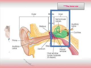
The inner ear contains the cochlea, which has two fluid-filled ducts separated by the cochlear partition. Within the cochlear partition is the organ of Corti, which contains hair cells that detect sound vibrations. Low frequency sounds cause maximum vibration at the apex of the basilar membrane, while high frequencies cause vibration at the base. The hair cells transduce these vibrations into neural signals that travel to the brainstem and auditory cortex, where pitch and other sound properties are processed. Damage to hair cells or auditory nerves can cause hearing loss.


Perception- Auditory
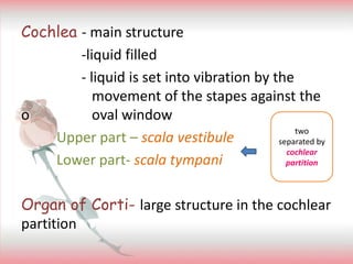
- 2. Cochlea - main structure -liquid filled - liquid is set into vibration by the movement of the stapes against the o oval window Upper part â scala vestibule Lower part- scala tympani Organ of Corti- large structure in the cochlear partition two separated by cochlear partition
- 3. Main structures Hair cells- Bending of cilia in the inner hair cells are responsible for transduction. 2 types of hair cells inner hair cells- 3,500- bends, and does transduction process. outer hair cells- 12,000- increase the vibration of the basilar membrane
- 4. Basilar membrane- supports the organ of corti and vibrates in response to sound Tectorial membrane- extends over the hair cells
- 5. Why inner hair cell bends? Ans: because the in and out movement of the stapes creates pressure changes in the liquid inside the cochlea that sets the cochlear partition into up and down motion
- 6. Movement in one direction of the cilia ï opens channel in the membraneï ions flow into the cell Movement opposite of that direction ï ion channels closeï no electrical signals generated ** we have low threshold for hearing Video Clip
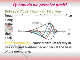
- 7. Q: how do we perceive pitch? Bekesyâs Place Theory of Hearing- frequency of a sound us indicated by the place along the cochlea at which nerve firing is highest. Low frequency- maximum activity in the in the hair cells and auditory nerve fibers at the apex end of the basilar membrane High frequency- cause maximum activity in hair cells and auditory nerve fibers at the base of the membrane.
- 8. Tonotropic map- an orderly map of frequencies along the length of the cochlea. Frequency tuning curve- This curve is determined by presenting pure tones of different frequencies and measuring how many decibels are necessary to cause the neuron to fire. This decibel level is the threshold for that frequency characteristic frequency- frequency to which the neuron is most sensitive Auditory masking- ability to hear a sound is decrease by the presence of other sounds
- 9. Q: Why does the basilar membrane vibrate more sharply in healthy cochleas? The outer hair cells expand and contract in response to the vibration of the basilar membrane, and this expansion and contraction, whichonly occurs in live cochleas, amplifies and sharpens the vibration of the basilar membrane. **Cochlear amplifier
- 10. Frequency- is signaled by which fibers in the cochlea fire to a tone as well as how the fibers are fired. Phase locking- property of firing at the same place in the sound stimulus Temporal coding- connection between the frequency of the sound stimulus and the timing of the auditory nerve firing; measurements of the pattern of firing for auditory nerve fibers indicate that phase locking occurs up to a frequency of about 4,000 Hz.
- 11. Causes of hearing loss âĒ Conductive hearing loss- Blockage of sound from reaching the receptors âĒ sensorineural hearing loss -Damage to the hair cells -Damage to the auditory nerve or the brain Presbycusis- caused by old age - Greatest at higher frequencies -Prevalent on males than females
- 12. âĒ Noise-induced Hearing loss -Occurs when loud noises cause degeneration of the hair cells. -damage to the organ of corti
- 13. WHERE AND WHAT STREAM ** a 45-year-old man with temporal lobe damage caused by a head injury ** a 64-year-old woman with parietal and frontal lobe damage caused by a stroke
- 14. Pathway of the Cochlea to the cortex Inner hair cells Auditory receiving area in the cortex Cochlear Nucleus Super Olivary Nucleus Inferior colliculus Medial Geniculate Nucleus Auditory receiving area (A1) Temporal lobe of the Cortex
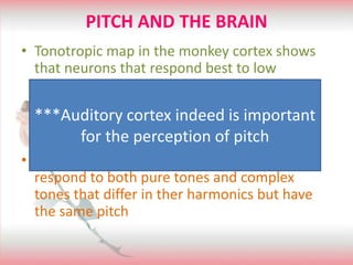
- 17. PITCH AND THE BRAIN âĒ Tonotropic map in the monkey cortex shows that neurons that respond best to low frequencies are located to the left, and neurons that respond best to higher frequencies are located to the right âĒ Studies found neurons in the brain that respond to both pure tones and complex tones that differ in ther harmonics but have the same pitch ***Auditory cortex indeed is important for the perception of pitch
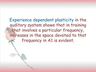
- 18. Experience dependent plasticity in the auditory system shows that in training that involves a particular frequency, increases in the space devoted to that frequency in A1 is evident.