際際滷

際際滷Share a Scribd company logo
Instructions
 This self assessment module on chest radiography consisting 30
  questions related to chest radiographs and explanations for each.

 You can either try all questions first and then check your answers or
  check the answer to each question after every attempt.

 Marks allocated to each component is given next to the question within
  brackets.

 Your suggestions to improve this module may be emailed to
  thilakpw@yahoo.com

Thilak Weerarathna,
Department of Medicine,
Faculty of Medicine, Galle
                                                              Questions
1
    This chest radiograph was taken from a
          patient after a road traffic accident
                            a) What is the abnormality


                            b) Give two other causes
                               for this radiological
                               abnormality


                               Next question   Answer
2


    a) What abnormality
      do you see in this
      chest radiograph


    b) List two complications
      of this condition


          Next question    Answer
3


    (a ) List two abnormalities
       in this chest
       radiograph


    (b) What is the most likely
       diagnosis based on
       your findings in 3 .a


         Next question    Answer
4

    a. Describe the abnormality
       seen in this chest
       radiograph


    b) Give two likely causes
      for this appearance


          Next question     Answer
5


    a) Describe the
      abnormality
      seen in this
      chest radiograph


    b) List two likely causes
      for the abnormality
      noted


      Next question   Answer
This radiograph was taken from a person
6   with hemoptysis

                           a) Describe two abnormalities


                           b) Give a likely diagnosis


                               Next question   Answer
This chest radiograph was taken from a 35 year old
7   man with 5 days of fever and cough.

                           a. What is the radiological
                              diagnosis


                           b. List two complications
                              of the condition
                              mentioned in 7. a


                                 Next Question   Answer
8


    a. What is the radiological
       diagnosis


    b. Give one association
       of this condition


         Next Question   Answer
This chest radiograph was taken from 35 year old
9
    man with productive cough of 6 months

                        a. List two abnormalities


                        b. What is the likely
                          diagnosis


                        c. List two complications


                              Next Question     Answer
10

     a. Describe the radiological
        abnormality


     b. What segment of the
        lung is involved


     c. Give reasons for your
        answer to ( b)


          Next question   Answer
11


     a) Describe the
       radiological
       abnormality


     b) List two investigations
        that would provide
        etiological diagnosis
        of this condition


          Next question   Answer
12   This chest radiograph is taken from a man with
     severe haemoptysis

                            a). What is the radiological
                             diagnosis


                                 Next question   Answer
13

     a) Describe the
        radiological
        abnormalities


     b) List two therapeutic
        measures in the
        management of
        this condition


        Next question   Answer
This chest radiograph was taken from a 45 year old
14
     male with recent onset finger clubbing

                         a) Describe the
                           abnormality seen.


                         b) List two extra pulmonary
                            complications of this
                            condition


                                Next question   Answer
This chest radiograph was taken from a man with
15   progressive breathlessness over one week

                            a) What is the radiological
                               diagnosis?


                            b) List the most useful
                               investigation for
                               arriving at a diagnosis
                               of the condition you
                               mentioned in (a)


                                 Next question   Answer
This chest radiograph was taken from a man with
16
     facial puffiness and engorged neck veins

                        a) What is the likely cause
                           for above findings
                           in this patient


                        b) Give two likely causes for
                           the condition you
                           mentioned in (a)


                                Next question   Answer
This chest radiograph was taken from a 40 year
17   old man with pyrexia of unknown origin

                             a) Describe the radiological
                                abnormality


                             b) What investigation
                               would confirm the
                                diagnosis


                                 Next question   Answer
This chest radiograph is taken from a 18 year old
18   boy with sudden onset chest pain and breathlessness

                            a) Right down the complete
                               radiological diagnosis


                            b) What is the immediate
                               therapeutic measure?


                                  Next question   Answer
19

     a) What is the radiological
        diagnosis


     b) What specific feature
        of the condition you
        mentioned above
        is present in this
        radiograph ?


         Next question   Answer
20


     a) What is the radiological
       diagnosis?


     b) Name one specific
        physical sign that
        can be elicited in this
        condition


       Next question   Answer
A patient with following chest radiograph presented
21
     with progressive breathlessness over 6 months

                           a) What condition would account
                              for the breathlessness in
                              this patient ?


                           b) List 3 radiological features
                              in this radiograph in favor of
                              the condition you mentioned
                              in (a)


                                 Next question   Answer
A 56 year old man presented with 3 weeks of fever.
22   His chest radiograph is shown below.

                          a. Name the abnormality in this
                             chest radiograph.


                          b. What is the next investigation
                             that would help in arriving
                             at a specific diagnosis ?


                                 Next question   Answer
This chest radiograph was taken from a 55 year
23   old male with fever, chest pain and breathlessness
     over 2 weeks

                             a) What is the radiological
                                diagnosis?


                             b) What investigation would
                                confirm your diagnosis
                                mentioned in ( a ) ?


                                 Next question   Answer
Following chest radiograph was taken from a
24   60 year old male with chest pain and breathlessness
     over 4 hours.
                               a. What is the radiological
                                  diagnosis?


                               b. List 4 immediate
                                  measures you take in this
                                  patients if the blood
                                   pressure
                                  is 130/ 85 mm Hg


                                  Next question   Answer
25

     a. What is the radiological
        diagnosis ?


     b. List 3 radiological features
        in favor of the diagnosis
        mentioned in (a).


            Next question   Answer
26

     a. Name the abnormality in this
        chest radiograph


     b. List 3 causes for the
        abnormality you mentioned
        in (a)


          Next question   Answer
27

     a) Write down the
         radiological
        abnormality


     b) List two causes
        for the abnormality
        you mentioned in (a)


        Next question   Answer
This chest radiograph was taken from a previously
28   healthy male presenting with cough and fever of
     one week

                          a) What is the radiological
                             diagnosis?


                          b) List two antibiotics you
                             select to treat this patient


                               Next question   Answer
29


     a) List two abnormalities in
        this chest radiograph


     b) Give a likely underlying cause
        for the abnormalities
        mentioned in (a)


           Next question   Answer
30


     a) Name two abnormalities
        in this chest radiograph


          Introductions   Answer
Answer 1
a) air ( gas ) under the diaphragm / ruptured
  viscus organ ( 06 marks )
b) following laporoscopy ( 04 marks)

Notes  perforation of bowel or stomach
 following trauma or other pathology such as
 peptic ulcer can give rise to gas under the
 diaphragm sign.
 Inflation of air at laporoscopy also can give
 rise to this radiological sign.


                                 Back    Next question
Answer 2

a) cystic shadows in right lung field/ pulmonary cysts (
   06 marks), Lung abscess (02 marks)

b) infection (02), rupture , pneumothorax (02)

Notes  cysts in lung are characterized by rounded,
  thin walled shadows. When are filled with fluid due
  to infection, they are called abscess. Abscesses are
  characterized by an air  fluid level. Sometimes
  these cysts can rupture giving rise to
  pneumothorax.

                                        Back     Next question
Answer 3

a) 1. right sided massive pleural effusion (3),
        pleural effusion (1),
   2. lytic lesions in clavicles and ribs (3)
b) malignancy (4), secondary deposits (4),

Notes  homogenous opacification of a hemithorax is a
  sign of pleural effusion. Usually large effusions causes
  displacement of trachea and if trachea is central,
  possibility of right sided lung collapse with effusion is
  more likely. Combination of collapse and large
  effusion is amore likely be due to malignancy. lytic
  lesions in ribs and clavicles also favor malignancy

                                     Back         Next question
Answer - 4


a) air- fliud level in the left hemithorax (6)
b) hydropneumothorax(2),
   pyopneumothorax (2)

Notes- collection of fluid in the pleural space with an air- fluid level
  is seen with lung abscess and hydro/ pyo pneumothorax. Lung
  abscess is characterized by a such collection with a well
  demarcated capsule and when the air fluid level is seen across
  the hemi thorax without a capsule it is due to hydro or
  pyopneumothorax. These can arise as a rupture of lung abscess
  (pyopneumothorax) or following trauma ( hemopneumothorax)


                                       Back           Next question
Answer 5


a) abnormal elevation of the right dome of the
     diaphragm(6)
b) paralysis of right phrenic nerve(2)
     collection of pus under the diaphragm(2) or
     any cause for the above
Notes  abnormal elevation of the right dome of the
   diaphragm can occur as a result of phrenic nerve
   paralysis due to causes such as malignant infiltration.
   The same appearance is seen when there is collection
   of pus under the diaphragm following ruptured liver
   abscess.
                                  Back      Next question
Answer 6

a) two opacities in the upper zones of the left lung(3),
   cavitations with thick walls in the left upper zone(3)
b) malignant lesions with central cavitations(4)

Notes-
  Lung opacities with irregular margins are features of
  either primary or secondary malignancy. Cavitations
  within the lesion also favor this. Cavitations with thick
  irregular walls are more in favor of a malignant lesions.
  Always look for erosion of ribs, hilar lymh node
  enlargement (which are not present in this case) when
  there is suspicious malignant lesions in the lung.

                                Back        Next question
Answer- 9

a) thickened bronchial markings(2), bilateral cystic
   shadows(2)
b) Bronchiectasis (4)
c) hemoptysis(1), secondary infection (consolidataion)(1),
   fibrosis(1), lung abscesse(1)

Notes- Bronchiectataic changes are often seen in bases of
  lung fields, but in this film the whole of the lung is
  affected. The characteristic features include thickened
  bronchial walls appearing as rail road sign and multiple
  small cystic shadows some filled with fluid especially in
  bases. Tuberculosis is a more likely etiology in upper
  lobe bronchiectais
                                    Back      Next question
Answer 7

a) consolidation of the lingular lobe (6)
  consolidation of left middle lobe(5)
  left sided pneumonia(4)
b) pleural effusion(2), lung abscess(2), myocarditis(2),
  septiciemia(2)

Notes- Diffuse opacification of the left mid zone of the lung
  obliterating the left heart border is characteristic of left
  lingular consolidation.As the left heart border and the left
  middle lobe are in the same plane the former is
  obliterated in consolidation of the later. Complication of
  pneumonia at any site include pleural effusion, abscess ,
  fibrosis, empyema, septicemia and myocarditis and rarely
   respiratory and renal failure

                                         Back    Next question
Answer 8


a) situs inversus(8) / dexrocardia (4)
b) Kartergeners syndrome, absent frontal sinuses ,
   infertility

Notes- Although the entire lung fields and cardiac shadow
  looks normal careful inspection would reveal the mark
  of left side on the top right corner. As the gastric air
  bubble is also in the same side of the heart this is not
  only dextrocardia but situs inversus. Be aware or the
  causes for apparent normal chest radiograph (small
  pneumothorax, rib notching )

                                       Back    Next question
Answer-10


a) consolidation of left lower lobe/ left sided
   pneumonia(4)
b) left lower lobe (2)
c) left heart border is well seen. But, the left hemi
   diaphragm is not seen (4).

Notes- consolidation of the left lower zone can occur as a
  result of involvement of ;left lingular lobe or left lower
  lobe. In the former left heart border is not well seen
  and in the later the left hemi diaphragm is obscured.


                                      Back      Next question
Answer-11


a) well demarcated , round, opacities of different sizes in
   both lung fields (canon ball appearance) (6).
b) brobchoscopy guided biopsy, brushings or
   washings(2), CT guided biopsy(2), Serum PSA level, or
   any specific tumor markers (Beta Hcg)

Notes- Canon balls in lung fields are very often due to
  secondary deposited from thyroid, renal, testicular or
  bony malignancies. The specific etiological diagnosis
  can be revealed in bronchoscopical biopsy, or
  brushings or elevated specific tumor markers such as
  PSA for prostatic and beta hcg for testicular
  malignancies etc.
                                    Back      Next question
Answer-12


a) Mycetoma on the left upper lobe (10), fibrosis of left
   upper lobe (6), bronchiectaisis of LUL (5). Abscess in
   LUL (2)

Notes- Mycetoma comprises of a ball of fungal tissue
  entrapped in a cavity surrounded by lung fibrosis and
  has a characteristic radiological appearance. A halo of
  air at the top giving rise to crescentic air shadow with
  an irregular opacity with avrying density within the
  fibrosed lung tissue. It differs from a lung abscess by
  not having a air fluid level and bronchiectasis may be
  an added finding
                                      Back     Next question
Answer-13


a) lung abscesses of different sizes in the right upper, left
   middle and left lower zones.
b) Antibiotic therapy comprising anaerobic
   (metronidazole) and staphylococal cover (cloxacillin)
   for a longer duration (2- 6 weeks) and postural drainage
   and chest physiotherapy

Notes- Thin walled cavities with air fluid level are
  characteristic of lung abscesses due to non malignant
  causes( suppurative pneumonia). Management
  protocols should comprise appropriate antibiotic
  therapy for a longer duration and chest physiotherapy
  including postural drainage.
                                      Back      Next question
Answer-14


a) rounded opacity in the left middle zone/ mass lesion in
   the lung (6)
b) hypercalcemia, effects due to secondary deposits
   ( convulsions, hepatomegaly, obstructive jaundice),
   neurological or endocrine para malignant syndromes (2
   each)

Notes- rounded lung lesions with the given history is
  more likely to be due to a malignant lung lesion.
  Primary lung cancer can leads to a variety of extra
  pulmonary effects including neurological, endocrine
  and other systemic effects.

                                    Back     Next question
Answer-15


a) right sided massive pleural effusion(6)/ pleural
   effusion(4)
b) pleural biopsy(4)/ pleural fluid analysis(1)

Notes- homogenous opaccification of the whole lung field
  with obliteration of costo phrenic angle and S shaped
  curve upward at the rib margin is characteristic of
  pleural effusion. As the effusion is massive the later
  feature is not obvious in this film. Massive effusion with
  no mediastinal displacement is a feature of collapse of
  the ipsilateral lung and more likely to be secondary to a
  malignant etiology
                                     Back      Next question
Answer-16

a) SVC obstruction due to a seperior mediastinal mass(6),
   SVC obstruction(4), Superior mediastinal mass(3),
   mediatinal mass(2)
b) retro sternal goiter(2), thymoma (2)

Notes- engorgement of neck veins with facial puffiness
  with given findings in chest radiograph confims SVC
  obstruction. Any superior mediatinal mass can give rise
  to this picture. I life threatning situations urgent
  radiotherapy may be reqiuredto relive upper air way
  obstruction. Steriod therapy is also used as a measure of
  reducing tissue oedema in this condition.

                                    Back      Next question
Answer-17

a) milliary mottling (6)
b) Liver biopsy for typical granulaoma (4)

Notes- Characteristic milliary mottling of this
 chest radiograph with given history is more
 likely to be due to milliary tuberculosis. Other
 causes for milliary mottling include sarcoidosis
 and histoplasmosis. Typically less than 2 mm
 mottlings are seen in these conditions. Liver
 biopsy would show characteristic caseating
 granulomas in milliary tuberculosis.

                                Back    Next question
Answer-18

a) Right sided complete tension pneumothorax (6) /
   Pneumothorax (3)
b) intercostal tube drainage(4) / aspiration (2)

Notes  absent lung markings in the right hemithorax
  with mediastinal displacement is characteristic of
  tension pneumothorax. Urgent IC tube drainage is
  indicated to avoid severe hypoxia. Small
  pneumothoracies may sometimes be left alone to be
  reabsorbed with time if there is no respiratory
  embarrassment.

                                  Back      Next question
Answer-19

a) Consolidation of the right upper lobe(6)/
  consolidation(3)
b) Airbronchogram (4)

Notes  homogenous opacification of a segment
 or a lobe of lung with clear demarcation with
 air bronchogram sign is almost
 pathognomonic of lung consolidation. Air
 bronchogram is due to the air trapped in the
 patent airways with consolidation of
 surrounding lung tissue.
                                Back    Next question
Answer-20

a) Subcutaneous emphysema (6)
b) crepitus elicited over chest wall (4)

Notes  rupture of alveoli or airways can cause
 leaking of air outside the pleural cavity
 through tissue planes in the neck. This will
 show thin layers of air trapped along tissue
 planes and muscle layers and produce
 characteristic physical sign of crepitations on
 palpation. Common causes for SC emphysema
 are ruptures bulle or in paraquat poisoning.

                                 Back      Next question
Answer-21

a) Emphysema (6)
b) flat diaphragms, horizontal ribs, elongated
  heart shadow, bilateral hyper translucent lungs

Notes Pulmonary emphysema is characterized
 by increased radiolucency of lung fiels( due to
 more air trapped in distended alveoli) and
 features of air trapping such as horizontal ribs,
 flat diaphragms. Horizontal elongated heart
 shadow is also a radiological feature in this
 condition
                               Back    Next question
Answer-22

a) Encysted pleural effusion(6), pleural based tumor(6),
   pleural effusion(3)
b) Ultra sound examination of thorax (4), CT scan of
   thorax (4)

Notes- Encysment of fluid between the two layers of
  pleura with fibrous adhesions would produce encysted
  effusions. Due to holding up of fluid in the closed
  cavity general appearance of an effusion is not seen.
  Once the nature of the material encysted is determined
  by the ultrasound scan and locate the best position to
  aspirate , diagnostic aspirations can be done.

                                     Back     Next question
Answer-23


a) Pericardial effusion(6) / cardiomegaly(2)
b) 2-D echocardiogram (4)

Notes- enlarged cardiac silhouette with sharply
  demarcated borders with appearance of a water bag is
  due to pericardial effusion. There may be features of
  cardiac failure. 2-D echocardiogram is the
  confirmatory investigation. Etiology could be primary
  cardiac pathology ( pericarditis, severe CCF or non
  cardiac, infections, such as tuberculosis


                                    Back       Next question
Answer-24

a) Pulmonary oedema (6) / left heart failure(3)
b) Prop-up the patient, Oxygen , intravenous fruesemide,
   Intravenous morphine,

Notes - enlarged heart shadow, pulmonary venous
  congestion with dilated upper lobar veins, hilar
  congestion and appearance of kerlys lines with or
  without small pleural effusion are features of
  pulmonary oedema. In some cases of pulmonary
  oedema heart size can be normal


                                   Back     Next question
Answer-25
a) right upper lobe collapse/ fibrosis (6)
b) elevation of right hilum, displacement of trachea
   to right side, elevation of the inter lobar fissure

Notes- fibrosis or collapse of the right upper lobe
  would cause pulling up of lung tissue and other
  surrounding organs towards it. In fibrosis the
  streaky shadows will be seen and in collapse, its
  cause such as a foreign body or a tumor/ lymph
  node may be evident.


                                    Back     Next question
Answer-26

a) Cardiomegaly with left ventricular type enlargement( 6)
   / cardiomegaly (4)
b) systemic hypertension, aortic stenosis, hypertrophic
   cardiomyppathy( 2 each)

Notes - enlargement of cardiac silhopute above the half
  of the trans thoracic diameter
 ( Cardio thoracic ratio >0.5) is seen here. As the cardiac
  apex is directed upwards in this film there is a
  possibility of left ventricular enlargement although this
  is not a very reliable sign.

                                    Back      Next question
Answer-27

a) Bilateral hilar lymph node enlargement( BHL)
  (6), Hilar lymphadenopathy (5)
b) Sarcoidosis, Hodgkins lymphoma( 3 each)

Notes- enlargement of hilar lymph nodes in both
 sides give rise to this appearance. Sarcoidosis
 is the commenest cause in the west and
 lymphomas esp Hodgkins is the other case.
 Look for other radiological features of
 sarcoidosis such as pulmonary fibrosis,
 milliary mottling and small pleural effusions in
 typical cases.
                                Back    Next question
Answer-28

a) right middle lobe consolidation(6)
b) Oral Erythromycin (or any macrolide), Oral
   amoxycillin.

Notes- consolidation of a segment of the right middle
  (RML) lobe is seen here. In complete consolidation of
  RML right heart border is not seen. As the patient
  involved is said to be previously healthy, Community
  acquired pneumonia is the likely diagnosis. Oral
  Macrolides with or without Amoxycillin are the
  recommended antibiotics for uncomplicated CAP in
  immune competent hosts.
                                    Back        Next question
Answer-29

a) opacification of left hemi thorax with tracheal and
   mediastinal displacement, hyper translucency
   (compensatory emphysema) of the right hemi thorax.
   3 each )
b) surgical removal of the left lung (due to a tumor),
   collapse of the left lung due to obstruction of left main
   bronchus( 6 for any of the two)

Notes- ipsilaterel opacification with contra lateral
  emphysema with tracheal displacement towards
  opaque side is characteristic of complete removal or
  collapse of left lung.
                                      Back      Next question
Answer- 30

a) hydro/pyopneumothorax (5), with IC tube in
  situ (5)

Notes- Opacity with air fluid level at top are
 features of air and fluid within the thorax. As
 there is no clear capsule seen and there is a
 intercostal tube in place this film shows a
 probable hydro/ pyo or hemo pneumothorax.


                               Back     Instructions

More Related Content

Similar to Ppcxrcd (20)

23204992
2320499223204992
23204992
radgirl
23204989
2320498923204989
23204989
radgirl
Surgery 209 krok 2
Surgery 209 krok 2Surgery 209 krok 2
Surgery 209 krok 2
Raj Twix
Critical chest radiographs cant miss
Critical chest radiographs cant missCritical chest radiographs cant miss
Critical chest radiographs cant miss
MEEQAT HOSPITAL
Mcq in cardiology 2015 magdi sasi
Mcq  in cardiology  2015  magdi  sasiMcq  in cardiology  2015  magdi  sasi
Mcq in cardiology 2015 magdi sasi
cardilogy
Drs. Milam and Thomas's CMC X-Ray Mastery Project: July cases
Drs. Milam and Thomas's CMC X-Ray Mastery Project: July casesDrs. Milam and Thomas's CMC X-Ray Mastery Project: July cases
Drs. Milam and Thomas's CMC X-Ray Mastery Project: July cases
Sean M. Fox
23204910
2320491023204910
23204910
radgirl
Emergency cadiopulmonary imaging
Emergency cadiopulmonary imagingEmergency cadiopulmonary imaging
Emergency cadiopulmonary imaging
Amir Mahmoud
Pleural effusion
Pleural effusionPleural effusion
Pleural effusion
AR Muhamad Na'im
23205036
2320503623205036
23205036
radgirl
OSCE FOR DNB.pptx
OSCE FOR DNB.pptxOSCE FOR DNB.pptx
OSCE FOR DNB.pptx
DileepRedemption
Clinical materials for medicine I
Clinical materials for medicine IClinical materials for medicine I
Clinical materials for medicine I
Dr Ajith Karawita
際際滷 Presentaton/ Quiz Israr bashir
際際滷 Presentaton/ Quiz Israr bashir際際滷 Presentaton/ Quiz Israr bashir
際際滷 Presentaton/ Quiz Israr bashir
Israr Bashir
Chest X-rays Basic Interpretation
Chest X-rays Basic InterpretationChest X-rays Basic Interpretation
Chest X-rays Basic Interpretation
Mohamed M.A. Zaitoun
Guide to Radiology
Guide to RadiologyGuide to Radiology
Guide to Radiology
Khurram Wazir
Radiation Associated Cardiac Disease
Radiation Associated Cardiac DiseaseRadiation Associated Cardiac Disease
Radiation Associated Cardiac Disease
magdy elmasry
Basic interpretation of cxr
Basic interpretation of cxrBasic interpretation of cxr
Basic interpretation of cxr
Kochi Chia
Cardiothoracic radiology review
Cardiothoracic radiology reviewCardiothoracic radiology review
Cardiothoracic radiology review
Shivankan Kakkar
Acute_chest_pain.pptx educational systamic
Acute_chest_pain.pptx educational systamicAcute_chest_pain.pptx educational systamic
Acute_chest_pain.pptx educational systamic
ssuser9083fe
5-Differential diagnosis chest pains.pdf
5-Differential diagnosis chest pains.pdf5-Differential diagnosis chest pains.pdf
5-Differential diagnosis chest pains.pdf
shibashaikh104
23204992
2320499223204992
23204992
radgirl
23204989
2320498923204989
23204989
radgirl
Surgery 209 krok 2
Surgery 209 krok 2Surgery 209 krok 2
Surgery 209 krok 2
Raj Twix
Critical chest radiographs cant miss
Critical chest radiographs cant missCritical chest radiographs cant miss
Critical chest radiographs cant miss
MEEQAT HOSPITAL
Mcq in cardiology 2015 magdi sasi
Mcq  in cardiology  2015  magdi  sasiMcq  in cardiology  2015  magdi  sasi
Mcq in cardiology 2015 magdi sasi
cardilogy
Drs. Milam and Thomas's CMC X-Ray Mastery Project: July cases
Drs. Milam and Thomas's CMC X-Ray Mastery Project: July casesDrs. Milam and Thomas's CMC X-Ray Mastery Project: July cases
Drs. Milam and Thomas's CMC X-Ray Mastery Project: July cases
Sean M. Fox
23204910
2320491023204910
23204910
radgirl
Emergency cadiopulmonary imaging
Emergency cadiopulmonary imagingEmergency cadiopulmonary imaging
Emergency cadiopulmonary imaging
Amir Mahmoud
23205036
2320503623205036
23205036
radgirl
Clinical materials for medicine I
Clinical materials for medicine IClinical materials for medicine I
Clinical materials for medicine I
Dr Ajith Karawita
際際滷 Presentaton/ Quiz Israr bashir
際際滷 Presentaton/ Quiz Israr bashir際際滷 Presentaton/ Quiz Israr bashir
際際滷 Presentaton/ Quiz Israr bashir
Israr Bashir
Chest X-rays Basic Interpretation
Chest X-rays Basic InterpretationChest X-rays Basic Interpretation
Chest X-rays Basic Interpretation
Mohamed M.A. Zaitoun
Guide to Radiology
Guide to RadiologyGuide to Radiology
Guide to Radiology
Khurram Wazir
Radiation Associated Cardiac Disease
Radiation Associated Cardiac DiseaseRadiation Associated Cardiac Disease
Radiation Associated Cardiac Disease
magdy elmasry
Basic interpretation of cxr
Basic interpretation of cxrBasic interpretation of cxr
Basic interpretation of cxr
Kochi Chia
Cardiothoracic radiology review
Cardiothoracic radiology reviewCardiothoracic radiology review
Cardiothoracic radiology review
Shivankan Kakkar
Acute_chest_pain.pptx educational systamic
Acute_chest_pain.pptx educational systamicAcute_chest_pain.pptx educational systamic
Acute_chest_pain.pptx educational systamic
ssuser9083fe
5-Differential diagnosis chest pains.pdf
5-Differential diagnosis chest pains.pdf5-Differential diagnosis chest pains.pdf
5-Differential diagnosis chest pains.pdf
shibashaikh104

Ppcxrcd

  • 1. Instructions This self assessment module on chest radiography consisting 30 questions related to chest radiographs and explanations for each. You can either try all questions first and then check your answers or check the answer to each question after every attempt. Marks allocated to each component is given next to the question within brackets. Your suggestions to improve this module may be emailed to thilakpw@yahoo.com Thilak Weerarathna, Department of Medicine, Faculty of Medicine, Galle Questions
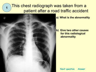
  • 2. 1 This chest radiograph was taken from a patient after a road traffic accident a) What is the abnormality b) Give two other causes for this radiological abnormality Next question Answer
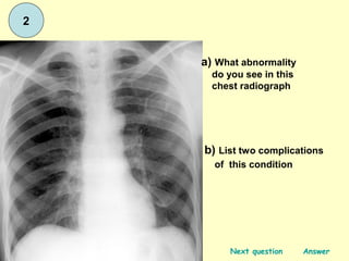
  • 3. 2 a) What abnormality do you see in this chest radiograph b) List two complications of this condition Next question Answer
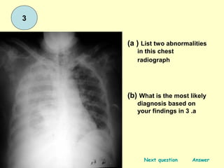
  • 4. 3 (a ) List two abnormalities in this chest radiograph (b) What is the most likely diagnosis based on your findings in 3 .a Next question Answer
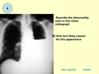
  • 5. 4 a. Describe the abnormality seen in this chest radiograph b) Give two likely causes for this appearance Next question Answer
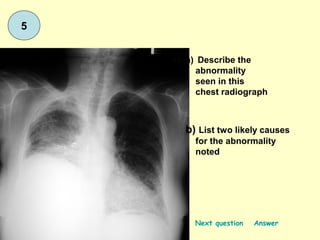
  • 6. 5 a) Describe the abnormality seen in this chest radiograph b) List two likely causes for the abnormality noted Next question Answer
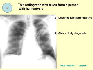
  • 7. This radiograph was taken from a person 6 with hemoptysis a) Describe two abnormalities b) Give a likely diagnosis Next question Answer
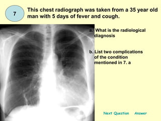
  • 8. This chest radiograph was taken from a 35 year old 7 man with 5 days of fever and cough. a. What is the radiological diagnosis b. List two complications of the condition mentioned in 7. a Next Question Answer
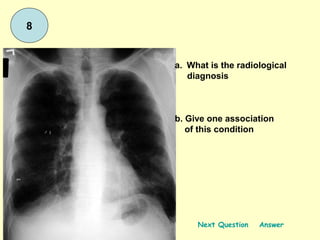
  • 9. 8 a. What is the radiological diagnosis b. Give one association of this condition Next Question Answer
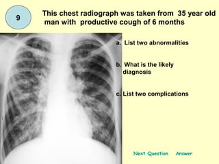
  • 10. This chest radiograph was taken from 35 year old 9 man with productive cough of 6 months a. List two abnormalities b. What is the likely diagnosis c. List two complications Next Question Answer
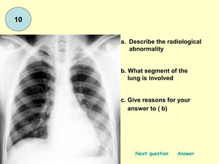
  • 11. 10 a. Describe the radiological abnormality b. What segment of the lung is involved c. Give reasons for your answer to ( b) Next question Answer
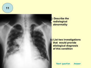
  • 12. 11 a) Describe the radiological abnormality b) List two investigations that would provide etiological diagnosis of this condition Next question Answer
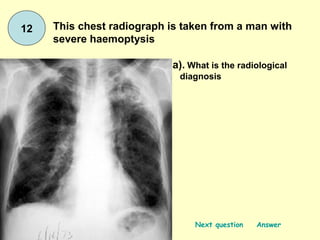
  • 13. 12 This chest radiograph is taken from a man with severe haemoptysis a). What is the radiological diagnosis Next question Answer
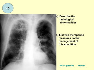
  • 14. 13 a) Describe the radiological abnormalities b) List two therapeutic measures in the management of this condition Next question Answer
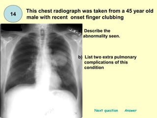
  • 15. This chest radiograph was taken from a 45 year old 14 male with recent onset finger clubbing a) Describe the abnormality seen. b) List two extra pulmonary complications of this condition Next question Answer
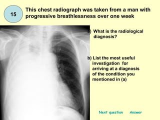
  • 16. This chest radiograph was taken from a man with 15 progressive breathlessness over one week a) What is the radiological diagnosis? b) List the most useful investigation for arriving at a diagnosis of the condition you mentioned in (a) Next question Answer
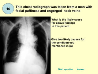
  • 17. This chest radiograph was taken from a man with 16 facial puffiness and engorged neck veins a) What is the likely cause for above findings in this patient b) Give two likely causes for the condition you mentioned in (a) Next question Answer
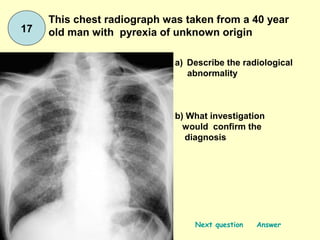
  • 18. This chest radiograph was taken from a 40 year 17 old man with pyrexia of unknown origin a) Describe the radiological abnormality b) What investigation would confirm the diagnosis Next question Answer
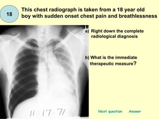
  • 19. This chest radiograph is taken from a 18 year old 18 boy with sudden onset chest pain and breathlessness a) Right down the complete radiological diagnosis b) What is the immediate therapeutic measure? Next question Answer
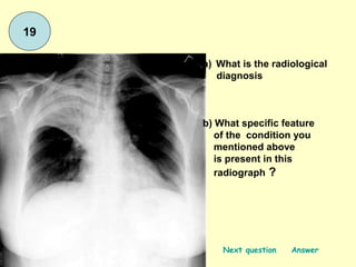
  • 20. 19 a) What is the radiological diagnosis b) What specific feature of the condition you mentioned above is present in this radiograph ? Next question Answer
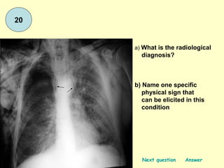
  • 21. 20 a) What is the radiological diagnosis? b) Name one specific physical sign that can be elicited in this condition Next question Answer
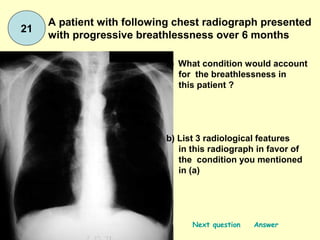
  • 22. A patient with following chest radiograph presented 21 with progressive breathlessness over 6 months a) What condition would account for the breathlessness in this patient ? b) List 3 radiological features in this radiograph in favor of the condition you mentioned in (a) Next question Answer
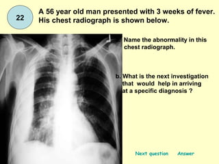
  • 23. A 56 year old man presented with 3 weeks of fever. 22 His chest radiograph is shown below. a. Name the abnormality in this chest radiograph. b. What is the next investigation that would help in arriving at a specific diagnosis ? Next question Answer
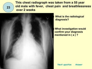
  • 24. This chest radiograph was taken from a 55 year 23 old male with fever, chest pain and breathlessness over 2 weeks a) What is the radiological diagnosis? b) What investigation would confirm your diagnosis mentioned in ( a ) ? Next question Answer
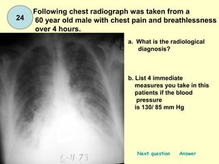
  • 25. Following chest radiograph was taken from a 24 60 year old male with chest pain and breathlessness over 4 hours. a. What is the radiological diagnosis? b. List 4 immediate measures you take in this patients if the blood pressure is 130/ 85 mm Hg Next question Answer
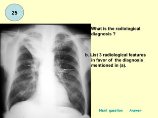
  • 26. 25 a. What is the radiological diagnosis ? b. List 3 radiological features in favor of the diagnosis mentioned in (a). Next question Answer
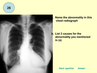
  • 27. 26 a. Name the abnormality in this chest radiograph b. List 3 causes for the abnormality you mentioned in (a) Next question Answer
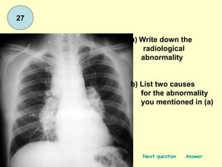
  • 28. 27 a) Write down the radiological abnormality b) List two causes for the abnormality you mentioned in (a) Next question Answer
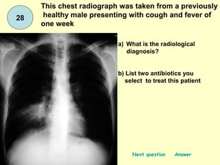
  • 29. This chest radiograph was taken from a previously 28 healthy male presenting with cough and fever of one week a) What is the radiological diagnosis? b) List two antibiotics you select to treat this patient Next question Answer
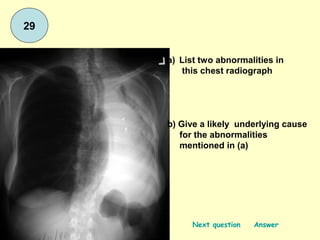
  • 30. 29 a) List two abnormalities in this chest radiograph b) Give a likely underlying cause for the abnormalities mentioned in (a) Next question Answer
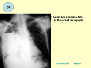
  • 31. 30 a) Name two abnormalities in this chest radiograph Introductions Answer
  • 32. This chest radiograph was taken from a 35 year old 7 man with 5 days of fever and cough. a. What is the radiological diagnosis b. List two complications of the condition mentioned in 7. a Next Question Answer
  • 33. 8 a. What is the radiological diagnosis b. Give one association of this condition Next Question Answer
  • 34. This chest radiograph was taken from 35 year old 9 man with productive cough of 6 months a. List two abnormalities b. What is the likely diagnosis c. List two complications Next Question Answer
  • 35. 10 a. Describe the radiological abnormality b. What segment of the lung is involved c. Give reasons for your answer to ( b) Next question Answer
  • 36. 11 a) Describe the radiological abnormality b) List two investigations that would provide etiological diagnosis of this condition Next question Answer
  • 37. 12 This chest radiograph is taken from a man with severe haemoptysis a). What is the radiological diagnosis Next question Answer
  • 38. 13 a) Describe the radiological abnormalities b) List two therapeutic measures in the management of this condition Next question Answer
  • 39. This chest radiograph was taken from a 45 year old 14 male with recent onset finger clubbing a) Describe the abnormality seen. b) List two extra pulmonary complications of this condition Next question Answer
  • 40. This chest radiograph was taken from a man with 15 progressive breathlessness over one week a) What is the radiological diagnosis? b) List the most useful investigation for arriving at a diagnosis of the condition you mentioned in (a) Next question Answer
  • 41. This chest radiograph was taken from a man with 16 facial puffiness and engorged neck veins a) What is the likely cause for above findings in this patient b) Give two likely causes for the condition you mentioned in (a) Next question Answer
  • 42. This chest radiograph was taken from a 40 year 17 old man with pyrexia of unknown origin a) Describe the radiological abnormality b) What investigation would confirm the diagnosis Next question Answer
  • 43. This chest radiograph is taken from a 18 year old 18 boy with sudden onset chest pain and breathlessness a) Right down the complete radiological diagnosis b) What is the immediate therapeutic measure? Next question Answer
  • 44. 19 a) What is the radiological diagnosis b) What specific feature of the condition you mentioned above is present in this radiograph ? Next question Answer
  • 45. 20 a) What is the radiological diagnosis? b) Name one specific physical sign that can be elicited in this condition Next question Answer
  • 46. A patient with following chest radiograph presented 21 with progressive breathlessness over 6 months a) What condition would account for the breathlessness in this patient ? b) List 3 radiological features in this radiograph in favor of the condition you mentioned in (a) Next question Answer
  • 47. A 56 year old man presented with 3 weeks of fever. 22 His chest radiograph is shown below. a. Name the abnormality in this chest radiograph. b. What is the next investigation that would help in arriving at a specific diagnosis ? Next question Answer
  • 48. This chest radiograph was taken from a 55 year 23 old male with fever, chest pain and breathlessness over 2 weeks a) What is the radiological diagnosis? b) What investigation would confirm your diagnosis mentioned in ( a ) ? Next question Answer
  • 49. Following chest radiograph was taken from a 24 60 year old male with chest pain and breathlessness over 4 hours. a. What is the radiological diagnosis? b. List 4 immediate measures you take in this patients if the blood pressure is 130/ 85 mm Hg Next question Answer
  • 50. 25 a. What is the radiological diagnosis ? b. List 3 radiological features in favor of the diagnosis mentioned in (a). Next question Answer
  • 51. 26 a. Name the abnormality in this chest radiograph b. List 3 causes for the abnormality you mentioned in (a) Next question Answer
  • 52. 27 a) Write down the radiological abnormality b) List two causes for the abnormality you mentioned in (a) Next question Answer
  • 53. This chest radiograph was taken from a previously 28 healthy male presenting with cough and fever of one week a) What is the radiological diagnosis? b) List two antibiotics you select to treat this patient Next question Answer
  • 54. 29 a) List two abnormalities in this chest radiograph b) Give a likely underlying cause for the abnormalities mentioned in (a) Next question Answer
  • 55. 30 a) Name two abnormalities in this chest radiograph Introductions Answer
  • 56. Answer 1 a) air ( gas ) under the diaphragm / ruptured viscus organ ( 06 marks ) b) following laporoscopy ( 04 marks) Notes perforation of bowel or stomach following trauma or other pathology such as peptic ulcer can give rise to gas under the diaphragm sign. Inflation of air at laporoscopy also can give rise to this radiological sign. Back Next question
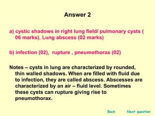
  • 57. Answer 2 a) cystic shadows in right lung field/ pulmonary cysts ( 06 marks), Lung abscess (02 marks) b) infection (02), rupture , pneumothorax (02) Notes cysts in lung are characterized by rounded, thin walled shadows. When are filled with fluid due to infection, they are called abscess. Abscesses are characterized by an air fluid level. Sometimes these cysts can rupture giving rise to pneumothorax. Back Next question
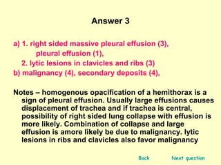
  • 58. Answer 3 a) 1. right sided massive pleural effusion (3), pleural effusion (1), 2. lytic lesions in clavicles and ribs (3) b) malignancy (4), secondary deposits (4), Notes homogenous opacification of a hemithorax is a sign of pleural effusion. Usually large effusions causes displacement of trachea and if trachea is central, possibility of right sided lung collapse with effusion is more likely. Combination of collapse and large effusion is amore likely be due to malignancy. lytic lesions in ribs and clavicles also favor malignancy Back Next question
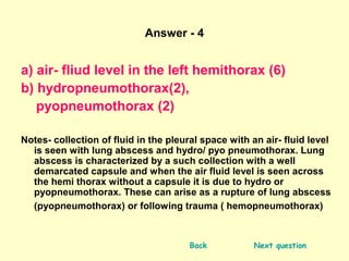
  • 59. Answer - 4 a) air- fliud level in the left hemithorax (6) b) hydropneumothorax(2), pyopneumothorax (2) Notes- collection of fluid in the pleural space with an air- fluid level is seen with lung abscess and hydro/ pyo pneumothorax. Lung abscess is characterized by a such collection with a well demarcated capsule and when the air fluid level is seen across the hemi thorax without a capsule it is due to hydro or pyopneumothorax. These can arise as a rupture of lung abscess (pyopneumothorax) or following trauma ( hemopneumothorax) Back Next question
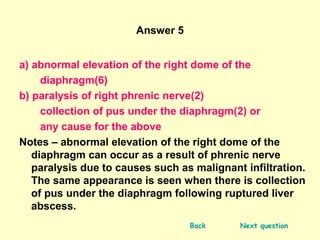
  • 60. Answer 5 a) abnormal elevation of the right dome of the diaphragm(6) b) paralysis of right phrenic nerve(2) collection of pus under the diaphragm(2) or any cause for the above Notes abnormal elevation of the right dome of the diaphragm can occur as a result of phrenic nerve paralysis due to causes such as malignant infiltration. The same appearance is seen when there is collection of pus under the diaphragm following ruptured liver abscess. Back Next question
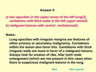
  • 61. Answer 6 a) two opacities in the upper zones of the left lung(3), cavitations with thick walls in the left upper zone(3) b) malignant lesions with central cavitations(4) Notes- Lung opacities with irregular margins are features of either primary or secondary malignancy. Cavitations within the lesion also favor this. Cavitations with thick irregular walls are more in favor of a malignant lesions. Always look for erosion of ribs, hilar lymh node enlargement (which are not present in this case) when there is suspicious malignant lesions in the lung. Back Next question
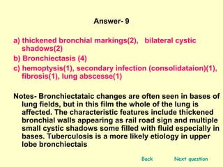
  • 62. Answer- 9 a) thickened bronchial markings(2), bilateral cystic shadows(2) b) Bronchiectasis (4) c) hemoptysis(1), secondary infection (consolidataion)(1), fibrosis(1), lung abscesse(1) Notes- Bronchiectataic changes are often seen in bases of lung fields, but in this film the whole of the lung is affected. The characteristic features include thickened bronchial walls appearing as rail road sign and multiple small cystic shadows some filled with fluid especially in bases. Tuberculosis is a more likely etiology in upper lobe bronchiectais Back Next question
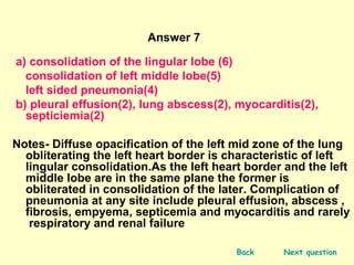
  • 63. Answer 7 a) consolidation of the lingular lobe (6) consolidation of left middle lobe(5) left sided pneumonia(4) b) pleural effusion(2), lung abscess(2), myocarditis(2), septiciemia(2) Notes- Diffuse opacification of the left mid zone of the lung obliterating the left heart border is characteristic of left lingular consolidation.As the left heart border and the left middle lobe are in the same plane the former is obliterated in consolidation of the later. Complication of pneumonia at any site include pleural effusion, abscess , fibrosis, empyema, septicemia and myocarditis and rarely respiratory and renal failure Back Next question
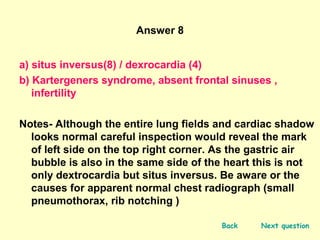
  • 64. Answer 8 a) situs inversus(8) / dexrocardia (4) b) Kartergeners syndrome, absent frontal sinuses , infertility Notes- Although the entire lung fields and cardiac shadow looks normal careful inspection would reveal the mark of left side on the top right corner. As the gastric air bubble is also in the same side of the heart this is not only dextrocardia but situs inversus. Be aware or the causes for apparent normal chest radiograph (small pneumothorax, rib notching ) Back Next question
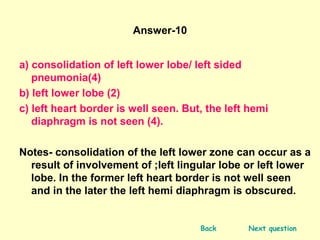
  • 65. Answer-10 a) consolidation of left lower lobe/ left sided pneumonia(4) b) left lower lobe (2) c) left heart border is well seen. But, the left hemi diaphragm is not seen (4). Notes- consolidation of the left lower zone can occur as a result of involvement of ;left lingular lobe or left lower lobe. In the former left heart border is not well seen and in the later the left hemi diaphragm is obscured. Back Next question
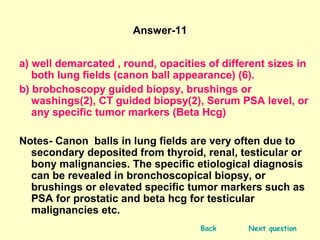
  • 66. Answer-11 a) well demarcated , round, opacities of different sizes in both lung fields (canon ball appearance) (6). b) brobchoscopy guided biopsy, brushings or washings(2), CT guided biopsy(2), Serum PSA level, or any specific tumor markers (Beta Hcg) Notes- Canon balls in lung fields are very often due to secondary deposited from thyroid, renal, testicular or bony malignancies. The specific etiological diagnosis can be revealed in bronchoscopical biopsy, or brushings or elevated specific tumor markers such as PSA for prostatic and beta hcg for testicular malignancies etc. Back Next question
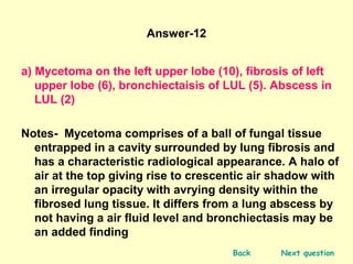
  • 67. Answer-12 a) Mycetoma on the left upper lobe (10), fibrosis of left upper lobe (6), bronchiectaisis of LUL (5). Abscess in LUL (2) Notes- Mycetoma comprises of a ball of fungal tissue entrapped in a cavity surrounded by lung fibrosis and has a characteristic radiological appearance. A halo of air at the top giving rise to crescentic air shadow with an irregular opacity with avrying density within the fibrosed lung tissue. It differs from a lung abscess by not having a air fluid level and bronchiectasis may be an added finding Back Next question
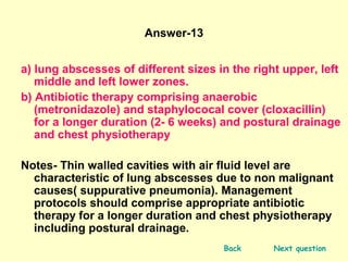
  • 68. Answer-13 a) lung abscesses of different sizes in the right upper, left middle and left lower zones. b) Antibiotic therapy comprising anaerobic (metronidazole) and staphylococal cover (cloxacillin) for a longer duration (2- 6 weeks) and postural drainage and chest physiotherapy Notes- Thin walled cavities with air fluid level are characteristic of lung abscesses due to non malignant causes( suppurative pneumonia). Management protocols should comprise appropriate antibiotic therapy for a longer duration and chest physiotherapy including postural drainage. Back Next question
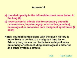
  • 69. Answer-14 a) rounded opacity in the left middle zone/ mass lesion in the lung (6) b) hypercalcemia, effects due to secondary deposits ( convulsions, hepatomegaly, obstructive jaundice), neurological or endocrine para malignant syndromes (2 each) Notes- rounded lung lesions with the given history is more likely to be due to a malignant lung lesion. Primary lung cancer can leads to a variety of extra pulmonary effects including neurological, endocrine and other systemic effects. Back Next question
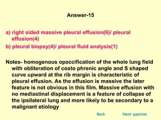
  • 70. Answer-15 a) right sided massive pleural effusion(6)/ pleural effusion(4) b) pleural biopsy(4)/ pleural fluid analysis(1) Notes- homogenous opaccification of the whole lung field with obliteration of costo phrenic angle and S shaped curve upward at the rib margin is characteristic of pleural effusion. As the effusion is massive the later feature is not obvious in this film. Massive effusion with no mediastinal displacement is a feature of collapse of the ipsilateral lung and more likely to be secondary to a malignant etiology Back Next question
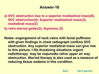
  • 71. Answer-16 a) SVC obstruction due to a seperior mediastinal mass(6), SVC obstruction(4), Superior mediastinal mass(3), mediatinal mass(2) b) retro sternal goiter(2), thymoma (2) Notes- engorgement of neck veins with facial puffiness with given findings in chest radiograph confims SVC obstruction. Any superior mediatinal mass can give rise to this picture. I life threatning situations urgent radiotherapy may be reqiuredto relive upper air way obstruction. Steriod therapy is also used as a measure of reducing tissue oedema in this condition. Back Next question
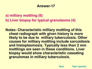
  • 72. Answer-17 a) milliary mottling (6) b) Liver biopsy for typical granulaoma (4) Notes- Characteristic milliary mottling of this chest radiograph with given history is more likely to be due to milliary tuberculosis. Other causes for milliary mottling include sarcoidosis and histoplasmosis. Typically less than 2 mm mottlings are seen in these conditions. Liver biopsy would show characteristic caseating granulomas in milliary tuberculosis. Back Next question
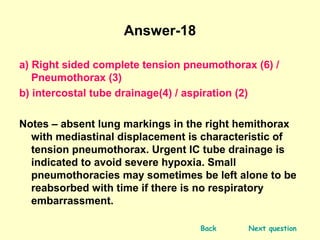
  • 73. Answer-18 a) Right sided complete tension pneumothorax (6) / Pneumothorax (3) b) intercostal tube drainage(4) / aspiration (2) Notes absent lung markings in the right hemithorax with mediastinal displacement is characteristic of tension pneumothorax. Urgent IC tube drainage is indicated to avoid severe hypoxia. Small pneumothoracies may sometimes be left alone to be reabsorbed with time if there is no respiratory embarrassment. Back Next question
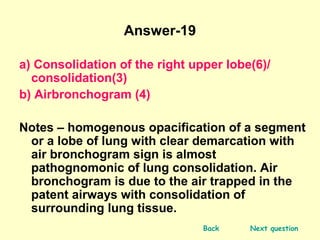
  • 74. Answer-19 a) Consolidation of the right upper lobe(6)/ consolidation(3) b) Airbronchogram (4) Notes homogenous opacification of a segment or a lobe of lung with clear demarcation with air bronchogram sign is almost pathognomonic of lung consolidation. Air bronchogram is due to the air trapped in the patent airways with consolidation of surrounding lung tissue. Back Next question
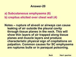
  • 75. Answer-20 a) Subcutaneous emphysema (6) b) crepitus elicited over chest wall (4) Notes rupture of alveoli or airways can cause leaking of air outside the pleural cavity through tissue planes in the neck. This will show thin layers of air trapped along tissue planes and muscle layers and produce characteristic physical sign of crepitations on palpation. Common causes for SC emphysema are ruptures bulle or in paraquat poisoning. Back Next question
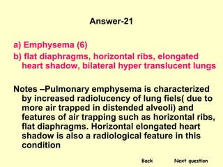
  • 76. Answer-21 a) Emphysema (6) b) flat diaphragms, horizontal ribs, elongated heart shadow, bilateral hyper translucent lungs Notes Pulmonary emphysema is characterized by increased radiolucency of lung fiels( due to more air trapped in distended alveoli) and features of air trapping such as horizontal ribs, flat diaphragms. Horizontal elongated heart shadow is also a radiological feature in this condition Back Next question
  • 77. Answer-22 a) Encysted pleural effusion(6), pleural based tumor(6), pleural effusion(3) b) Ultra sound examination of thorax (4), CT scan of thorax (4) Notes- Encysment of fluid between the two layers of pleura with fibrous adhesions would produce encysted effusions. Due to holding up of fluid in the closed cavity general appearance of an effusion is not seen. Once the nature of the material encysted is determined by the ultrasound scan and locate the best position to aspirate , diagnostic aspirations can be done. Back Next question
  • 78. Answer-23 a) Pericardial effusion(6) / cardiomegaly(2) b) 2-D echocardiogram (4) Notes- enlarged cardiac silhouette with sharply demarcated borders with appearance of a water bag is due to pericardial effusion. There may be features of cardiac failure. 2-D echocardiogram is the confirmatory investigation. Etiology could be primary cardiac pathology ( pericarditis, severe CCF or non cardiac, infections, such as tuberculosis Back Next question
  • 79. Answer-24 a) Pulmonary oedema (6) / left heart failure(3) b) Prop-up the patient, Oxygen , intravenous fruesemide, Intravenous morphine, Notes - enlarged heart shadow, pulmonary venous congestion with dilated upper lobar veins, hilar congestion and appearance of kerlys lines with or without small pleural effusion are features of pulmonary oedema. In some cases of pulmonary oedema heart size can be normal Back Next question
  • 80. Answer-25 a) right upper lobe collapse/ fibrosis (6) b) elevation of right hilum, displacement of trachea to right side, elevation of the inter lobar fissure Notes- fibrosis or collapse of the right upper lobe would cause pulling up of lung tissue and other surrounding organs towards it. In fibrosis the streaky shadows will be seen and in collapse, its cause such as a foreign body or a tumor/ lymph node may be evident. Back Next question
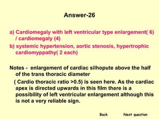
  • 81. Answer-26 a) Cardiomegaly with left ventricular type enlargement( 6) / cardiomegaly (4) b) systemic hypertension, aortic stenosis, hypertrophic cardiomyppathy( 2 each) Notes - enlargement of cardiac silhopute above the half of the trans thoracic diameter ( Cardio thoracic ratio >0.5) is seen here. As the cardiac apex is directed upwards in this film there is a possibility of left ventricular enlargement although this is not a very reliable sign. Back Next question
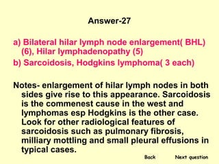
  • 82. Answer-27 a) Bilateral hilar lymph node enlargement( BHL) (6), Hilar lymphadenopathy (5) b) Sarcoidosis, Hodgkins lymphoma( 3 each) Notes- enlargement of hilar lymph nodes in both sides give rise to this appearance. Sarcoidosis is the commenest cause in the west and lymphomas esp Hodgkins is the other case. Look for other radiological features of sarcoidosis such as pulmonary fibrosis, milliary mottling and small pleural effusions in typical cases. Back Next question
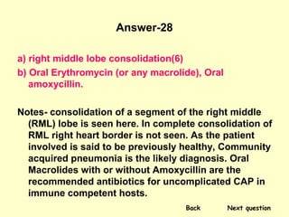
  • 83. Answer-28 a) right middle lobe consolidation(6) b) Oral Erythromycin (or any macrolide), Oral amoxycillin. Notes- consolidation of a segment of the right middle (RML) lobe is seen here. In complete consolidation of RML right heart border is not seen. As the patient involved is said to be previously healthy, Community acquired pneumonia is the likely diagnosis. Oral Macrolides with or without Amoxycillin are the recommended antibiotics for uncomplicated CAP in immune competent hosts. Back Next question
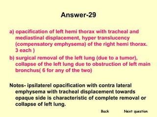
  • 84. Answer-29 a) opacification of left hemi thorax with tracheal and mediastinal displacement, hyper translucency (compensatory emphysema) of the right hemi thorax. 3 each ) b) surgical removal of the left lung (due to a tumor), collapse of the left lung due to obstruction of left main bronchus( 6 for any of the two) Notes- ipsilaterel opacification with contra lateral emphysema with tracheal displacement towards opaque side is characteristic of complete removal or collapse of left lung. Back Next question
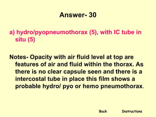
  • 85. Answer- 30 a) hydro/pyopneumothorax (5), with IC tube in situ (5) Notes- Opacity with air fluid level at top are features of air and fluid within the thorax. As there is no clear capsule seen and there is a intercostal tube in place this film shows a probable hydro/ pyo or hemo pneumothorax. Back Instructions