Principles of new attachment
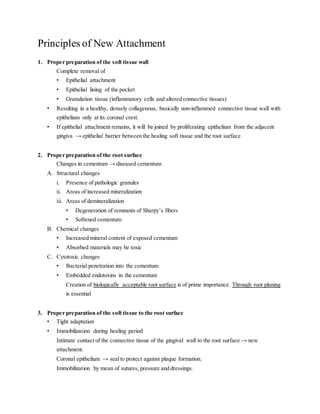
- 1. Principles of New Attachment 1. Proper preparation of the soft tissue wall Complete removal of ŌĆó Epithelial attachment ŌĆó Epithelial lining of the pocket ŌĆó Granulation tissue (inflammatory cells and altered connective tissues) ŌĆó Resulting in a healthy, densely collagenous, basically non-inflammed connective tissue wall with epithelium only at its coronal crest. ŌĆó If epithelial attachment remains, it will be joined by proliferating epithelium from the adjacent gingiva ŌåÆ epithelial barrier between the healing soft tissue and the root surface 2. Proper preparation of the root surface Changes in cementum ŌåÆ diseased cementum A. Structural changes i. Presence of pathologic granules ii. Areas of increased mineralization iii. Areas of demineralization ŌĆó Degeneration of remnants of SharpyŌĆÖs fibers ŌĆó Softened cementum B. Chemical changes ŌĆó Increased mineral content of exposed cementum ŌĆó Absorbed materials may be toxic C. Cytotoxic changes ŌĆó Bacterial penetration into the cementum ŌĆó Embedded endotoxins in the cementum Creation of biologically acceptable root surface is of prime importance. Through root planing is essential 3. Proper preparation of the soft tissue to the root surface ŌĆó Tight adaptation ŌĆó Immobilization during healing period Intimate contact of the connective tissue of the gingival wall to the root surface ŌåÆ new attachment. Coronal epithelium ŌåÆ sealto protect against plaque formation. Immobilization by mean of sutures, pressure and dressings.
- 2. 4. Effective etiology control pre-surgically and post-surgically Effective plaque control ŌĆö essential for optimal healing leading to maximum new attachment. Periodontal procedures to achieve newattachment 1. Subgingival curettage 2. GTR 3. Excisional new attachment procedure 4. Modified excisional new attachment procedure 5. Access flap operation


