Purvi shah anatomy of fibula ppt
- 1. Anatomy Of Fibula LL Bone Part-5 BY PURVI SHAH B.P.T
- 2. Fibula
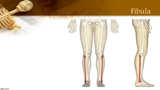
- 3. Fibula ’āśIt is the lateral and smaller bone of the leg. ’āśIt is very thin as compared to the tibia. ’āśIt is homologous with the ulna of the upper limb.
- 4. Side Determination ’āśUpper end and head is expanded in all directions. ’āśLower end or lateral malleolus is expanded AP direction and flattened from side to side. ’āśMedial side of lower end bears a triangular articular facet anteriorly and a deep or malleolar fossa posteriorly.
- 5. Features ’āśFibula has an upper end, shaft and lower end. ’āśUPPER END: Expanded ’ā╝ Superior surfaces bears a circular articulate facet; which articulate with lateral condyle of tibia. ’ā╝ The Apex of head styloid process projects upwards from its posterolateral aspect. ’ā╝ Constriction below head is known as neck of the fibula.
- 7. Features ’āśSHAFT: Three Borders 1.Anterior 2.posterior 3.interosseous ’ā╝Three Surfaces 1.Medial (it is very narrow measuring 1 mm or less) 2.Lateral 3.Posterior
- 9. Features ’āśLOWER END: The tips of lateral malleolus is 0.5cm lower than medial malleolus. ’ā╝ Anterior surface is 1.5 cm posterior to the that of the medial malleolus. ’ā╝ It has four surfaces: Anterior (rough) Posterior Lateral (subcutaneous) Medial ( bears triangular articular facets)
- 10. Functions ’āśThe fibula's role is to act as and attachment for muscles, as well as providing stability of the ankle joint. ’āśThe fibula is a non-weight-bearing bone.
- 11. Attachments ’āśProximally, ’ā╝Extensor digitorum longus Superior 3/4 of medial border of fibula, ’ā╝Extensor hallucis longus Middle of anterior surface ’ā╝Fibularis tertius Inferior 1/3 of anterior surface ’ā╝Fibularis longus Fibular head and superior 2/3 of lateral surface
- 12. ’ā╝Fibularis brevis Inferior 2/3 of lateral surface ’ā╝Soleus Fibular head (posterior) and superior 1/4 of posterior surface ’ā╝Flexor hallucis longus: Inferior 2/3 of posterior surface ’ā╝Flexor digitorum longus Via tendon ’ā╝Tibialis posterior Posterior surface Attachments
- 14. Attachments
- 15. Blood Supply ’āśPeroneal artery gives off nutrient artery for fibula, which enters bone on its posterior surface.
- 16. Articulations ’āśProximal tibiofibular joint ’āśDistal tibiofibular joint
- 17. Ossifications ’āśThe fibula ossifies from the one primary and two secondary centres. ’āśThe primary centre for the shaft appears during 8th week of intrauterine life. ’āśA secondary centre for lower end appears during first year and fuse with shaft by about 16 years.
- 18. Clinical Anatomy ’āśThe upper and lower ends of the fibula are subcutaneous and palpable ’āśCommon peroneal nerve can be rolled against the neck of fibula; this nerve commonly injured here. ’āśIt leads of foot drop ’āśThe fibula is an ideal spare bone for a bone graft.
- 19. ’āśThe most common area for fibula fractures are 2-6 cm proximal to distal end of fibula. ’āśIt is associated with ankle fracture.
- 21. Thank You


