Radiographic positioning of Upper limb (ELBOW & HUMERUS)
- 1. RADIOGRAPHIC ANATOMY & POSITIONING 0F UPPER LIMB 1.ELBOW 2.HUMERUS Prepared by: NAASIR Wani
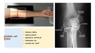
- 2. ELBOW- AP ? INDICATION: Trauma, Dislocation, Pathological condition. ? PATIENT POSITION: Patient sits beside the X ray table with the patient face directed away from the primary beam. ? POSITION OF THE ELBOW: The arm and forearm are fully extended, the elbow in question is placed in supine position in the middle of the half of cassette. Arm and forearm should be in one level. Identification marker must be present. ? CENTERING POINT AND THE DIRECTION OF THE X TUBE: The vertical radiation beam is directed towards 01cm below the epicondyles of the humerus.
- 3. ELBOW-->AP VIEW ? Distance: 100cm ? Exposure factor: ? MAS 08-10 KVP 45-50 ? Collimation: Yes ? Cassette size- 8x10''
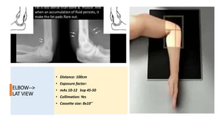
- 4. ELBOW- LATERAL ? INDICATION: Trauma, Arthritis, Pathological condition. ? PATIENT POSITION: The patient sits beside the X Table with the face directed away from the primary beam. ? POSITION OF THE ELBOW: The elbow is fixed making an right angle btw the arm and forearm. The elbow is placed in the middle of the other half of the cassette. Sand bag is placed on the arm for immobilization. Identification marks must be present. ? CENTERING AND THE DIRECTION OF THE X TUBE: The vertical beam of radiation is directed to the lateral epicondyle of the humerus.
- 5. ELBOW--> LAT VIEW ? Distance: 100cm ? Exposure factor: ? mAs 10-12 kvp 45-50 ? Collimation: Yes ? Cassette size: 8x10''
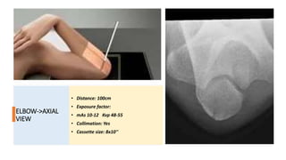
- 6. ELBOW- AXIAL ? INDICATION: Its an additional projection to check the healing process of supracondylar fracture or trauma to the olecranon process. ? PATIENT POSITION; The patient sits beside the X ray table with the face of the patient directed away from the radiation. ? POSITION OF THE ELBOW; the elbow is fully flexed and the palm of the hand facing the shoulder, the posterior side of the palm rest on the film. ? CENTERING POINT AND THE DIRECTION OF THE X TUBE; The vertical beam of radiation is directed to a point 5cm distal to the olecranon process. The beam is angled roughly 45degree towards the long axis of humerus
- 7. ELBOW->AXIAL VIEW ? Distance: 100cm ? Exposure factor: ? mAs 10-12 Kvp 48-55 ? Collimation: Yes ? Cassette size: 8x10''
- 8. AP POSITION OF ELBOW WITH ARM IN CONTACT ? INDICATION: Supra-condylar fracture. ? Since this type of fracture needs careful handling because of the importance of vessels and nerves which passes in the elbow region. ? Four projections are taken to demonstrate supra-condylar fracture: ? A) Elbow arm in contact with the film. ? B) Elbow forearm in contact with the film. ? PATIENT POSITION: the patient sits beside X table, with face directed away from the prim X beam. ? POSITIONING OF THE ELBOW: Arm is placed in contact with the film. Identification marker must be present. ? CENTERING POINT AND THE DIRECTION OF THE X TUBE: The vertical central X beam is directed 1cm below the condyles of the humerus.
- 9. ELBOW->AP ARM IN CONTACT ? Distance: 100cm ? Exposure factor: ? MAS 10-12 KVP 48-55 ? Collimation- Yes ? Cassette size: 10x12''
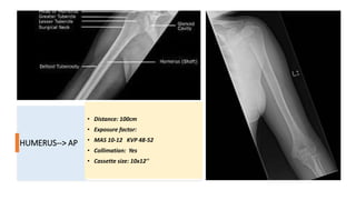
- 10. HUMERUS-AP ? INDICATION; trauma or any pathological condition. ? PATIENT POSITION; the patient should be seated upright or in the standing position with the back of the shoulder resting on the buckey. The patient should suspend respiration for the exposure. ? POSITION OF HUMERUS; slightly abduct the arm and supinate the hand. The coronal planes of the epicondyles and the large receptor plane should be parallel. ? DIRECTION AND CENTERING OF X RAY BEAM: Perpendicular to the image receptor and directed to the middle portion of the humerus and centre to the large receptor.
- 11. HUMERUS--> AP ? Distance: 100cm ? Exposure factor: ? MAS 10-12 KVP 48-52 ? Collimation: Yes ? Cassette size: 10x12''
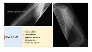
- 12. HUMERUS- LATERAL ? INDICATION; To exclude fracture or displaced fracture. ? PATIENT POSITION; The patient should be seated upright or in a standing position with the back of their shoulder resting on the buckey. ? PART POSITION; Internally rotate the arm and then flex the elbow about 90degree. Place the hand over hip unless contraindicated due to pain or fracture. ? DIRECTION AND CENTERING OF X BEAM; Perpendicular to the image receptor at the middle portion of the humerus and centre of the image receptor.
- 13. HUMERUS-LAT ? Distance- 100cm ? Exposure Factor: ? MAS 10-12 KVP 52-55 ? Collimation- Yes ? Cassette size¨C 10x12''
- 14. THANKYOU NAASIR MOHIDIN WANI BSC MEDICAL IMAGING TECHNOLOGY (Radio) STUDENT OF SHARDA UNIVERSITY (2018014297)

