際際滷

際際滷Share a Scribd company logo
Faculty In-Charge:
Dr. R. P Tripathi
Mentor:
Dr. Priyadarshini
Presented By:
Dr. Tarun Goyal
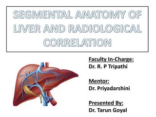
Functionally, can be divided into three lobes: right, left, and caudate.
The right lobe of the liver is separated from the left by the main lobar
fissure, which passes through the gallbladder fossa to the inferior vena
cava (IVC).
The right lobe of the liver can be further divided into anterior and
posterior segments by the right intersegmental fissure.
The left intersegmental fissure divides the left lobe into medial and
lateral segments.
The caudate lobe is situated on the posterior aspect of the liver, with the
IVC as its posterior border and the fissure for the ligamentum venosum
as its anterior border.
The major hepatic veins course between the lobes and segments (interlobar
and intersegmental).
The middle hepatic vein courses within the main lobar fissure and separates
the anterior segment of the right lobe from the medial segment of the left.
The right hepatic vein runs within the right intersegmental fissure and divides
the right lobe into anterior and posterior segments.
The major branches of the right and left portal veins run centrally within the
segments (intrasegmental), with the exception of the ascending portion of the
left portal vein, which runs in the left intersegmental fissure.
The left intersegmental fissure, which separates the medial segment of the left
lobe from the lateral segment, can be divided into cranial, middle, and caudal
sections.
The left hepatic vein forms the boundary of the cranial third, the ascending
branch of the left portal vein represents the middle third, and the fissure for
the ligamentum teres acts as the most caudal division of the left lobe.
Couinaud classification
It divides the liver into eight functionally independent segments.
Each segment has its own vascular inflow, outflow and biliary drainage.
In the centre of each segment there is a branch of the portal vein, hepatic
artery and bile duct.
In the periphery of each segment there is vascular outflow through the
hepatic veins.
Right hepatic vein divides the right lobe into anterior and posterior
segments.
Middle hepatic vein divides the liver into right and left lobes (or right and
left hemi liver). This plane runs from the inferior vena cava to the gallbladder
fossa.
The Falciform ligament divides the left lobe into a medial- segment IV and
a lateral part - segment II and III.
The portal vein divides the liver into upper and lower segments.
The left and right portal veins branch superiorly and inferiorly to project into
the centre of each segment.
SEGMENTAL
ANATOMY
Radiological anatomy of liver segments
遺看顎庄稼温顎糸s壊艶乙馨艶稼岳壊
Radiological anatomy of liver segments
Couinaud divided the liver into a
functional left and right liver by a
main portal scissurae containing the
middle hepatic vein.
This is known as Cantlie's line.
Cantlie's line runs from the middle of
the gallbladder fossa anteriorly to the
inferior vena cava posteriorly.
This figure is a transverse image
through the superior liver segments,
that are divided by the right and
middle hepatic veins and the
falciform ligament.
This is a transverse image at the level of
the left portal vein.
At this level the left portal vein divides the
left lobe into the superior segments (II
and IVa) and the inferior segments (III
and IVb).
The left portal vein is at a higher level than
the right portal vein
This image is at the level of the right
portal vein.
At this level the right portal vein divides
the right lobe of the liver into superior
segments (VII and VIII) and the
inferior segments (V and VI).
The level of the right portal vein is
inferior to the level of the left portal
vein.
At the level of the splenic vein, which
is below the level of the right portal
vein, only the inferior segments are
visible
Radiological anatomy of liver segments
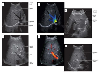
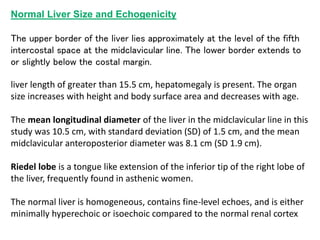
Normal Liver Size and Echogenicity
The upper border of the liver lies approximately at the level of the fifth
intercostal space at the midclavicular line. The lower border extends to
or slightly below the costal margin.
liver length of greater than 15.5 cm, hepatomegaly is present. The organ
size increases with height and body surface area and decreases with age.
The mean longitudinal diameter of the liver in the midclavicular line in this
study was 10.5 cm, with standard deviation (SD) of 1.5 cm, and the mean
midclavicular anteroposterior diameter was 8.1 cm (SD 1.9 cm).
Riedel lobe is a tongue like extension of the inferior tip of the right lobe of
the liver, frequently found in asthenic women.
The normal liver is homogeneous, contains fine-level echoes, and is either
minimally hyperechoic or isoechoic compared to the normal renal cortex
Cross-
Section
Imaging
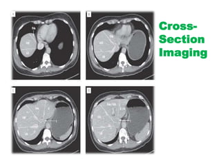
How to separate liver segments
on cross sectional imaging
Left lobe: lateral(II/III) vs
medial segment (IVA/B)
Extrapolate a line along the falciform
ligament superiorly to the confluence
of the left and middle hepatic veins at
the IVC (blue line).
Left vs Right lobe: IVA/B vs
V/VIII
Extrapolate a line from the
gallbladder fossa superiorly along the
middle hepatic vein to the IVC (red
line).
Right lobe: anterior (V/VIII) vs
posterior segment (VI/VII)
Extrapolate a line along the right
hepatic vein from the IVC inferiorly to
the lateral liver margin (green line).
Radiological anatomy of liver segments

More Related Content

Radiological anatomy of liver segments

  • 1. Faculty In-Charge: Dr. R. P Tripathi Mentor: Dr. Priyadarshini Presented By: Dr. Tarun Goyal
  • 2. Functionally, can be divided into three lobes: right, left, and caudate. The right lobe of the liver is separated from the left by the main lobar fissure, which passes through the gallbladder fossa to the inferior vena cava (IVC). The right lobe of the liver can be further divided into anterior and posterior segments by the right intersegmental fissure. The left intersegmental fissure divides the left lobe into medial and lateral segments. The caudate lobe is situated on the posterior aspect of the liver, with the IVC as its posterior border and the fissure for the ligamentum venosum as its anterior border.
  • 3. The major hepatic veins course between the lobes and segments (interlobar and intersegmental). The middle hepatic vein courses within the main lobar fissure and separates the anterior segment of the right lobe from the medial segment of the left. The right hepatic vein runs within the right intersegmental fissure and divides the right lobe into anterior and posterior segments. The major branches of the right and left portal veins run centrally within the segments (intrasegmental), with the exception of the ascending portion of the left portal vein, which runs in the left intersegmental fissure. The left intersegmental fissure, which separates the medial segment of the left lobe from the lateral segment, can be divided into cranial, middle, and caudal sections. The left hepatic vein forms the boundary of the cranial third, the ascending branch of the left portal vein represents the middle third, and the fissure for the ligamentum teres acts as the most caudal division of the left lobe.
  • 4. Couinaud classification It divides the liver into eight functionally independent segments. Each segment has its own vascular inflow, outflow and biliary drainage. In the centre of each segment there is a branch of the portal vein, hepatic artery and bile duct. In the periphery of each segment there is vascular outflow through the hepatic veins. Right hepatic vein divides the right lobe into anterior and posterior segments. Middle hepatic vein divides the liver into right and left lobes (or right and left hemi liver). This plane runs from the inferior vena cava to the gallbladder fossa. The Falciform ligament divides the left lobe into a medial- segment IV and a lateral part - segment II and III. The portal vein divides the liver into upper and lower segments. The left and right portal veins branch superiorly and inferiorly to project into the centre of each segment. SEGMENTAL ANATOMY
  • 16. Couinaud divided the liver into a functional left and right liver by a main portal scissurae containing the middle hepatic vein. This is known as Cantlie's line. Cantlie's line runs from the middle of the gallbladder fossa anteriorly to the inferior vena cava posteriorly. This figure is a transverse image through the superior liver segments, that are divided by the right and middle hepatic veins and the falciform ligament.
  • 17. This is a transverse image at the level of the left portal vein. At this level the left portal vein divides the left lobe into the superior segments (II and IVa) and the inferior segments (III and IVb). The left portal vein is at a higher level than the right portal vein This image is at the level of the right portal vein. At this level the right portal vein divides the right lobe of the liver into superior segments (VII and VIII) and the inferior segments (V and VI). The level of the right portal vein is inferior to the level of the left portal vein.
  • 18. At the level of the splenic vein, which is below the level of the right portal vein, only the inferior segments are visible
  • 21. Normal Liver Size and Echogenicity The upper border of the liver lies approximately at the level of the fifth intercostal space at the midclavicular line. The lower border extends to or slightly below the costal margin. liver length of greater than 15.5 cm, hepatomegaly is present. The organ size increases with height and body surface area and decreases with age. The mean longitudinal diameter of the liver in the midclavicular line in this study was 10.5 cm, with standard deviation (SD) of 1.5 cm, and the mean midclavicular anteroposterior diameter was 8.1 cm (SD 1.9 cm). Riedel lobe is a tongue like extension of the inferior tip of the right lobe of the liver, frequently found in asthenic women. The normal liver is homogeneous, contains fine-level echoes, and is either minimally hyperechoic or isoechoic compared to the normal renal cortex
  • 23. How to separate liver segments on cross sectional imaging Left lobe: lateral(II/III) vs medial segment (IVA/B) Extrapolate a line along the falciform ligament superiorly to the confluence of the left and middle hepatic veins at the IVC (blue line). Left vs Right lobe: IVA/B vs V/VIII Extrapolate a line from the gallbladder fossa superiorly along the middle hepatic vein to the IVC (red line). Right lobe: anterior (V/VIII) vs posterior segment (VI/VII) Extrapolate a line along the right hepatic vein from the IVC inferiorly to the lateral liver margin (green line).