This document discusses the history and techniques of radiotherapy in ENT. It begins with the discovery of x-rays in 1895 and progresses to modern technologies like IMRT, IGRT, proton beam therapy and SBRT. It covers the physics, biology and mechanisms of radiation therapy. Key aspects of radiotherapy for head and neck cancers like dosimetry, fractionation schedules, acute and chronic toxicities are summarized. Newer conformal techniques aim to reduce normal tissue damage while adequately treating tumors.
Role of radiotherapy and chemotherapy in oral cavity cancerDr.Rashmi Yadav
油
Radiotherapy and chemotherapy play important roles in the treatment of oral cavity cancer alongside surgery. Radiotherapy is often used as the primary treatment for early stage cancers or as an adjuvant treatment with surgery for more advanced cancers. Chemotherapy is commonly used neoadjuvantly or concurrently with radiotherapy to improve treatment outcomes, especially for advanced cancers. Brachytherapy can also be used as a radiation boost for early stage oral cavity cancers. The goals of treatment are maximizing local tumor control while preserving function and minimizing side effects through a multidisciplinary approach.
Radiotherapy is used in the management of oral cancer for curative and palliative purposes. It can be delivered as primary treatment combined with chemotherapy for organ preservation or after surgery as adjuvant treatment. Newer radiotherapy techniques like IMRT allow higher doses to be delivered to tumors while reducing damage to nearby organs. Side effects depend on treatment dose and area irradiated, and may include mucositis, xerostomia, skin changes, osteoradionecrosis and rare complications like carotid rupture. Ongoing research aims to reduce toxicity through altered fractionation schedules and novel delivery methods.
This document discusses various methods of altered fractionation in radiation therapy. It begins with an introduction to the history of fractionation based on experiments in the 1920s-1930s. It then covers radiobiological principles of fractionation including repair of sublethal damage and reoxygenation. Various methods of altered fractionation are classified and described, including hyperfractionation, accelerated fractionation using different schedules, hypofractionation, and continuous hyperfractionated accelerated radiotherapy (CHART). Comparisons are made between conventional and altered fractionation schedules.
This document discusses hypofractionation in the treatment of head and neck cancers. It begins by outlining outcomes for different stages of disease, then discusses how fraction size, total dose, and treatment time impact treatment. Hypofractionation can counter tumor repopulation and improve local control. Studies show hypofractionation is effective for early disease, palliative cases, and can be safely delivered using simultaneous integrated boost with IMRT. Severe toxicity is low while disease control remains high. Extreme hypofractionation with SBRT also provides good local control with acceptable toxicity.
The document discusses staging of cervical cancer according to FIGO staging criteria, with stages ranging from 0 to IVB. It then summarizes guidelines from ESMO on indications for adjuvant treatment, which include chemoradiation as the best option for stages IB2 to IVA. Finally, it reviews several studies that have investigated neoadjuvant chemotherapy followed by radiation therapy versus radiation therapy alone in advanced cervical cancer.
This document discusses the management of oropharyngeal cancer. It begins by stating the goals of treatment are functional organ preservation and minimizing treatment-induced morbidity while maintaining cure rates. For early stage disease, single modality radiotherapy or surgery is usually sufficient. For advanced stages, surgery plus radiation or chemoradiation are recommended based on risk factors. It then discusses treatment options and outcomes for different subsites within the oropharynx and the benefits of adjuvant therapy or altered fractionation schedules for radiotherapy.
Smart radiotherapy aims to precisely target tumor cells while sparing healthy cells. New techniques described in the document include using hypoxic cell sensitizers to target hypoxic tumor regions, anti-angiogenic agents to inhibit tumor blood vessels, and nanoparticles to enhance radiation dose and selectively deliver drugs. Molecular imaging helps optimize treatment by identifying tumor characteristics. Combining radiotherapy with immunotherapy or targeted depletion of host cells may also improve outcomes. Overall, the document discusses developing more precise radiation approaches through better understanding of tumor biology and microenvironment.
This document discusses radiotherapy for head and neck malignancies. It covers the history, mechanisms of action including direct and indirect damage to cells, characteristics of radiotherapy such as its effects on dividing cells, and the quantitative difference in response between malignant and normal cells. Fractions of radiotherapy doses are discussed including hypofractionation and hyperfractionation. Factors such as oxygen levels, cell cycle phases, and radiobiology are also summarized. The document then discusses various radiotherapy techniques including external beam, brachytherapy, and unsealed radioactive sources. Treatment planning and quality control of radiotherapy are also briefly mentioned.
1) Radioiodine treatment (RAI) utilizes the ability of thyroid cancer cells to absorb iodine to effectively treat well-differentiated thyroid cancer. However, less differentiated cancers may not absorb iodine and become resistant to RAI treatment.
2) The document discusses the physics and physiology of radioactive iodine treatment, factors that influence treatment effectiveness like stunning, and guidelines around patient preparation and follow up including dosimetry approaches and activity levels for treatment.
3) Optimal RAI treatment requires differentiating cancer cells to absorb sufficient iodine doses without exceeding radiation safety limits, and the document discusses approaches and considerations for individualizing safe and effective treatment.
Role of radiotherapy in oral ca ppt for csmsailesh kumar
油
Radiotherapy plays an important role in the management of oral cancer. It uses ionizing radiation to deliver tumoricidal doses to cancer while limiting dose to surrounding normal tissues. There are several techniques of radiotherapy including external beam therapy and brachytherapy. Factors like total radiation dose, chemotherapy combination, treatment delays and interruptions can influence effectiveness. Complications include both early side effects like mucositis and late effects like osteoradionecrosis. Advances in radiotherapy techniques aim to improve targeting accuracy and reduce side effects.
The document discusses various radiation fractionation schedules used in cancer treatment. It begins with an overview of conventional fractionation, which divides the total radiation dose into smaller daily doses to allow healthy cells to repair sublethal damage between fractions. It then explores the radiobiological rationale of the 5 R's of fractionation - repair, redistribution, reoxygenation, repopulation, and radiosensitivity. The document discusses various altered fractionation schedules including hyperfractionation, accelerated fractionation, split-course, and hypofractionation, explaining how each schedule aims to improve the therapeutic ratio for cancer patients.
This document discusses the use of radioactive iodine (131I) for diagnosis and treatment of thyroid cancer. Some key points:
- 131I localizes in thyroid tissue and can be used to ablate thyroid remnants after surgery or treat thyroid cancer metastases. It emits beta and gamma radiation.
- For remnant ablation, lower doses (30-100 mCi) are usually sufficient while higher doses (100-200 mCi) may be needed for more aggressive cancers. Success rates are similar between low vs high doses and thyroid hormone withdrawal vs rhTSH.
- Post-therapy scans 2-10 days after treatment can identify additional metastases not seen on diagnostic scans in 10-26% of
Fractionation refers to dividing the total radiation dose into smaller doses given over multiple treatment sessions. The standard fractionation schedule gives doses of 1.8-2 Gy daily, 5 days a week, to the total prescribed dose. Altered fractionation schedules like hyperfractionation, accelerated fractionation, and hypofractionation aim to improve tumor control by modifying dose, dose per fraction, and treatment time based on differences in radiation response between tumors and normal tissues. Hyperfractionation gives smaller doses more times to allow a higher total dose within normal tissue tolerance. Accelerated fractionation reduces time to decrease tumor repopulation but maintains standard dose per fraction. Hypofractionation gives higher doses fewer times per week to shorten treatment time.
Radiation therapy involves using radiation to treat cancer and other diseases. It works by damaging malignant cells' DNA to stop their growth and reproduction. There are several types of radiation therapy including external beam radiation, brachytherapy, and systemic radioisotope therapy. The dose and fractionation of radiation is tailored to each patient's situation. Factors like tumor type and location, patient health, and treatment intent are considered. Radiation can be used curatively, adjuvantly, palliatively, or therapeutically depending on the case. Both malignant and some non-malignant conditions can be treated with radiation.
Radiation Therapy of cancer patients _2013.pptBaljeet Kaur
油
Radiation therapy involves using radiation to treat cancer and other diseases. It works by damaging cancer cell DNA to stop tumor growth. There are several types of radiation therapy including external beam radiation from a machine, brachytherapy using radioactive sources inside the body, and systemic radioisotope therapy giving radioactive substances. The total dose is divided over multiple sessions to allow normal cells time to recover while continuing to damage cancer cells. Factors like tumor type, size, location and patient health determine the treatment intent of being curative, adjuvant, or palliative. Radiation therapy has risks but is effective for treating many cancer types when used appropriately.
Common Types of Cancer Treatment
Surgery: An operation where doctors cut out tissue with cancer cells. Chemotherapy: Special medicines that shrink or kill cancer cells that we cannot see. Radiation therapy: Using high-energy rays (similar to X-rays) to kill cancer cells.
Treatment of Advanced stage of Carcinoma Cervix & Ca cervix in Pregnancy.pptxMuthuRamanK3
油
1. Treatment of advanced stage of carcinoma cervix: Radiotherapy (including brachytherapy, teletherapy and adjuvant radiotherapy), Chemotherapy and Chemoradiotherapy;
2. Ca Cervix in Pregnancy: Includes flowchart for screening and management
HDR brachytherapy for Non-Melanoma Skin cancersAli Bagheri
油
This document discusses the use of interventional radiotherapy called brachytherapy for the treatment of non-melanoma skin cancers. Brachytherapy involves placing radioactive sources close to or inside the tumor to deliver a high radiation dose. It has advantages over surgery or external beam radiation including a steep dose gradient that limits dose to nearby tissues, shorter treatment time, and a higher biological effect. The document outlines indications, evidence, techniques, dosimetry, and the author's experiences using brachytherapy for various skin cancers and keloids.
Radiosensitizers and Biological modifiers in RadiotherapySubhash Thakur
油
This document discusses various agents that can be used as radiosensitizers to increase the lethal effects of radiation therapy on tumor cells. It describes radiosensitization as using a physical, chemical, or pharmacological intervention to differentially increase the effects of radiation on tumor cells over normal tissues. Some key radiosensitizers mentioned include hyperthermia, carbogen with nicotinamide to overcome tumor hypoxia, and chemotherapeutic drugs that inhibit DNA repair pathways in tumor cells. The ideal radiosensitizer lacks toxicity, has a potent sensitizing effect in both normoxic and hypoxic cells, and can be conveniently administered in an outpatient setting.
Nasopharyngeal carcinoma has unique features including association with Epstein-Barr virus and a high risk of distant metastases. Definitive radiotherapy is the primary treatment, with intensity-modulated radiotherapy improving outcomes. Concurrent chemoradiotherapy provides significantly improved progression-free and overall survival compared to radiotherapy alone for locally advanced disease based on a landmark randomized trial. Brachytherapy may be used as a boost for early-stage tumors following external beam radiotherapy.
This document discusses various strategies for radiosensitization and radioprotection in radiation oncology. It describes how radiation directly damages DNA and how free radicals generated can be fixed by oxygen, leading to cell death. It then discusses chronic and acute hypoxia in tumors and various mechanisms and agents that can be used for radiosensitization, including hypoxic cell sensitizers like misonidazole and nimorazole, hypoxic cytotoxins like mitomycin C, and strategies like blood transfusion, hyperbaric oxygen, and carbogen inhalation. It also discusses the radioprotector amifostine and its mechanisms of action and administration. Glutamine is mentioned as a potential protector against radiation-induced
Radioimmunotherapy involves coupling a radionuclide to monoclonal antibodies targeting tumor antigens. This allows targeted delivery of radiation to cancer cells while sparing healthy cells. Radionuclides like Yttrium-90 and Iodine-131 are attached to antibodies using chelators or chemical reactions. Low dose radiation is continuously delivered over days, inducing cell death through various radiobiological effects like the crossfire effect. Radioimmunotherapy has been used to treat hematologic malignancies like NHL and solid tumors. Clinical trials show radioimmunotherapy improves progression-free survival when used as consolidation after chemotherapy for follicular lymphoma.
1) The document discusses optimal practice in radiation treatment for head and neck cancer in the 21st century, focusing on balancing treatment targets and sparing normal tissues using available technology and expertise.
2) It reviews treatment options and approaches for different stages of head and neck cancer, highlighting evidence that altered fractionation and chemoradiation can improve outcomes over standard radiation alone.
3) Challenges of implementing intensity-modulated radiation therapy (IMRT) for head and neck cancer are discussed, as well as examples of how IMRT can improve target coverage and tissue sparing compared to conventional techniques.
This chapter discusses fractionated radiation and the dose-rate effect. It covers operational classifications of radiation damage including potentially lethal damage and sublethal damage. Fractionation allows for repair of sublethal damage through processes like reassortment and repopulation. The dose-rate effect results from increased repair at lower dose rates. Examples are provided for both in vitro and in vivo models. Brachytherapy techniques like intracavitary and interstitial brachytherapy are also summarized.
This document discusses radiation therapy for head and neck tumors. It covers the biologic effects of radiation, different modalities like external beam radiation therapy and brachytherapy, and fractionation schedules. Changes in radiation therapy over time are also reviewed, including increased doses, use of chemotherapy with radiation (chemoRT), and intensity modulated radiation therapy (IMRT). The document discusses both acute and long term tissue effects of radiation therapy and increasing post-treatment morbidity as techniques have advanced.
Prelims of Kaun TALHA : a Travel, Architecture, Lifestyle, Heritage and Activism quiz, organized by Conquiztadors, the Quiz society of Sri Venkateswara College under their annual quizzing fest El Dorado 2025.
More Related Content
Similar to Radiotherapy indications and complications (20)
This document discusses the management of oropharyngeal cancer. It begins by stating the goals of treatment are functional organ preservation and minimizing treatment-induced morbidity while maintaining cure rates. For early stage disease, single modality radiotherapy or surgery is usually sufficient. For advanced stages, surgery plus radiation or chemoradiation are recommended based on risk factors. It then discusses treatment options and outcomes for different subsites within the oropharynx and the benefits of adjuvant therapy or altered fractionation schedules for radiotherapy.
Smart radiotherapy aims to precisely target tumor cells while sparing healthy cells. New techniques described in the document include using hypoxic cell sensitizers to target hypoxic tumor regions, anti-angiogenic agents to inhibit tumor blood vessels, and nanoparticles to enhance radiation dose and selectively deliver drugs. Molecular imaging helps optimize treatment by identifying tumor characteristics. Combining radiotherapy with immunotherapy or targeted depletion of host cells may also improve outcomes. Overall, the document discusses developing more precise radiation approaches through better understanding of tumor biology and microenvironment.
This document discusses radiotherapy for head and neck malignancies. It covers the history, mechanisms of action including direct and indirect damage to cells, characteristics of radiotherapy such as its effects on dividing cells, and the quantitative difference in response between malignant and normal cells. Fractions of radiotherapy doses are discussed including hypofractionation and hyperfractionation. Factors such as oxygen levels, cell cycle phases, and radiobiology are also summarized. The document then discusses various radiotherapy techniques including external beam, brachytherapy, and unsealed radioactive sources. Treatment planning and quality control of radiotherapy are also briefly mentioned.
1) Radioiodine treatment (RAI) utilizes the ability of thyroid cancer cells to absorb iodine to effectively treat well-differentiated thyroid cancer. However, less differentiated cancers may not absorb iodine and become resistant to RAI treatment.
2) The document discusses the physics and physiology of radioactive iodine treatment, factors that influence treatment effectiveness like stunning, and guidelines around patient preparation and follow up including dosimetry approaches and activity levels for treatment.
3) Optimal RAI treatment requires differentiating cancer cells to absorb sufficient iodine doses without exceeding radiation safety limits, and the document discusses approaches and considerations for individualizing safe and effective treatment.
Role of radiotherapy in oral ca ppt for csmsailesh kumar
油
Radiotherapy plays an important role in the management of oral cancer. It uses ionizing radiation to deliver tumoricidal doses to cancer while limiting dose to surrounding normal tissues. There are several techniques of radiotherapy including external beam therapy and brachytherapy. Factors like total radiation dose, chemotherapy combination, treatment delays and interruptions can influence effectiveness. Complications include both early side effects like mucositis and late effects like osteoradionecrosis. Advances in radiotherapy techniques aim to improve targeting accuracy and reduce side effects.
The document discusses various radiation fractionation schedules used in cancer treatment. It begins with an overview of conventional fractionation, which divides the total radiation dose into smaller daily doses to allow healthy cells to repair sublethal damage between fractions. It then explores the radiobiological rationale of the 5 R's of fractionation - repair, redistribution, reoxygenation, repopulation, and radiosensitivity. The document discusses various altered fractionation schedules including hyperfractionation, accelerated fractionation, split-course, and hypofractionation, explaining how each schedule aims to improve the therapeutic ratio for cancer patients.
This document discusses the use of radioactive iodine (131I) for diagnosis and treatment of thyroid cancer. Some key points:
- 131I localizes in thyroid tissue and can be used to ablate thyroid remnants after surgery or treat thyroid cancer metastases. It emits beta and gamma radiation.
- For remnant ablation, lower doses (30-100 mCi) are usually sufficient while higher doses (100-200 mCi) may be needed for more aggressive cancers. Success rates are similar between low vs high doses and thyroid hormone withdrawal vs rhTSH.
- Post-therapy scans 2-10 days after treatment can identify additional metastases not seen on diagnostic scans in 10-26% of
Fractionation refers to dividing the total radiation dose into smaller doses given over multiple treatment sessions. The standard fractionation schedule gives doses of 1.8-2 Gy daily, 5 days a week, to the total prescribed dose. Altered fractionation schedules like hyperfractionation, accelerated fractionation, and hypofractionation aim to improve tumor control by modifying dose, dose per fraction, and treatment time based on differences in radiation response between tumors and normal tissues. Hyperfractionation gives smaller doses more times to allow a higher total dose within normal tissue tolerance. Accelerated fractionation reduces time to decrease tumor repopulation but maintains standard dose per fraction. Hypofractionation gives higher doses fewer times per week to shorten treatment time.
Radiation therapy involves using radiation to treat cancer and other diseases. It works by damaging malignant cells' DNA to stop their growth and reproduction. There are several types of radiation therapy including external beam radiation, brachytherapy, and systemic radioisotope therapy. The dose and fractionation of radiation is tailored to each patient's situation. Factors like tumor type and location, patient health, and treatment intent are considered. Radiation can be used curatively, adjuvantly, palliatively, or therapeutically depending on the case. Both malignant and some non-malignant conditions can be treated with radiation.
Radiation Therapy of cancer patients _2013.pptBaljeet Kaur
油
Radiation therapy involves using radiation to treat cancer and other diseases. It works by damaging cancer cell DNA to stop tumor growth. There are several types of radiation therapy including external beam radiation from a machine, brachytherapy using radioactive sources inside the body, and systemic radioisotope therapy giving radioactive substances. The total dose is divided over multiple sessions to allow normal cells time to recover while continuing to damage cancer cells. Factors like tumor type, size, location and patient health determine the treatment intent of being curative, adjuvant, or palliative. Radiation therapy has risks but is effective for treating many cancer types when used appropriately.
Common Types of Cancer Treatment
Surgery: An operation where doctors cut out tissue with cancer cells. Chemotherapy: Special medicines that shrink or kill cancer cells that we cannot see. Radiation therapy: Using high-energy rays (similar to X-rays) to kill cancer cells.
Treatment of Advanced stage of Carcinoma Cervix & Ca cervix in Pregnancy.pptxMuthuRamanK3
油
1. Treatment of advanced stage of carcinoma cervix: Radiotherapy (including brachytherapy, teletherapy and adjuvant radiotherapy), Chemotherapy and Chemoradiotherapy;
2. Ca Cervix in Pregnancy: Includes flowchart for screening and management
HDR brachytherapy for Non-Melanoma Skin cancersAli Bagheri
油
This document discusses the use of interventional radiotherapy called brachytherapy for the treatment of non-melanoma skin cancers. Brachytherapy involves placing radioactive sources close to or inside the tumor to deliver a high radiation dose. It has advantages over surgery or external beam radiation including a steep dose gradient that limits dose to nearby tissues, shorter treatment time, and a higher biological effect. The document outlines indications, evidence, techniques, dosimetry, and the author's experiences using brachytherapy for various skin cancers and keloids.
Radiosensitizers and Biological modifiers in RadiotherapySubhash Thakur
油
This document discusses various agents that can be used as radiosensitizers to increase the lethal effects of radiation therapy on tumor cells. It describes radiosensitization as using a physical, chemical, or pharmacological intervention to differentially increase the effects of radiation on tumor cells over normal tissues. Some key radiosensitizers mentioned include hyperthermia, carbogen with nicotinamide to overcome tumor hypoxia, and chemotherapeutic drugs that inhibit DNA repair pathways in tumor cells. The ideal radiosensitizer lacks toxicity, has a potent sensitizing effect in both normoxic and hypoxic cells, and can be conveniently administered in an outpatient setting.
Nasopharyngeal carcinoma has unique features including association with Epstein-Barr virus and a high risk of distant metastases. Definitive radiotherapy is the primary treatment, with intensity-modulated radiotherapy improving outcomes. Concurrent chemoradiotherapy provides significantly improved progression-free and overall survival compared to radiotherapy alone for locally advanced disease based on a landmark randomized trial. Brachytherapy may be used as a boost for early-stage tumors following external beam radiotherapy.
This document discusses various strategies for radiosensitization and radioprotection in radiation oncology. It describes how radiation directly damages DNA and how free radicals generated can be fixed by oxygen, leading to cell death. It then discusses chronic and acute hypoxia in tumors and various mechanisms and agents that can be used for radiosensitization, including hypoxic cell sensitizers like misonidazole and nimorazole, hypoxic cytotoxins like mitomycin C, and strategies like blood transfusion, hyperbaric oxygen, and carbogen inhalation. It also discusses the radioprotector amifostine and its mechanisms of action and administration. Glutamine is mentioned as a potential protector against radiation-induced
Radioimmunotherapy involves coupling a radionuclide to monoclonal antibodies targeting tumor antigens. This allows targeted delivery of radiation to cancer cells while sparing healthy cells. Radionuclides like Yttrium-90 and Iodine-131 are attached to antibodies using chelators or chemical reactions. Low dose radiation is continuously delivered over days, inducing cell death through various radiobiological effects like the crossfire effect. Radioimmunotherapy has been used to treat hematologic malignancies like NHL and solid tumors. Clinical trials show radioimmunotherapy improves progression-free survival when used as consolidation after chemotherapy for follicular lymphoma.
1) The document discusses optimal practice in radiation treatment for head and neck cancer in the 21st century, focusing on balancing treatment targets and sparing normal tissues using available technology and expertise.
2) It reviews treatment options and approaches for different stages of head and neck cancer, highlighting evidence that altered fractionation and chemoradiation can improve outcomes over standard radiation alone.
3) Challenges of implementing intensity-modulated radiation therapy (IMRT) for head and neck cancer are discussed, as well as examples of how IMRT can improve target coverage and tissue sparing compared to conventional techniques.
This chapter discusses fractionated radiation and the dose-rate effect. It covers operational classifications of radiation damage including potentially lethal damage and sublethal damage. Fractionation allows for repair of sublethal damage through processes like reassortment and repopulation. The dose-rate effect results from increased repair at lower dose rates. Examples are provided for both in vitro and in vivo models. Brachytherapy techniques like intracavitary and interstitial brachytherapy are also summarized.
This document discusses radiation therapy for head and neck tumors. It covers the biologic effects of radiation, different modalities like external beam radiation therapy and brachytherapy, and fractionation schedules. Changes in radiation therapy over time are also reviewed, including increased doses, use of chemotherapy with radiation (chemoRT), and intensity modulated radiation therapy (IMRT). The document discusses both acute and long term tissue effects of radiation therapy and increasing post-treatment morbidity as techniques have advanced.
Prelims of Kaun TALHA : a Travel, Architecture, Lifestyle, Heritage and Activism quiz, organized by Conquiztadors, the Quiz society of Sri Venkateswara College under their annual quizzing fest El Dorado 2025.
Blind Spots in AI and Formulation Science Knowledge Pyramid (Updated Perspect...Ajaz Hussain
油
This presentation delves into the systemic blind spots within pharmaceutical science and regulatory systems, emphasizing the significance of "inactive ingredients" and their influence on therapeutic equivalence. These blind spots, indicative of normalized systemic failures, go beyond mere chance occurrences and are ingrained deeply enough to compromise decision-making processes and erode trust.
Historical instances like the 1938 FD&C Act and the Generic Drug Scandals underscore how crisis-triggered reforms often fail to address the fundamental issues, perpetuating inefficiencies and hazards.
The narrative advocates a shift from reactive crisis management to proactive, adaptable systems prioritizing continuous enhancement. Key hurdles involve challenging outdated assumptions regarding bioavailability, inadequately funded research ventures, and the impact of vague language in regulatory frameworks.
The rise of large language models (LLMs) presents promising solutions, albeit with accompanying risks necessitating thorough validation and seamless integration.
Tackling these blind spots demands a holistic approach, embracing adaptive learning and a steadfast commitment to self-improvement. By nurturing curiosity, refining regulatory terminology, and judiciously harnessing new technologies, the pharmaceutical sector can progress towards better public health service delivery and ensure the safety, efficacy, and real-world impact of drug products.
APM event hosted by the South Wales and West of England Network (SWWE Network)
Speaker: Aalok Sonawala
The SWWE Regional Network were very pleased to welcome Aalok Sonawala, Head of PMO, National Programmes, Rider Levett Bucknall on 26 February, to BAWA for our first face to face event of 2025. Aalok is a member of APMs Thames Valley Regional Network and also speaks to members of APMs PMO Interest Network, which aims to facilitate collaboration and learning, offer unbiased advice and guidance.
Tonight, Aalok planned to discuss the importance of a PMO within project-based organisations, the different types of PMO and their key elements, PMO governance and centres of excellence.
PMOs within an organisation can be centralised, hub and spoke with a central PMO with satellite PMOs globally, or embedded within projects. The appropriate structure will be determined by the specific business needs of the organisation. The PMO sits above PM delivery and the supply chain delivery teams.
For further information about the event please click here.
Digital Tools with AI for e-Content Development.pptxDr. Sarita Anand
油
This ppt is useful for not only for B.Ed., M.Ed., M.A. (Education) or any other PG level students or Ph.D. scholars but also for the school, college and university teachers who are interested to prepare an e-content with AI for their students and others.
Mate, a short story by Kate Grenvile.pptxLiny Jenifer
油
A powerpoint presentation on the short story Mate by Kate Greenville. This presentation provides information on Kate Greenville, a character list, plot summary and critical analysis of the short story.
Information Technology for class X CBSE skill SubjectVEENAKSHI PATHAK
油
These questions are based on cbse booklet for 10th class information technology subject code 402. these questions are sufficient for exam for first lesion. This subject give benefit to students and good marks. if any student weak in one main subject it can replace with these marks.
Finals of Rass MELAI : a Music, Entertainment, Literature, Arts and Internet Culture Quiz organized by Conquiztadors, the Quiz society of Sri Venkateswara College under their annual quizzing fest El Dorado 2025.
Research & Research Methods: Basic Concepts and Types.pptxDr. Sarita Anand
油
This ppt has been made for the students pursuing PG in social science and humanities like M.Ed., M.A. (Education), Ph.D. Scholars. It will be also beneficial for the teachers and other faculty members interested in research and teaching research concepts.
How to attach file using upload button Odoo 18Celine George
油
In this slide, well discuss on how to attach file using upload button Odoo 18. Odoo features a dedicated model, 'ir.attachments,' designed for storing attachments submitted by end users. We can see the process of utilizing the 'ir.attachments' model to enable file uploads through web forms in this slide.
APM People Interest Network Conference 2025
- Autonomy, Teams and Tension
- Oliver Randall & David Bovis
- Own Your Autonomy
Oliver Randall
Consultant, Tribe365
Oliver is a career project professional since 2011 and started volunteering with APM in 2016 and has since chaired the People Interest Network and the North East Regional Network. Oliver has been consulting in culture, leadership and behaviours since 2019 and co-developed HPTM速an off the shelf high performance framework for teams and organisations and is currently working with SAS (Stellenbosch Academy for Sport) developing the culture, leadership and behaviours framework for future elite sportspeople whilst also holding down work as a project manager in the NHS at North Tees and Hartlepool Foundation Trust.
David Bovis
Consultant, Duxinaroe
A Leadership and Culture Change expert, David is the originator of BTFA and The Dux Model.
With a Masters in Applied Neuroscience from the Institute of Organisational Neuroscience, he is widely regarded as the Go-To expert in the field, recognised as an inspiring keynote speaker and change strategist.
He has an industrial engineering background, majoring in TPS / Lean. David worked his way up from his apprenticeship to earn his seat at the C-suite table. His career spans several industries, including Automotive, Aerospace, Defence, Space, Heavy Industries and Elec-Mech / polymer contract manufacture.
Published in Londons Evening Standard quarterly business supplement, James Caans Your business Magazine, Quality World, the Lean Management Journal and Cambridge Universities PMA, he works as comfortably with leaders from FTSE and Fortune 100 companies as he does owner-managers in SMEs. He is passionate about helping leaders understand the neurological root cause of a high-performance culture and sustainable change, in business.
Session | Own Your Autonomy The Importance of Autonomy in Project Management
#OwnYourAutonomy is aiming to be a global APM initiative to position everyone to take a more conscious role in their decision making process leading to increased outcomes for everyone and contribute to a world in which all projects succeed.
We want everyone to join the journey.
#OwnYourAutonomy is the culmination of 3 years of collaborative exploration within the Leadership Focus Group which is part of the APM People Interest Network. The work has been pulled together using the 5 HPTM速 Systems and the BTFA neuroscience leadership programme.
https://www.linkedin.com/showcase/apm-people-network/about/
2. DEFINITION
The application of radiation for the purpose of
therapeutic gain.
Radiation is the process of emitting radiant energy in
the form of waves or particle.
Aim of Radiotherapy
Delivering tumoricidal dose to a defined target
volume
Respecting the normal tissue tolerance
Trying to achieve an optimum therapeutic ratio
Improvement of quality of life
3. MECHANISM OF RADIOTHERAPY
Radiations act on biological cells by ionization
which can be direct or indirect.
Direct ionization(Electron, Heavy Ion
radiations)
photons hit matter
produce secondary electrons
damage DNA directly
4. Indirect ionization( X-ray, Neutrons )
photons hit matter
produce secondary electrons
with water and oxygen free radicals damage DNA
Indirect ionization is more common ( > 2/3rd )
Radiations also causes cell reproduction failure by
inhibiting mitosis.
5. BIOLOGICAL EFFECTS OF RADIATIONS
Affects cell by breaking strands of DNA
Single strand breaks - repairable than double strand
breaks which are lethal.
Sub-lethal damage refers to DNA damage that can be
repaired if given the right cellular conditions and
sufficient time.
Sub-lethal damage is less efficient in tumor cells,
hypoxia or low pH.
DNA damage is repairable with low radiation dose
and the relation is proportional. (Absorbable dose )
6. TUMOR CELL KINETICS AND RESPONSE TO RT
CELL PROLIFERATION AND LOSS
radiosensitive in the G(2)-M phase,
less sensitive in the G(1) phase
least sensitive during the latter part of the S phase
INTRINSIC RADIOSENSITIVITY
Lesser the SF2 better the radiosensitivity/prognosis.
X-rays are more effective on cells which have a
greater reproductive activity
HYPOXIA - radioresistant
7. The response of a tumour to radiotherapy is dependent
upon
Inherent Radiosensitivity
Tumour cell Repopulation
Redistribution through the cell cycle (G2/M is the
most sensitive phase of the cell cycle, late-S phase is
the most radioresistant)
Repair of radiation induced damage- atleast 6hrs to
complete.
Reoxygenation of tumour tissues between fractions
i.e. Hypoxic cells re-oxygenate and become
radiosensitive.
8. FACTORS INFLUENCING THE EFFECTIVENESS OF RT
FACTOR CHANGE IN LOCAL CONTROL
CONCURRENT CHEMOTHERAPY
OR BIOLOGICAL
AGENTS
10% INCREASE
70Gy rather than 66Gy 5% INCREASE
DELAYS IN STARTING
RADIOTHERAPY
15% DECREASE PER MONTH
TREATMENT INTERRUPTIONS 1.4% DECREASE PER EXTRA
DAY
ANEMIA 10-15% DECREASE
SMOKING 10-15% DECREASE
9. TYPES OF RADIOTHERAPY
External Beam Radiotherapy or Teletherapy
Brachytherapy or Sealed Source Radiotherapy
Systemic Radioisotope therapy or Unsealed Source
Radiotherapy
10. EXTERNAL BEAM RADIOTHERAPY
Radiation beam is directed from a machine placed
outside the patient to a treatment volume located
within.
SOURCE OF RADIATIONS
X-Ray Machines Particle accelerators
Gamma rays i) LINAC
(Cobalt 60) ii) Betatron
iii)Cyclotron
iv) Nuclear Reactors
v) Radionuclides
11. COBALT 60 MACHINE
Gamma rays
Beam is weak
Cheap
Disadvantage : Decaying source causing reduced
output & requires change of source every 5-7 years
Does not give ideal depth dose and require
complicated plans to deliver the effective tumor dose
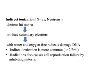
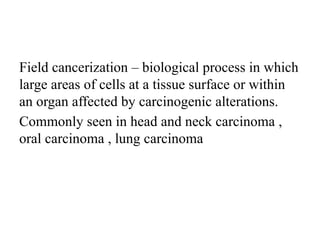
13. LINAC MACHINE[ Linear accelerator]
High, Medium , Low energy X-rays, Electrons
Beam is much superior.
Expensive
No decaying source
Good penetration
14. BRACHYTHERAPY
Radioactive material is introduced directly to within
the tumor or tumor bearing area.
Radium needles were first to be used.
Iridium (Ir-192) wire is the source of choice for
modern head and neck brachytherapy.
Brachytherapy can be temporary or permanent.
15. INDICATIONS OF BRACHYTHERAPY
Site oral cavity , base of tongue
Localized disease
Small (T1) lesion
Accessible site
Substantial local recurrence rate
Lesions away from bone
16. The temporary sources
are usually placed by a
technique called
Afterloading.
In afterloading a hollow
tube or applicator is
placed surgically in the
organ to be treated, and
the sources are loaded
into the applicator after
the applicator is
implanted.
17. This minimizes radiation exposure to health care
personnel.
Advantage - high dose in small area
Limitation - need for adequate radiation protection
and cant be done in cases where wider field
irradiation is required.
18. Doses in Brachytherapy
Low dose rate = <2 Gy/hr ( used in oral, lip, tongue
CA )
High dose rate = >12 Gy/hr
Pulsed dose rate (uncommon) = 2-12 Gy/hr
19. SYSTEMIC RADIOISOTOPE THERAPY
Systemic radioisotope therapy (RIT) is a
form of targeted therapy.
The radioisotopes are delivered through
infusion (into the bloodstream) or ingestion.
Examples : Infusion of meta-
iodobenzylguanidine (MIBG) to treat
neuroblastoma,
Oral iodine-131 to treat thyroid cancer or
thyrotoxicosis.
20. RADIOTHERAPY IN HEAD AND
NECK CANCER
More than 70% of oncological cases receive RT
Modalities of RT used in head and neck cases are
Definitive/Curative/Radical RT- Stage I-IV A
Palliative RT Stage IV B- IV C, unresectable /
metastatic disease with good general condition
Neo-Adjuvant RT- adjuvant treatment given prior to
definitive treatment
Concurrent treatment : Chemoradiation
21. PRE-RT ASSESSMENT
Examination under anesthesia
Tumor biopsy
Imaging CT/MRI of head and neck
CXR/ CT thorax
Full Blood count
Urea and electrolytes
LFT
Dietician assessment
Dental assessment
Speech Therapist
22. CONTRA-INDICATIONS OF RT
Cachexia
Anemia
Leukopenia
Acute septic states
Decompensated states of heart, liver, kidneys
Active tuberculosis
Extension of tumors to adjacent hollow organs
Growth into great blood vessels.
An inflammatory process
23. DOSIMETRY
It is the measurement, calculation and assessment of
the ionizing radiation dose absorbed by the human
body.
It can be internal or external dosimetry.
Medical dosimetry is the calculation of absorbed
dose and optimization of dose delivery in radiation
therapy.
Grays (Gy) is the S.I unit of absorbed dose
Sieverts is the S.I unit of dose equivalence.
1 Gy = 100 rads = 1J/kg
24. Dose required for disease control
Subclinical disease : 45-50Gy
Microscopic disease : 60-65Gy
Gross disease : 65-80Gy
25. Selection total radiation dose in head and neck cancer
depends on
Primary tumor
Neck node size
Fractionation
Clinical circumstances
Concurrent systemic therapy used or not
26. To ensure accurate radiation
therapy, the following
measures are taken
Positioning and immobilization
of patient ( moulded
thermoplastic shield, custom
made cabulite)
27. Target definition and delineation by imaging
(Gross tumor volume(GTV) , Clinical target
volume(CTV), Planning target volume(PTV)
Coverage of Organ at risk
Field arrangement ( Lateral parallel opposed
fields and a wedged pair field are the most
common used)
Beam Modification ( wedges and shielding )
28. RADIOTHERAPY FRACTIONATION
CONVENTIONAL FRACTIONATION
1.8-2Gy/day for 5 days/week.
Curative doses 66-70Gy in 33-35
Fractions over 6.5-7weeks
HYPERFRACTIONATION
<1.8Gy/day
Increases time
Reduces risk for late damage
Reduce effectiveness
29. HYPOFRACTIONATION
> 2Gy/day
Shorter duration
Limits tumor repopulation
Increases risk for late damage
ACCELERATED FRACTIONATION
Treatment time reduced
Hyperfractionation treating two-three times each day
Time between fractions should be minimum 6 hours
Reduced risk of normal tissue damage
31. DEFINITIVE /CURATIVE
RADIOTHERAPY
Dose of curative radiotherapy by EBRT is dependent on
the sites
The maximum dose limits are 70 Gy (2Gy/day) for the
following sites
Lip, oral cavity, oropharynx, hypopharynx, glottic
larynx, supraglottic larynx, occult primary, salivary
gland tumors
32. Cancer pharynx with high subclinical risk should be
given 2.12Gy/day radiations for total dose of 69.96
Gy for 6-7 weeks ( Mon-Fri ).
3D-Conformal RT and sequential planned IMRT 44-
50 Gy.
For IMRT, some suggest 54-63 Gy.
33. PALLIATIVE RADIOTHERAPY
Palliative RT considered in advanced cancer when
curative intent is not appropriate.
Basic principle of palliative RT is to avoid any
regimen that causes severe toxicities.
Hypofractionated regimen should be preferred in end
stage diseases.
34. Recommended RT regimens
50 Gy in 20 fractions
37.5 Gy in 5 fractions
30 Gy in 10 fractions
44.4 Gy in 12 fractions, in 3 cycles
35. POST-OPERATIVE RT
Post-operative RT is recommended based on
T-stage T3/T4 disease
Close or positive margins of excision
Depth of invasion
Multiple positive nodes ( without extracapsular nodal
spread)
Perineural/lymphatic/vascular invasion.
36. Higher doses of post-operative RT alone (60-66 Gy) or
with systemic therapy are recommended for the high-
risk features of extracapsular disease and/ or positive
margins.
The preferred interval is 6 weeks or less, between
resection and commencement of postoperative RT
37. FOLLOW-UPAFTER RT
Assessment of thyroid function (i.e. TSH test ever 6-
12 months)
Clinical assessment at 4-8 weeks or CT-evaluation
with or without contrast at 8 weeks after completion
of RT alone or chemoradiation.
Follow up 3-6 monthly
38. REIRRADIATION
Repeating course of RT carries greater risk of damage
to normal tissue.
Reirradiation has to be weighed against other
treatment options and against the risk and
consequences of normal tissue damage.
General rule-
Minimize dose to critical normal tissues esp. spinal
cord and optic tracts. ( IMRT, IGRT etc )
6 months gap between previous RT and reirradiation.
Addition of chemotherapy increases the effectiveness
of reirradiation without increasing late morbidity.
39. Field cancerization biological process in which
large areas of cells at a tissue surface or within
an organ affected by carcinogenic alterations.
Commonly seen in head and neck carcinoma ,
oral carcinoma , lung carcinoma
40. ORAL CAVITY
Buccal mucosa , floor of mouth , retromolar trigone
皰 T1 and small T2 tumours- Radical implantation
皰 Larger lesions- EBRT and implantation
皰 In case of bone invasion surgery followed by EBRT
皰 In case of retromolar trigone surgery followed by
postoperative EBRT or EBRT alone
41. LIP
T1, T2 surgery (wide excision)
RT (radical radiotherapy/ brachytherapy)
T3, T4 surgery + postoperative radiotherapy
N0 observe or Supraomohyoid neck dissection
N+ - Modified neck dissection
42. Tongue
皰 SmallLarger T2 and T3 lesion EBRT followed by
interstitial implantation
皰 Early tumors with mobile lymph nodes Surgery/
interstitial implant for primary and neck dissection
for lymph nodes
皰 superficial tumors - Surgery
皰 T1 and small T2 tumors- Interstitial implants
44. DOSE IN ORAL CAVITY
Brachytherapy
Radical treatment-65-70 Gy to the 85% reference iso
doseusing Paris system
Boost treatment:25-30 Gy to the 85% reference iso
doseusing Paris system
EBRT
Radical treatment:66-70 Gy in 33-35 fractions over
6.5-7 weeks
Preimplantation:40-50 Gy in 20-25 fractions given 4-
5 weeks
45. OROPHARYNX
Early T1-T2 tumours: Radiotherapy alone
T3-T4 tumours:Concomitant chemotherapy +
Radiotherapy
T1-T2,N2-N3:Concomitant chemotherapy +
Radiotherapy followed by neck salvage if residual
nodes are present
46. In node negative patients- Elective neck irradiation is
done
Mobile unilateral nodes: block dissection followed by
radiotherapy
Fixed bilateral nodes:Radical radiotherapy if residual
nodes- Surgery
47. Dose for Oropharynx:
66 Gy in 33 fractions over 6.5 weeks for T1-T2
nonbulky
70 Gy in 35 fraction over 7 weeks for T2(bulky)-T4
48. LARYNX
Choice of treatment depends upon
Voice preservation
Local control rate
Fitness for surgery
Reliability of follow up
49. Supraglottic tumours
T1-T2- radiotherapy or partial laryngectomy
T3-T4-laryngectomy and post op radiotherapy
N0 disease target volume includes primary tumor,
upper deep cervical and midjugular lymphnodes.
50. Glottic tumour
T1 and T2 radical radiotherapy; 5yr survival rate
being 80-95%
T3 radiotherapy and surgery; 5 yr survival rate
being 50%
Target volume
T1 T2 5x5 field size , center lies below the
promontory of thyroid cartilage
51. If T2 disease with supraglottic or subglottic
extension; volume larger to include the extension and
margins
T3 and T4 radiotherapy n postoperative setting and
target volume is to cover the potential sites of
recurrence.
52. Subglottic tumour
T1 and T2- radiotherapy or partial laryngectomy
T3 and T4 laryngectomy and postoperative
radiotherapy
Target volume primay tumour , pre and paratracheal
lymph nodes, lower jugular lymph nodes and superior
mediastinum
Dose 66Gy in 33 fractions over 6.5 weeks
In postoperative , 58-60Gy is required
53. Hypopharynx
Pyriform fossa radiotherapy in postoperative to
reduce the local recurrence
Postcricoid tumors without lymphadenopathy or with
mobile lymph node laryngopharyngectomy .
Radical RT is given in palliative settings for advanced
cases
Posterior pharyngeal wall tumors radical radiotherapy
is treatment o choice
54. Ear
Given in postoperative settings
Palliative settings
Inoperable cases
Assessment of disease
Clinical examination- parotid region, ear, facial nerve ,
mastoid region, regional lymphatics
Otoscopy
Ct scan
55. Indications for radiotherapy in Glomus
tumor
Larger tumors
Inoperable sites: glomus jugulare , glomus
tympanicum
Extensive bone destruction
Intracranial involvement
Jugular foramen syndrome
Dose : 45-55 Gy in 5 weeks
56. Nasal cavity and Ethmoid sinuses
Limited disease : target volume includes medial
maxillary sinus , ethmoid sinus , medial portion of
orbit , nasopharynx , sphenoid sinus and base of skull
57. Nasopharynx
For all stages radiotherapy is treatment of choice
and intent is radical
Bilateral neck irradiation mandatory even it is
unilateral involvement
T3 , T4 with any N status- concurrent chemotherapy
and radiotherapy
59. Patients with lymphadenopathy
Large lateral fields 40Gy in 20 fractions over 4
weeks
Nasopharyngeal field 26 Gy in 13 fractions over
2.5 weeks
Neck field boost 26Gy in 13 fractions over 2.5
weeks
60. TREATMENT MORBIDITY OF RT IN
HEAD AND NECK CA
ACUTE TOXICITY
Dose-response relationship
Starts from 3rd week onwards
61. TOXICITIES MANAGEMENT
Xerostomia ( damage to the plasma
membrane of
secretory granules both in Parotid &
Submandible gland)
Non alcoholic Anti-septic mouth wash,
narcotic analgesic, anti-fungals for oral
candidiasis, saliva
replacement gel
Ageusia ( Increased by Xerostomia )
Mucositis ( erythemapatchy
mucositisconfluent
mucositis)
Analgesics, Zinc supplementation,
Amifostine
Odynophagia/dysphagia AnalgesicNG tubePEG
Skin erythemaMoist desquamation Aqueous creamHydrocolloid
dressing
Lethargy Resolves itself in 6months
62. CHRONIC TOXICITY
Toxicity is based on dose and latency of
tissues at risk.
64. OSTEORADIONECROSIS
Inflammatory condition of bone(osteomyelitis) due to
high doses of radiation given for malignancy of head
and neck region.
Mandible is particularly susceptible( microanatomy
and less vasculature)
Dose above 50Gy cause irreversible damage
Hallmark : Loss of mucosal covering and exposed
bone
65. Pain +/-
Swelling present and drainage extraorally
Necrosis of bone- result of loss of vasularity from
periosteum and sequestra
Radiologically
Early changes : well defined area of bone resorption
Later changes : lytic or sclerotic or mixture
66. Management
Administration of antibiotics , rinsing
Use of narcotic analgesics, hydration , nutrition
Ultrasound therapy
RADICAL METHOD
Hyerbaric O2 therapy reduces hypoxia and
increases healing
Sequestrectomy , local debridement
67. NEWER TECHNIQUES OF RT
3D conformal Radiotherapy
Intensity Modulated Radiotherapy ( IMRT )
Volumetric Modulated Arc Radiotherapy ( VMAT )
TomoTherapy
Image Guided Radiotherapy (IGRT)
Proton Beam Therapy (PBT)
Stereotactic Body Radiation Therapy (SBRT)
Advanced radiation technologies mostly offer the
advantage of sparing of important organs at risk and
tight conformal doses to cancer targets
68. INTENSITY MODULATED RADIOTHERAPY
The intensity of radiation beam can
be modulated to decrease doses
to normal tissue without
compromising the doses to the
cancer targets
Advanced form of 3D-conformal RT
IMRT dose painting refers to the
medthod of assigning different
dose levels to different structures
within the same treatment
fraction resulting in different total
doses to different targets.
Useful in oropharyngeal, paranasal
sinus and nasopharyngeal cancers
Xerostomia decreased greatly
69. Volumetric modulated arc radiotherapy (VMAT)
VMAT is a new type of IMRT
technique.
The radiotherapy machine
rotates around the patient
during treatment. The
machine continuously
reshapes and changes the
intensity of the radiation
beam as it moves around the
body
70. IMAGE GUIDED RADIOTHERAPY
Images of each beam are
obtained, stored and reviewed
electronically
Used to confirm that set up is
within tolerance i.e. 3mm for
head and neck cancer and no
unacceptable deviation from the
original treatment plan.
71. IGRT involves both fitting the linear accelerator with
CT capability so that patient position may be
confirmed prior to treatment and programming the
linear accelerator to carry out any necessary shifts in
treatment position.
Helical Tomotherapy is advanced technique of IGRT
72. PET- BASED RT PLANNING
Potential to improve
accuracy of target
definition.
PET images obtained
using hypoxia marker
offers possibility of
delivering a greater
does to hypoxic areas in
order to overcome their
relative radioresistance.
73. PROTON BEAM THERAPY
Uses Proton particle as
radiation
Highly Conformal RT
Lower mean doses.
Typically used in patients
with most challenging
disease for which other
RT options were not safe
or of any benefit.
74. PBT for treatment of sinonasal cancer is associated with
good locoregional control, freedom from distant
metastasis and acceptable toxicity.
Serious toxicities encountered in trials but in less rate.
75. STEREOTACTIC BODY RADIATION THERAPY (SBRT)
Advanced technique of
EBRT.
Delivers large ablative doses
of radiation.
Shorter treatment time,
promising local control rates
and acceptable toxicities.
Less evidence for treatment
of Head and neck cancers.
Beneficial for palliation or
older adults.
76. PRINCIPLE
Uses 3D imaging to target high dose of radiation
Minimal impact to the surrounding tissue
Works by damaging the DNA of the targeted cells
Delivery of radiation is accurate to within 1-2mm
Radiation is delivered only when the outer and inner
collimators are aligned.
78. RADIOSURGERY/CYBERKNIFE THERAPY
Radiosurgery is a form of
radiation therapy that uses
precisely targeted radiation
to destroy tumors.
Radiosurgery is non-invasive
there is no cutting
involved.
The CyberKnife System is a
unique, robotic system
designed to deliver high-
precision radiosurgical and
SBRT procedures.
79. Patient lies comfortably on a treatment table while the
machine's robotic arm moves around him/her, aiming
and firing targeted radiation beams from numerous
angles.
The cumulative dose of radiation kills tumor cells while
minimizing exposure to the surrounding healthy
tissue.
Single high dose radiation fraction.
Used in acoustic neuroma, skull base tumors.