


![’üĮ When the ovum is not fertilised, the corpus
luteum starts to degenerate. (In the event of
pregnancy, the corpus luteum is supported by
human chorionic gonadotrophin [hCG] secreted
by the developing embryo.) Progesterone and
oestrogen levels therefore fall, and the functional
layer of the endometrium, which is dependent on
high levels of these ovarian hormones, is shed in
menstruation (Fig. 18.10C). The menstrual flow
consists of the secretions from endometrial
glands, endometrial cells, blood from the
degenerating capillaries and the unfertilised
ovum.](https://image.slidesharecdn.com/reproductivesystemanatomy-221113134955-5ac4fd64/85/reproductive-system-anatomy-pptx-45-320.jpg)


More Related Content
Similar to reproductive system anatomy.pptx (20)
Recently uploaded (20)


reproductive system anatomy.pptx
- 1. Ms mitali solanki Assistant professor.
- 2. The functions of the female reproductive system are: ’üĮ formation of ova ’üĮ reception of spermatozoa ’üĮ provision of suitable environments for fertilization and fetal development ’üĮ parturition (childbirth) ’üĮ lactation, the production of breast milk, which provides complete nourishment for the baby in its early life.
- 3. ’üĮ External genitalia (vulva) The external genitalia are known collectively as the vulva, and consist of the labia majora and labia minora, the clitoris, the vaginal orifice, the vestibule, the hymen and the vestibular glands (BartholinŌĆÖs glands). ’üĮ Labia majora : These are the two large folds forming the boundary of the vulva. They are composed of skin, fibrous tissue and fat and contain large numbers of sebaceous glands. Anteriorly the folds join in front of the symphysis pubis, and posteriorly they merge with the skin of the perineum. At puberty, hair grows on the mons pubis and on the lateral surfaces of the labia majora
- 5. ’üĮ Labia minora These are two smaller folds of skin between the labia majora, containing numerous sebaceous glands. The cleft between the labia minora is the vestibule. The vagina, urethra and ducts of the greater vestibular glands open into the vestibule. ’üĮ Clitoris The clitoris corresponds to the penis in the male and contains sensory nerve endings and erectile tissue, but it has no reproductive significance.
- 6. ’üĮ Hymen The hymen is a thin layer of mucous membrane that partially occludes the opening of the vagina. It is normally incomplete to allow for passage of menstrual flow. ’üĮ Vestibular glands The vestibular glands (BartholinŌĆÖs glands) are situated one on each side near the vaginal opening. They are about the size of a small pea and have ducts, opening into the vestibule immediately lateral to the attachment of the hymen. They secrete mucus that keeps the vulva moist.
- 7. ’üĮ Arterial supply This is by branches from the internal pudendal arteries that branch from the internal iliac arteries and by external pudendal arteries that branch from the femoral arteries. ’üĮ Venous drainage This forms a large plexus which eventually drains into the internal iliac veins. ’üĮ Lymph drainage This is through the superficial inguinal nodes. ’üĮ Nerve supply This is by branches from pudendal nerves.
- 8. ’üĮ Perineum The perineum is the area extending from the base of the labia minora to the anal canal. It is roughly triangular and consists of connective tissue, muscle and fat. It gives attachment to the muscles of the pelvic floor
- 9. ’üĮ The internal organs of the female reproductive system lie in the pelvic cavity and consist of the vagina, uterus, two uterine tubes and two ovaries.
- 11. ’üĮ The vagina is a fibromuscular tube lined with stratified squamous epithelium (Fig. 3.14, p. 35), connecting the external and internal organs of reproduction. ’üĮ It runs obliquely upwards and backwards at an angle of about 45┬░ between the bladder in front and rectum and anus behind. In the adult, the anterior wall is about 7.5 cm long and the posterior wall about 9 cm long. ’üĮ The difference is due to the angle of insertion of the cervix through the anterior wall.
- 12. ’üĮ The vagina has three layers: an outer covering of areolar tissue, a middle layer of smooth muscle and an inner lining of stratified squamous epithelium that forms ridges or rugae. ’üĮ It has no secretory glands but the surface is kept moist by cervical secretions. Between puberty and the menopause, Lactobacillus acidophilus bacteria are normally present, which secrete lactic acid, maintaining the pH between 4.9 and 3.5. The acidity inhibits the growth of most other micro-organisms that may enter the vagina from the perineum.
- 13. ’üĮ Arterial supply An arterial plexus is formed round the vagina, derived from the uterine and vaginal arteries, which are branches of the internal iliac arteries. ’üĮ Venous drainage A venous plexus, situated in the muscular wall, drains into the internal iliac veins. Lymph drainage This is through the deep and superficial iliac glands. ’üĮ Nerve supply This consists of parasympathetic fibres from the sacral outflow, sympathetic fibres from the lumbar outflow and somatic sensory fibres from the pudendal nerves.
- 14. ’üĮ The vagina acts as the receptacle for the penis during sexual intercourse (coitus), and provides an elastic passageway through which the baby passes during childbirth.
- 18. ’üĮ The uterus is a hollow muscular pear-shaped organ, flattened anteroposteriorly. It lies in the pelvic cavity between the urinary bladder and the rectum. ’üĮ In most women, it leans forward (anteversion), and is bent forward (anteflexion) almost at right angles to the vagina, so that its anterior wall rests partly against the bladder below, and forming the vesicouterine pouch between the two organs. ’üĮ When the body is upright, the uterus lies in an almost horizontal position. It is about 7.5 cm long, 5 cm wide and its walls are about 2.5 cm thick. It weighs from 30 to 40 grams. The parts of the uterus are the fundus, body and cervix
- 19. ’üĮ Fundus This is the dome-shaped part of the uterus above the openings of the uterine tubes. ’üĮ Body This is the main part. It is narrowest inferiorly at the internal os where it is continuous with the cervix. ’üĮ Cervix (ŌĆśneckŌĆÖ of the uterus) This protrudes through the anterior wall of the vagina, opening into it at the external os. ’üĮ Structure The walls of the uterus are composed of three layers of tissue: perimetrium, myometrium and endometrium
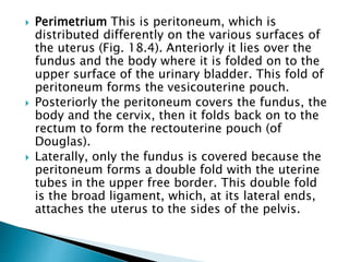
- 20. ’üĮ Perimetrium This is peritoneum, which is distributed differently on the various surfaces of the uterus (Fig. 18.4). Anteriorly it lies over the fundus and the body where it is folded on to the upper surface of the urinary bladder. This fold of peritoneum forms the vesicouterine pouch. ’üĮ Posteriorly the peritoneum covers the fundus, the body and the cervix, then it folds back on to the rectum to form the rectouterine pouch (of Douglas). ’üĮ Laterally, only the fundus is covered because the peritoneum forms a double fold with the uterine tubes in the upper free border. This double fold is the broad ligament, which, at its lateral ends, attaches the uterus to the sides of the pelvis.
- 21. ’üĮ MyometriumThis is the thickest layer of tissue in the uterine wall. It is a mass of smooth muscle fibres interlaced with areolar tissue, blood vessels and nerves. ’üĮ Endometrium This consists of columnar epithelium containing a large number of mucus-secreting tubular glands. ’üĮ It is divided functionally into two layers: The functional layer is the upper layer and it thickens and becomes rich in blood vessels in the first half of the menstrual cycle. If the ovum is not fertilised and does not implant, this layer is shed during menstruation. ’üĮ The basal layer lies next to the myometrium, and is not lost during menstruation. It is the layer fromwhich the fresh functional layer is regenerated during each cycle. The upper two-thirds of the cervical canal is lined with this mucous membrane. Lower down, however, the mucosa changes, becoming stratified squamous epithelium, which is continuous with the lining of the vagina itself
- 22. ’üĮ Arterial supply This is by the uterine arteries, branches of the internal iliac arteries. They pass up the lateral aspects of the uterus between the two layers of the broad ligaments. They supply the uterus and uterine tubes and join with the ovarian arteries to supply the ovaries. ’üĮ Venous drainage The veins follow the same route as the arteries and eventually drain into the internal iliac veins. ’üĮ Lymph drainage Deep and superficial lymph vessels drain lymph from the uterus and the uterine tubes to the aortic lymph nodes and groups of nodes associated with the iliac blood vessels. ’üĮ Nerve supply The nerves supplying the uterus and the uterine tubes consist of parasympathetic fibres from the sacral outflow and sympathetic fibres from the lumbar outflow.
- 23. ’üĮ The uterus is supported in the pelvic cavity by surrounding organs, muscles of the pelvic floor and ligaments that suspend it from the walls of the pelvis
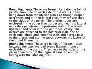
- 24. ’üĮ Broad ligaments These are formed by a double fold of peritoneum, one on each side of the uterus. They hang down from the uterine tubes as though draped over them and at their lateral ends they are attached to the sides of the pelvis. The uterine tubes are enclosed in the upper free border and near the lateral ends they penetrate the posterior wall of the broad ligament and open into the peritoneal cavity. The ovaries are attached to the posterior wall, one on each side. Blood and lymph vessels and nerves pass to the uterus and uterine tubes between the layers of the broad ligaments. ’üĮ Round ligaments These are bands of fibrous tissue between the two layers of broad ligament, one on each side of the uterus. They pass to the sides of the pelvis then through the inguinal canal to end by fusing with the labia majora..
- 25. ’üĮ Uterosacral ligaments These originate from the posterior walls of the cervix and vagina and extend backwards, one on each side of the rectum, to the sacrum. ’üĮ Transverse cervical (cardinal) ligaments These extend one from each side of the cervix and vagina to the side walls of the pelvis. ’üĮ Pubocervical fascia This extends forward from the transverse cervical ligaments on each side of the bladder and is attached to the posterior surface of the pubic bones
- 26. ’üĮ After puberty, the endometrium of the uterus goes through a regular monthly cycle of changes, the menstrual cycle, under the control of hypothalamic and anterior pituitary hormones (see Ch. 9). ’üĮ The purpose of the menstrual cycle is to prepare the uterus to receive, nourish and protect a fertilised ovum. The cycle is usually regular, lasting between 26 and 30 days. If the ovum is not fertilised a new cycle begins with a short period of bleeding (menstruation). ’üĮ If the ovum is fertilised the zygote embeds itself in the uterine wall. The uterine muscle grows to accommodate the developing baby, which is called an embryo during its first 8 weeks, and a fetus for the remainder of the pregnancy. ’üĮ Uterine secretions nourish the ovum before it implants in the endometrium, and after implantation the rapidly expanding ball of cells is nourished by the endometrial cells themselves. This is sufficient for only the first few weeks and the placenta is the organ that takes over thereafter (see Ch. 5).
- 27. ’üĮ The placenta, which is attached to the fetus by the umbilical cord, is also firmly attached to the wall of the uterus, and provides the route by which the growing baby receives oxygen and nutrients, and gets rid of its wastes. ’üĮ During pregnancy, which normally lasts about 40 weeks, the muscular walls of the uterus are prevented from contracting and expelling the baby early by high levels of the hormone progesterone secreted by the placenta. At the end of pregnancy (at term) the hormone oestrogen, which increases uterine contractility, becomes the predominant sex hormone in the blood. Additionally, oxytocin is released from the posterior pituitary, and also stimulates contraction of the uterine muscle. Control of oxytocin release is by positive feedback (see also Fig. 9.5, p. 212). During labour, the uterus forcefully expels the baby by means of powerful rhythmical contractions
- 28. ’üĮ The uterine (Fallopian) tubes (Fig. 18.4) are about 10 cm long and extend from the sides of the uterus between the body and the fundus. ’üĮ They lie in the upper free border of the broad ligament and their trumpet-shaped lateral ends penetrate the posterior wall, opening into the peritoneal cavity close to the ovaries. The end of each tube has fingerlike projections called fimbriae. ’üĮ The longest of these is the ovarian fimbria, which is in close association with the ovary.
- 29. ’üĮ The uterine tubes are covered with peritoneum (broad ligament), have a middle layer of smooth muscle and are lined with ciliated epithelium. ’üĮ Blood and nerve supply and lymphatic drainage are as for the uterus.
- 30. ’üĮ The uterine tubes propel the ovum from the ovary to the uterus by peristalsis and ciliary movement. ’üĮ The secretions of the uterine tube nourish both ovum and spermatozoa. ’üĮ Fertilisation of the ovum usually takes place in the uterine tube, and the zygote is propelled into the uterus for implantation.
- 31. ’üĮ The ovaries are the female gonads (glands producing sex hormones and the ova), and they lie in shallow fossa on the lateral walls of the pelvis. ’üĮ They are 2.5ŌĆō3.5 cm long, 2 cm wide and 1 cm thick. Each is attached to the upper part of the uterus by the ovarian ligament and to the back of the broad ligament by a broad band of tissue, the mesovarium. Blood vessels and nerves pass to the ovary through the mesovarium (Fig. 18.7).
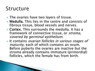
- 34. ’üĮ The ovaries have two layers of tissue. ’üĮ Medulla. This lies in the centre and consists of fibrous tissue, blood vessels and nerves. ’üĮ Cortex. This surrounds the medulla. It has a framework of connective tissue, or stroma, covered by germinal epithelium. ’üĮ It contains ovarian follicles in various stages of maturity, each of which contains an ovum. Before puberty the ovaries are inactive but the stroma already contains immature (primordial) follicles, which the female has from birth.
- 35. ’üĮ During the childbearing years, about every 28 days, one or more ovarian follicle (Graafian follicle) matures, ruptures and releases its ovum into the peritoneal cavity. This is called ovulation and it occurs during most menstrual cycles (Figs 18.7 and 18.8). ’üĮ Following ovulation, the ruptured follicle develops into the corpus luteum (meaning ŌĆśyellow bodyŌĆÖ), which in turn will leave a small permanent scar of fibrous tissue called the corpus albicans (meaning ŌĆśwhite bodyŌĆÖ) on the surfaceof the ovary.
- 36. ’üĮ Arterial supply. This is by the ovarian arteries, which branch from the abdominal aorta just below the renal arteries. ’üĮ Venous drainage. This is into a plexus of veins behind the uterus from which the ovarian veins arise. The right ovarian vein opens into the inferior vena cava and the left into the left renal vein. ’üĮ Lymph drainage. This is to the lateral aortic and preaortic lymph nodes. The lymph vessels follow the same route as the arteries. ’üĮ Nerve supply. The ovaries are supplied by parasympathetic nerves from the sacral outflow and sympathetic nerves from the lumbar outflow.
- 37. ’üĮ The ovary is the organ in which the female gametes are stored and develop prior to ovulation. ’üĮ Their maturation is controlled by the hypothalamus and the anterior pituitary gland, which releases gonadotrophins (follicle stimulating hormone, FSH, and luteinising hormone, LH), both of which act on the ovary. ’üĮ In addition, the ovary has endocrine functions, and releases hormones essential to the physiological changes during the reproductive cycle.
- 38. ’üĮ The source of these hormones, oestrogen, progesterone and inhibin, is the follicle itself. During the first half of the cycle, while the ovum is developing within the follicle, the follicle secretes increasing amounts of oestrogen. ’üĮ However, after ovulation, the corpus luteum secretes primarily progesterone, with some oestrogen and inhibin. The significance of this is discussed under the menstrual cycle.
- 39. HYPOTHALAMUS (LHRH) LUTEINISING HORMONE RELEASING HORMONES ANTERIOR PITUITARY FSH LH OVARIAN FOLLICLES CORPUS LUTEUM OESTROGEN PROGESTERONE
- 40. Puberty is the age at which the internal reproductive organs reach maturity, usually between the ages of 12 and 14. This is called the menarche, and marks the beginning of the childbearing period. The ovaries are stimulated by the gonadotrophins from the anterior pituitary: follicle stimulating hormone and luteinising hormone. A number of physical and psychological changes take place at puberty: ’üĮ the uterus, the uterine tubes and the ovaries reach maturity ’üĮ the menstrual cycle and ovulation begin (menarche) ’üĮ the breasts develop and enlarge
- 41. ’üĮ pubic and axillary hair begins to grow ’üĮ increase in height and widening of the pelvis ’üĮ increased fat deposited in the subcutaneous tissue, especially at the hips and breasts.
- 42. ’üĮ This is a series of events, occurring regularly in females every 26 to 30 days throughout the childbearing period between menarche and menopause (Fig. 18.10). The cycle consists of a series of changes taking place concurrently in the ovaries and uterine lining, stimulated by changes in blood concentrations of hormones (Fig. 18.10B and D). Hormones secreted during the cycle are regulated by negative feedback mechanisms.
- 43. ’üĮ The hypothalamus secretes luteinising hormone releasing hormone (LHRH), which stimulates the anterior pituitary to secrete (see Table 9.1): ŌĆó follicle stimulating hormone (FSH), which promotes the maturation of ovarian follicles and the secretion of oestrogen, leading to ovulation. FSH is therefore predominantly active in the first half of the cycle. Its secretion is suppressed once ovulation has taken place, to prevent other follicles maturing during the current cycle ’üĮ ŌĆó luteinising hormone (LH), which triggers ovulation, stimulates the development of the corpus luteum and the secretion of progesterone.
- 44. ’üĮ The hypothalamus responds to changes in the blood levels of oestrogen and progesterone. It is stimulated by high levels of oestrogen alone (as happens in the first half of the cycle) but suppressed by oestrogen and progesterone together (as happens in the second half of the cycle). ’üĮ The average length of the cycle is about 28 days. By convention the days of the cycle are numbered from the beginning of the menstrual phase, which usually lasts about 4 days. This is followed by the proliferative phase (approximately 10 days), then by the secretory phase (about 14 days).
- 45. ’üĮ When the ovum is not fertilised, the corpus luteum starts to degenerate. (In the event of pregnancy, the corpus luteum is supported by human chorionic gonadotrophin [hCG] secreted by the developing embryo.) Progesterone and oestrogen levels therefore fall, and the functional layer of the endometrium, which is dependent on high levels of these ovarian hormones, is shed in menstruation (Fig. 18.10C). The menstrual flow consists of the secretions from endometrial glands, endometrial cells, blood from the degenerating capillaries and the unfertilised ovum.
- 46. ’üĮ During the menstrual phase, levels of oestrogen and progesterone are very low because the corpus luteum that had been active during the second half of the previous cycle has degenerated. This means the hypothalamus and anterior pituitary can resume their cyclical activity, and levels of FSH begin to rise, initiating a new cycle. Proliferative phase ’üĮ At this stage an ovarian follicle, stimulated by FSH, is growing towards maturity and is producing oestrogen, which stimulates proliferation of the functional layer of the endometrium in preparation for the reception of a fertilised ovum. The endometrium thickens, becoming very vascular and rich in mucus-secreting glands. Rising levels of oestrogen are responsible for triggering a surge of LH approximately mid-cycle. This LH surge triggers ovulation, marking the end of the proliferative phase.
- 47. ’üĮ After ovulation, LH from the anterior pituitary stimulates development of the corpus luteum from the ruptured follicle, which produces progesterone, some oestrogen, and inhibin. Under the influence of progesterone, the endometrium becomes oedematous and the secretory glands produce increased amounts of watery mucus. This assists the passage of the spermatozoa through the uterus to the uterine tubes where the ovum is usually fertilised. There is a similar increase in secretion of watery mucus by the glands of the uterine tubes and by cervical glands that lubricate the vagina.
- 48. ’üĮ The ovum may survive in a fertilisable form for a very short time after ovulation, probably as little as 8 hours. The spermatozoa, deposited in the vagina during intercourse, may be capable of fertilising the ovum for only about 24 hours although they can survive for several days. This means that the period in each cycle during which fertilisation can occur is relatively short. Observable changes in the womanŌĆÖs body occur around the time of ovulation. Cervical mucus, normally thick and dry, becomes thin, elastic and watery, and body temperature rises by about 1┬░C immediately following ovulation. Some women experience abdominal discomfort in the middle of the cycle, thought to correspond to rupture of the follicle and release of its contents into the abdominal cavity. ’üĮ After ovulation, the combination of progesterone, oestrogen and inhibin from the corpus luteum suppresses the hypothalamus and anterior pituitary, so FSH and LH levels fall. Low FSH levels in the second half of the cycle prevent further follicular development in case a pregnancy results from the current cycle.
- 49. ’üĮ If the ovum is not fertilised, falling LH levels leads to degeneration and death of the corpus luteum, which is dependent on LH for survival. The resultant steady decline in circulating oestrogen, progesterone and inhibin leads to degeneration of the uterine lining and menstruation, with the initiation of a new cycle. ’üĮ If the ovum is fertilised there is no breakdown of the endometrium and no menstruation. The fertilised ovum (zygote) travels through the uterine tube to the uterus where it becomes embedded in the wall and produces human chorionic gonadotrophin (hCG), which is similar to anterior pituitary luteinising hormone. This hormone keeps the corpus luteum intact, enabling it to continue secreting progesterone and oestrogen for the first 3ŌĆō4 months of the pregnancy, inhibiting the maturation of further ovarian follicles (Figure 18.11). During that time the placenta develops and produces oestrogen, progesterone and gonadotrophins.
- 50. The menopause (climacteric) usually occurs between the ages of 45 and 55 years, marking the end of the period of years, sometimes as long as 10 years, and is caused by a progressive reduction in oestrogen levels, as the number of functional follicles in the ovaries declines with age. The ovaries gradually become less responsive to FSH and LH, and ovulation and the menstrual cycle become irregular, eventually ceasing. Several other phenomena may occur at the same time, including: ’üĮ short-term unpredictable vasodilation with flushing, sweating and palpitations, causing discomfort and disturbance of the normal sleep pattern ’üĮ shrinkage of the breasts ’üĮ axillary and pubic hair become sparse
- 51. ’üĮ atrophy of the sex organs ’üĮ episodes of uncharacteristic behaviour, e.g. irritability, mood changes ’üĮ gradual thinning of the skin ’üĮ loss of bone mass predisposing to osteoporosis ’üĮ slow increase in blood cholesterol levels that increase the risk of cardiovascular disease in postmenopausal women to that in males of the same age. ’üĮ Similar changes occur after bilateral irradiation or surgical removal of the ovaries.
- 52. ’üĮ The breasts or mammary glands are accessory glands of the female reproductive system. They exist also in the male, but in only a rudimentary form. ’üĮ Structure ’üĮ The mammary glands or breasts (Fig 18.12) consist of varying amounts of glandular tissue, responsible for milk production, supported by fatty tissue and fibrous connective tissue that anchor the breast to the chest wall. ’üĮ Each breast contains about 20 lobes, each of which contains a number of glandular structures called lobules, where milk is produced. Lobules open into tiny lactiferous ducts, which drain milk towards the nipple. Supporting fatty and connective tissues run through the breast, surrounding the lobules, and the breast itself is covered in subcutaneous fat. In the lactating breast, glandular tissue proliferates (hyperplasia, Fig. 3.41) to support milk production, and recedes again after lactation stops.
- 53. ’üĮ The nipple. This is a small conical eminence at the centre of the breast surrounded by a pigmented area, the areola. On the surface of the areola are numerous sebaceous glands (MontgomeryŌĆÖs tubercles), which lubricate the nipple during lactation. ’üĮ Blood supply, lymph drainage and nerve supply ’üĮ Arterial supply. The breasts are supplied with blood from the thoracic branches of the axillary arteries and from the internal mammary and intercostal arteries.
- 55. ’üĮ Venous drainage. This is formed by an anastomotic circle round the base of the nipple from which branches carry the venous blood to the circumference, and end in the axillary and mammary veins. ’üĮ Lymph drainage. This is mainly into the superficial axillary lymph vessels and nodes. Lymph may drain through the internal mammary nodes if the superficial route is obstructed. ’üĮ Nerve supply. The breasts are supplied by branches from the 4th, 5th and 6th thoracic nerves, which contain sympathetic fibres. There are numerous somatic sensory nerve endings in the breast, especially around the nipple. When these touch receptors are stimulated by sucking, impulses pass to the hypothalamus and secretion of the hormone oxytocin is increased, promoting the release of milk.
- 56. ’üĮ In the female, the breasts are small and immature until puberty. Thereafter they grow and develop under the influence of oestrogen and progesterone. During pregnancy these hormones stimulate further growth. After the baby is born the hormone prolactin from the anterior pituitary stimulates the production of milk, and oxytocin from the posterior pituitary stimulates the release of milk in response to the stimulation of the nipple by the sucking baby, by a positive feedback mechanism.