ROLE OF HIGH-RESOLUTION COMPUTER TOMOGRAPHY IN SPUTUM POSITIVE.pptx
- 1. ROLE OF HIGH-RESOLUTION COMPUTER TOMOGRAPHY IN SPUTUM POSITIVE AND NEGATIVE PULMONARY TUBERCULOSIS. THESIS TOPIC GUIDE: PROF. DR. V. VENKATARATHNAM BY: DR. G. SOWJANYA
- 2. ŌĆó Tuberculosis, caused by Mycobacterium tuberculosis, is a global health issue primarily affecting the lungs. ŌĆó Pulmonary tuberculosis accounts for the majority of tuberculosis cases worldwide, posing significant morbidity and mortality, particularly in developing countries. ŌĆó While chest radiography remains valuable, high- resolution computed tomography (HRCT) proves more sensitive, especially when other diagnostic methods are limited. INTRODUCTION
- 3. ŌĆó HRCT we use narrow xray beam collimation to obtain thin sections of <1.5mm helps in obtaining the high resolution images of lung parenchyma vessels and airspaces and interstitium even without giving contrast and it has diagnostic accuracy rates of up to 87.5% in sputum- positive and 81.67% in sputum-negative tuberculosis cases. ŌĆó Patients with diabetes mellitus and HIV are at higher risk of tuberculosis infection, with increasing trends in diabetes prevalence adding urgency to tuberculosis control measures.
- 4. ŌĆó Enhanced knowledge and evidence-based decisions are crucial for effective tuberculosis management and prevention, necessitating research into new diagnostic markers and treatment strategies ŌĆó Studying HRCT features in pulmonary tuberculosis holds potential to expand understanding and improve diagnostic capabilities in tuberculosis.
- 5. III. Objectives of the study: ŌĆó To study the HRCT pattern of pulmonary involvement in sputum positive pulmonary tuberculosis. ŌĆó Role of HRCT in sputum negative but clinically strongly suspected case of tuberculosis.
- 6. IV. Review of Literature: 1. Waqas Rasheed et., at., in 2020 described the HRCT findings of Pulmonary tuberculosis as the presence of consolidation, centrilobular nodules, branching nodules with tree in bud appearance with or without lymphadenopathy, and pleural effusion. He also conveyed the Diagnostic accuracy of HRCT in diagnosing PTB was found to be 84.26% with sensitivity, specificity, positive predictive value (PPV) and negative predictive value (NPV) of 89.09%, 79.25%, 81.67%, and 87.50%, respectivelyŌĆŗ(1)ŌĆŗ.
- 7. 1. 2. Harsh lathiya et., al ., in 2020 concluded that HRCT is a powerful and reliable investigation in the diagnosis of Pulmonary Tuberculosis and determination of itŌĆÖs disease activity, when other means of diagnosis such as sputum/BAL AFB test and Culture fail to settle the matter, are not available or time consuming and should be routinely indicated in sputum smear negative patients for prompt initiation of antitubercular treatment. He also described the HRCT findings of active Tuberculosis as centrilobular nodules, tree in bud opacities, and larger nodules. Miliary nodules and inactive Tuberculosis as fibrosis with or without traction bronchiectasis / bronchiolectasis and
- 8. 3. Himandri Harish Warbhe ., et., al., in 2022., defined HRCT findings of Pulmonary Tuberculosis as the presence of consolidation, centrilobular nodules, branching nodules with tree in bud appearance with or without lymphadenopathy and pleural effusion and concluded that diagnosing sputum smear-positive and sputum smear- negative PT, HRCT has high sensitivity. The specificity of HRCT was high in diagnosing sputum smear-positive PT, whereas in case of sputum smear-negative PT it was slightly lowŌĆŗ(5)ŌĆŗ.
- 9. 4. Siddhartha Sarma Biswas et., al., in 2022 .,Described that Consolidation was the most common HRCT finding in patients of pulmonary tuberculosis. These patients also showed other patterns such as tree in bud opacities, ill-defined patchy opacities, cavities, bronchiectasis. Additional features like pleural effusion, pneumothorax, tubercular spondylitis are also seen in many patients. HRCT thorax is a useful investigation modality used to diagnose as well as for follow up of patientsŌĆŗ(6)ŌĆŗ.
- 10. V. Materials & Methods (Methodology): 1.Study design: ŌĆó Diagnostic observational study. 2.Study setting: ŌĆó Department of Radiodiagnosis PESIMSR, KUPPAM. 3.Study period: 18 MONTHS.
- 11. 4. Study population: ŌĆó All the patients with sputum positive (both smear and culture positive) status subjected to HRCT chest ŌĆó All patients with sputum negative status of Tuberculosis but clinically strongly suspected case of tuberculosis subjected to HRCT chest. ŌĆó Age criteria:- ŌĆó 18to 80 years. 5. Sampling method: ŌĆó Purposive sampling.
- 12. ŌĆóSample size: Based on the reference Article(1). Sample size calculated for your study (84.26%) (min take =39)
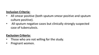
- 13. Inclusion Criteria: ŌĆó All smear positive (both sputum smear positive and sputum culture positive). ŌĆó All sputum negative cases but clinically strongly suspected case of tuberculosis. Exclusion Criteria: ŌĆó Those who are not willing for the study. ŌĆó Pregnant women.
- 14. 8.Tools to be used in the study: a.A proforma containing set of Questionnaire. b.Helical Computer Tomography from GE -16 reformatted 32 slice machine c. Open Epi d.SPSS e.Microsoft office 2019- Word & Excel.
- 15. 9.Procedure for data collection: Each subject referred to Radiology department with both sputum positive and sputum negative status will be contacted. Written consent will be obtained. Relevant clinical history in the form of standard questioner will be collected. After this HRCT will be planned and patient will be subjected to HRCT in Helical Computer Tomography from GE -16 reformatted 32 slice machine. The imaging features obtained in the HRCT will be studied and tabulated.
- 16. 10.Statistical Analysis of data: The data will be entered into MS Excel 2019 version and further analyzed using SPSS 20. For descriptive analysis, the categorical variables will be analyzed by using percentages and the continuous variables will be analyzed by calculating mean ┬▒ Standard Deviation. For inferential statistics, Chi-square test was used. Efficacy was expressed as Sensitivity, specificity, and predictive values. A probability value of less than 0.05 was considered statistically significant.
- 17. What are the expected outcome(s) of the study? The study helps in reviewing the HRCT diagnostic findings in sputum positive and negative cases of Pulmonary Tuberculosis. VIII. What is the impact of the study in the population or in science: HRCT findings of pulmonary tuberculosis patients gives additional information in the diagnostic criteria of Tuberculosis and have impact on its management of pulmonary tuberculosis even in sputum negative condition. Detection and early treatment of Pulmonary Kochs will significamtly reduces morbidity and mortality.
- 18. IX. Funding: By Self / Institute / Any other source: Specify: Nil. X. Conflict of Interest (if any) in the project: NIL. XI. How is the confidentiality of participants ensured? The name & identity will not be disclosed. Thus, personal data about every subject will be kept confidential. XII. Ethical clearance & approval: Ethical clearance will be obtained from the Institutional Human Ethics Committee, PESIMSR, Kuppam.
- 19. XIII. REFFRENCES: 1. Rasheed W, Qureshi R, Jabeen N, Shah HA, Naseem Khan R. Diagnostic Accuracy of High-Resolution Computed Tomography of Chest in Diagnosing Sputum Smear Positive and Sputum Smear Negative Pulmonary Tuberculosis. Cureus. 2020 Jun 5;12(6):e8467. doi: 10.7759/cureus.8467. PMID: 32642373; PMCID: PMC7336620. 2. Restrepo BI. Diabetes and Tuberculosis. Microbiol Spectr. 2016 Dec;4(6):10.1128/microbiolspec. TNMI7-0023-2016. doi: 10.1128/microbiolspec.TNMI7-0023-2016. PMID: 28084206; PMCID: PMC5240796.
- 20. 3. Yeh JJ, Yu JK, Teng WB, Chou CH, Hsieh SP, Lee TL, Wu MT. High-resolution CT for identify patients with smear- positive, active pulmonary tuberculosis. Eur J Radiol. 2012 Jan;81(1):195-201. doi: 10.1016/j.ejrad.2010.09.040. Epub 2010 Oct 27. PMID: 21030177; PMCID: PMC7127118. 4. HARSH LATHIYA, PRABHAT DEBBARMA, JAYBRATA RAY, ANJAN DAS. High Resolution Computed Tomography in the Diagnosis of Pulmonary Tuberculosis and its Correlation with Sputum/ Bronchoalveolar Lavage Analysis. International Journal of Anatomy, Radiology and Surgery 2020 Oct, Vol-9(4): RO24-RO28; DOI: 10.7860/IJARS/2020/45083:2579.
- 21. 5. Himandri Harish Warbhe, Himanshu Pophale, Pankaj Magar, Archana Kanavde, & Sudhanshu Sunil Tonpe. (2022). Diagnosis of the Sputum Smear Positive and Sputum Smear Negative Pulmonary Tuberculosis - The Role of High - Resolution Computed Tomography of Chest. Journal of Evolution of Medical and Dental Sciences, 11(12), 880ŌĆō883. DDOI:10.14260/jemds.v11i12.262. 6. Biswas SS, Borpatragohain D, Thoumoung C. Findings of HRCT Thorax in Patients of Sputum Positive Pulmonary Tuberculosis in a Tertiary Health Care Centre of North East India. Journal of Medical Sciences and Health ;Journal of Medical Sciences and Health; Year: -0001, Volume: 8, Issue: 1, Pages: 47- 51; DOI: 10.46347/jmsh.v8i1.21.43


