際際滷

際際滷Share a Scribd company logo
Skeletal
Physiology
Types of Bones
A) Long bones are easily identified by their extended
longitudinal axes and expanded and often uniquely shaped
articular ends. Examples are the femur of the thigh and the
humerus of the arm.
B) Short bones are often described as cube-or box-shaped
structures, which are about as broad as they are long.
Examples are the wrist (carpals) and ankle (tarsal) bones.
C) Flat bones are generally broad and thin with a flattened and
often curved surface. Examples are certain bones of the skull,
the shoulder blades (scapulae), ribs, and breastbone
(sternum).
D) Irregular bones are often clustered in groups and come in
various sizes and shapes. An example is the kneecap (patella).
The Long Bone
a) Diaphysis: main shaft like portion.
b) Epiphyses: both ends of a long bone.
c) Articular cartilage: thin layer of hyaline cartilage
that covers articular or joint surfaces of epiphyses.
d) Periosteum: dense, white fibrous membrane that
covers bone except at joint surfaces, where
articular cartilage forms the covering.
e) Medullary (or marrow) cavity: a tube like hollow
space in the diaphysis of a long bone.
f) Endosteum: a thin epithelial membrane that lines
the medullary cavity of long bones.
Bones Cont.
It consists of cells, fibers, and extracellular material
also known as matrix. Its extracellular components
are hard and calcified. In bone the extracellular
material, or matrix, predominates. The rigidity of
bone enables it to serve supportive and protective
functions. As a tissue, bone is ideally suited to its
functions, and the concept that structure and
function are interrelated is apparent in this tissue. It
has a tensile strength nearly equal to cast iron but
at less than one third the weight. Bone is organized
so that its great strength and minimal weight result
from the interrelationships of its structural
components.
The Haversian System
Lamellae- Concentric, cylinder-shaped layers of calcified
matrix.
Lacunae- Small spaced containing tissue fluid in which
bone cells lie imprisoned between the hard layer of the
lamellae.
Canaliculi- Ultrasmall canals radiating in all directions
from the lacunae and connecting them to each other and
into a larger canal (The Haversian Canal)
Haversian Canal- extends lengthwise through the center
or each Haversian System; contains blood vessels,
lymphatic vessels, and nerves from the haversian canal;
nutrients and oxygen move through canaliculi to the
lacunae and their bone cells
Cells in Bones
Osteoclasts: Osteoclasts are large cells that dissolve the bone. They
come from the bone marrow and are related to white blood cells. They
usually have more than one nucleus and are found on the surface of
the bone mineral.
Osteoblasts: Osteoblasts are cells that form new bones. They come
from bone marrow and are related to structural cells. They only have
one nucleus and they produce new bone called osteoid which is
made of bone collagen and other proteins. They also control calcium
and mineral deposition. They are found on the surface of new bone.
Old osteoblasts are called lining cells that regulate the passage of
calcium in and out of the bone. They also respond to hormones that
create protein that activate osteoclasts.
Osteocytes: These cells are found inside the bone that come from
osteoblasts. Some turn into osteocytes while the new bone is being
formed. These cells can sense pressures or cracks in the bone and
help direct where the osteoclasts will dissolve the bone.
Homeostatic Functions
1.Support: Bones are the supporting framework in the body and contribute to shape,
alignment, and positioning of the body parts. This involves homeostasis because without
support we would have nothing to hold our weight. We would be a glob of blood and
organs.
2.Protection: Bones protect the body from the external environment. Bones also protect
the structures that they enclose. Without protection from bones we would easily get hurt
because there would be nothing to prevent objects from hurting our organs or easily
crushing our internal parts. By having protection we can endure more difficult tasks and
participate in more rigorous activities.
3.Movement: Muscles are anchored to bones. As the muscles move, they pull on bones,
producing movement. Without movement it would be hard to maintain our body
temperature because we wouldn't be able to get warm when its cold or get cold when its
hot.
4.Mineral Storage: Bones serve as the major place for calcium, phosphorus, and other
minerals. Homeostasis of blood calcium concentration depends a lot on changes in the
rate of calcium movement between the blood and bones. For example, if blood calcium
moves faster out of the blood into bones and more slowly in the opposite direction, blood
calcium concentration decreases. This is essential for healthy survival.
5.Hematopoiesis: Hematopoiesis is another word for blood cell formation. This is the vital
process carried on by red bone marrow. This is important because it keeps the body in
balance. It helps regulate differences in the body such as infections or fevers.
Intramembranous and
Endochondral bone
Intramembranous Bone (flat bones): Membranous bones first
look like flat, membrane-like layers. These layers have a
continuing flow of nutrient blood supply from blood vessels
that are formed between the layers. In the beginning,
connective tissue cells situate themselves among the layers
and then form into osteoblasts which are bone-forming cells.
Osteoblasts remove calcium from the blood and put it in the
bone matrix. As a result, layers of spongy bone form around
the original cartilage. Later, spaces among the spongy bone fill
with bone matrix to become compact bone. The osteoblasts
keep depositing calcium supplements into the matrix until it is
completely surrounded by it. After, the osteoblasts are
considered to be in the lacunae and are called osteocytes.
Broad, flat skull bones are examples of intramembranous
ossification.
Intramembranous and
Endochondral bone
Endochondral bone(long bones). This ossification forms bone by
replacing a cartilaginous model that is earlier in embryonic
development. These models first undergo changes as the
connective tissue gets bigger, destroying surrounding matrix.
Soon after, connective tissue cells die. As the cells die, a
periosteum is formed on the outside of the developing structure.
Next, blood vessels and other cells rush into the dying tissue. Once
a significant amount of spongy bone is formed, growth in the
cartilage bones occurs. Thickness in cartilage bones is done by
intramembrous ossification. Beneath the layer of periosteum,
compact bone is formed and hardened with the help of
osteoblasts filling portions of the spongy bone with calcium
phosphate crystals. Sometimes the compact bone is formed on
the surfaces of existing bone tissue and osteoblasts must get rid
of them. The crystals are then delivered to blood and tissues.
Similarities
Similarities: Both processes create bone and
sometimes the creation of bone requires both
endochondral and intramembranous ossification.
When both processes are used, mesenchyme cells
develop into chondroblasts and increase in number
by cell division. The chondroblasts get bigger and
excrete a matrix which hardens due to presence or
inorganic minerals. Then, chambers form within the
matrix and osteoblasts and blood-forming cells
enter these chambers. The osteoblasts then
secrete minerals to form the bone matrix
Bone Fracture Repair

1. Fracture has
occured
2. Formation of a
fracture hematoma
3.Formation of an
internal and
external callus
4.Bone remodeling
completes
Types of Cartilage

Hyaline Cartilage- the most common type
of cartilage, it appears gelatinous and
glossy
Elastic Cartilage- cartilage with elastic, as
well as collagenous fibers; provides
elasticity and firmness
Fibrocartilage-cartilage with the greatest
number of collagenous fibers; strongest
and most durable type of cartilage
Types of Cartilage


                                     Elastic Cartilage
Hyaline Cartilage
                    Fibrocartilage
Ad

Recommended

Bone tissue
Bone tissue
Michael Wrock
Bone tissue
Bone tissue
rajkamble
Bone tissue
Bone tissue
abraml02
Histology of Bone
Histology of Bone
Eneutron
2401 lecture05 ch 6 bones and bone tissue for class and web
Abhishek Sharma
Histology of bone
Histology of bone
Najeebullah2016
General osteology
General osteology
Jamil Anwar
Bone : Basics
Bone : Basics
Navneet Randhawa
Lecture bone structure & markings
MissReith
Skeletal physiology
Skeletal physiology
aldana0942
LT4.2 Skeletal System
NeQuelle DeFord
Histology of bone
Histology of bone
TaimurKhan87
Introduction to osteology
AfTaab AfRaz
General osteology
General osteology
Jay Patel
Skeletal System
Skeletal System
circadian_wellness
Presentation on bones
Sam Shaikh
Chapter Six
Chapter Six
dquevedo1
Parts of adult & young bone, 123
Fati Naqvi
Bone or osseous tissue/ oral surgery courses
Indian dental academy
mbbs ims msu
mbbs ims msu
MBBS IMS MSU
Bone tissue to skeletal system ppt
Devron Jeko
Bone histology amir
Bone histology amir
Alam Zeb Amir
Bones
Bones
Shimaa Essa
Bones and cartilages
Shivani Rana
Artifact #2 skeletal physiology
cz0634bn
04 cartilages and bone
Richard Ayisa
Cartilages and bones
Helao Silas
Chapter 4
Chapter 4
ahoward
Tissues Proj
Tissues Proj
guest1b441b
Muscular Physiology
Muscular Physiology
Ashley

More Related Content

What's hot (19)

Lecture bone structure & markings
MissReith
Skeletal physiology
Skeletal physiology
aldana0942
LT4.2 Skeletal System
NeQuelle DeFord
Histology of bone
Histology of bone
TaimurKhan87
Introduction to osteology
AfTaab AfRaz
General osteology
General osteology
Jay Patel
Skeletal System
Skeletal System
circadian_wellness
Presentation on bones
Sam Shaikh
Chapter Six
Chapter Six
dquevedo1
Parts of adult & young bone, 123
Fati Naqvi
Bone or osseous tissue/ oral surgery courses
Indian dental academy
mbbs ims msu
mbbs ims msu
MBBS IMS MSU
Bone tissue to skeletal system ppt
Devron Jeko
Bone histology amir
Bone histology amir
Alam Zeb Amir
Bones
Bones
Shimaa Essa
Bones and cartilages
Shivani Rana
Artifact #2 skeletal physiology
cz0634bn
04 cartilages and bone
Richard Ayisa
Cartilages and bones
Helao Silas
Lecture bone structure & markings
MissReith
Skeletal physiology
Skeletal physiology
aldana0942
LT4.2 Skeletal System
NeQuelle DeFord
Histology of bone
Histology of bone
TaimurKhan87
Introduction to osteology
AfTaab AfRaz
General osteology
General osteology
Jay Patel
Presentation on bones
Sam Shaikh
Chapter Six
Chapter Six
dquevedo1
Parts of adult & young bone, 123
Fati Naqvi
Bone or osseous tissue/ oral surgery courses
Indian dental academy
Bone tissue to skeletal system ppt
Devron Jeko
Bone histology amir
Bone histology amir
Alam Zeb Amir
Bones and cartilages
Shivani Rana
Artifact #2 skeletal physiology
cz0634bn
04 cartilages and bone
Richard Ayisa
Cartilages and bones
Helao Silas

Viewers also liked (7)

Chapter 4
Chapter 4
ahoward
Tissues Proj
Tissues Proj
guest1b441b
Muscular Physiology
Muscular Physiology
Ashley
09.10.08: Connective Tissue
Open.Michigan
Blood typing analysis
Ashley
Experiments
Experiments
Ashley
Blood typing analysis
Ashley
Chapter 4
Chapter 4
ahoward
Muscular Physiology
Muscular Physiology
Ashley
09.10.08: Connective Tissue
Open.Michigan
Blood typing analysis
Ashley
Experiments
Experiments
Ashley
Blood typing analysis
Ashley
Ad

Similar to Skeletal Physiology (20)

Bone and Cartilage_ 6th May 2024,_RI.pdf
wasimnawaz10
Topic 2 & 3 Pj
Topic 2 & 3 Pj
zue5588
Skeletal physiology
Skeletal physiology
td0297bn
Bones and its structure in detail with two different form of bone formation
bhartisharma175
Introduction to osteology
AfTaab AfRaz
SWERNY SKELETAL 1.pdf
SwernyMumba
The musculoskeletal system Anatomy and physiology
kajal chandel
Anatomy and Physiology on the musculoskeletal system
DR .PALLAVI PATHANIA
THE ANATOMY AND PHYSIOLOGY - THE_SKELETAL_SYSTEM.pptx
ndixdo
The skeletal system
The skeletal system
Vanja Drljevic
1587723684_BC(H)-IV-Human_Physiology_Bone_Structure_and_formation-1 (1).pdf
ebonus324
1587723684_BC(H)-IV-Human_Physiology_Bone_Structure_and_formation-1 (1).pdf
ebonus324
Bones Tissue and Ossification of Bone.pptx
Sid Roy
Lecture on General Oteology: study of the structure and function of the skele...
Sanskar Virmani
Bone structure and type
4ugautam
A Lecture on Skeletal System: Components, Structures, and Functions.pptx
Erlison Lorenz Ognilla
Skeletal system
Skeletal system
WKUDistanceLearning
orthopedic nursing unit AHN II #1 complete.pptx
Saida Sadiq
Skeletal physiology 101
ag1430bn
Bone function and_formation_2009
Personal
Bone and Cartilage_ 6th May 2024,_RI.pdf
wasimnawaz10
Topic 2 & 3 Pj
Topic 2 & 3 Pj
zue5588
Skeletal physiology
Skeletal physiology
td0297bn
Bones and its structure in detail with two different form of bone formation
bhartisharma175
Introduction to osteology
AfTaab AfRaz
SWERNY SKELETAL 1.pdf
SwernyMumba
The musculoskeletal system Anatomy and physiology
kajal chandel
Anatomy and Physiology on the musculoskeletal system
DR .PALLAVI PATHANIA
THE ANATOMY AND PHYSIOLOGY - THE_SKELETAL_SYSTEM.pptx
ndixdo
The skeletal system
The skeletal system
Vanja Drljevic
1587723684_BC(H)-IV-Human_Physiology_Bone_Structure_and_formation-1 (1).pdf
ebonus324
1587723684_BC(H)-IV-Human_Physiology_Bone_Structure_and_formation-1 (1).pdf
ebonus324
Bones Tissue and Ossification of Bone.pptx
Sid Roy
Lecture on General Oteology: study of the structure and function of the skele...
Sanskar Virmani
Bone structure and type
4ugautam
A Lecture on Skeletal System: Components, Structures, and Functions.pptx
Erlison Lorenz Ognilla
orthopedic nursing unit AHN II #1 complete.pptx
Saida Sadiq
Skeletal physiology 101
ag1430bn
Bone function and_formation_2009
Personal
Ad

Skeletal Physiology

  • 2. Types of Bones A) Long bones are easily identified by their extended longitudinal axes and expanded and often uniquely shaped articular ends. Examples are the femur of the thigh and the humerus of the arm. B) Short bones are often described as cube-or box-shaped structures, which are about as broad as they are long. Examples are the wrist (carpals) and ankle (tarsal) bones. C) Flat bones are generally broad and thin with a flattened and often curved surface. Examples are certain bones of the skull, the shoulder blades (scapulae), ribs, and breastbone (sternum). D) Irregular bones are often clustered in groups and come in various sizes and shapes. An example is the kneecap (patella).
  • 3. The Long Bone a) Diaphysis: main shaft like portion. b) Epiphyses: both ends of a long bone. c) Articular cartilage: thin layer of hyaline cartilage that covers articular or joint surfaces of epiphyses. d) Periosteum: dense, white fibrous membrane that covers bone except at joint surfaces, where articular cartilage forms the covering. e) Medullary (or marrow) cavity: a tube like hollow space in the diaphysis of a long bone. f) Endosteum: a thin epithelial membrane that lines the medullary cavity of long bones.
  • 4. Bones Cont. It consists of cells, fibers, and extracellular material also known as matrix. Its extracellular components are hard and calcified. In bone the extracellular material, or matrix, predominates. The rigidity of bone enables it to serve supportive and protective functions. As a tissue, bone is ideally suited to its functions, and the concept that structure and function are interrelated is apparent in this tissue. It has a tensile strength nearly equal to cast iron but at less than one third the weight. Bone is organized so that its great strength and minimal weight result from the interrelationships of its structural components.
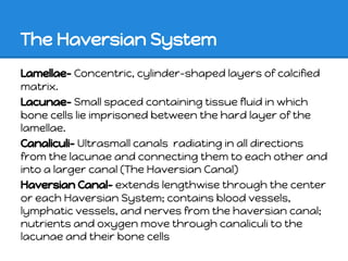
  • 5. The Haversian System Lamellae- Concentric, cylinder-shaped layers of calcified matrix. Lacunae- Small spaced containing tissue fluid in which bone cells lie imprisoned between the hard layer of the lamellae. Canaliculi- Ultrasmall canals radiating in all directions from the lacunae and connecting them to each other and into a larger canal (The Haversian Canal) Haversian Canal- extends lengthwise through the center or each Haversian System; contains blood vessels, lymphatic vessels, and nerves from the haversian canal; nutrients and oxygen move through canaliculi to the lacunae and their bone cells
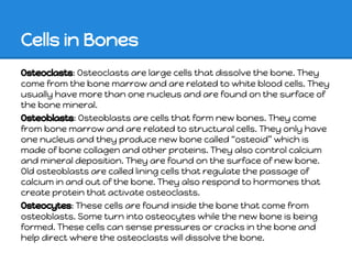
  • 6. Cells in Bones Osteoclasts: Osteoclasts are large cells that dissolve the bone. They come from the bone marrow and are related to white blood cells. They usually have more than one nucleus and are found on the surface of the bone mineral. Osteoblasts: Osteoblasts are cells that form new bones. They come from bone marrow and are related to structural cells. They only have one nucleus and they produce new bone called osteoid which is made of bone collagen and other proteins. They also control calcium and mineral deposition. They are found on the surface of new bone. Old osteoblasts are called lining cells that regulate the passage of calcium in and out of the bone. They also respond to hormones that create protein that activate osteoclasts. Osteocytes: These cells are found inside the bone that come from osteoblasts. Some turn into osteocytes while the new bone is being formed. These cells can sense pressures or cracks in the bone and help direct where the osteoclasts will dissolve the bone.
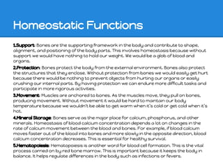
  • 7. Homeostatic Functions 1.Support: Bones are the supporting framework in the body and contribute to shape, alignment, and positioning of the body parts. This involves homeostasis because without support we would have nothing to hold our weight. We would be a glob of blood and organs. 2.Protection: Bones protect the body from the external environment. Bones also protect the structures that they enclose. Without protection from bones we would easily get hurt because there would be nothing to prevent objects from hurting our organs or easily crushing our internal parts. By having protection we can endure more difficult tasks and participate in more rigorous activities. 3.Movement: Muscles are anchored to bones. As the muscles move, they pull on bones, producing movement. Without movement it would be hard to maintain our body temperature because we wouldn't be able to get warm when its cold or get cold when its hot. 4.Mineral Storage: Bones serve as the major place for calcium, phosphorus, and other minerals. Homeostasis of blood calcium concentration depends a lot on changes in the rate of calcium movement between the blood and bones. For example, if blood calcium moves faster out of the blood into bones and more slowly in the opposite direction, blood calcium concentration decreases. This is essential for healthy survival. 5.Hematopoiesis: Hematopoiesis is another word for blood cell formation. This is the vital process carried on by red bone marrow. This is important because it keeps the body in balance. It helps regulate differences in the body such as infections or fevers.
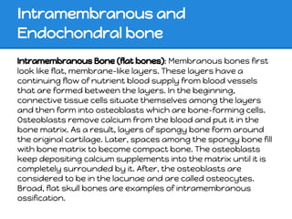
  • 8. Intramembranous and Endochondral bone Intramembranous Bone (flat bones): Membranous bones first look like flat, membrane-like layers. These layers have a continuing flow of nutrient blood supply from blood vessels that are formed between the layers. In the beginning, connective tissue cells situate themselves among the layers and then form into osteoblasts which are bone-forming cells. Osteoblasts remove calcium from the blood and put it in the bone matrix. As a result, layers of spongy bone form around the original cartilage. Later, spaces among the spongy bone fill with bone matrix to become compact bone. The osteoblasts keep depositing calcium supplements into the matrix until it is completely surrounded by it. After, the osteoblasts are considered to be in the lacunae and are called osteocytes. Broad, flat skull bones are examples of intramembranous ossification.
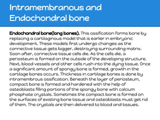
  • 9. Intramembranous and Endochondral bone Endochondral bone(long bones). This ossification forms bone by replacing a cartilaginous model that is earlier in embryonic development. These models first undergo changes as the connective tissue gets bigger, destroying surrounding matrix. Soon after, connective tissue cells die. As the cells die, a periosteum is formed on the outside of the developing structure. Next, blood vessels and other cells rush into the dying tissue. Once a significant amount of spongy bone is formed, growth in the cartilage bones occurs. Thickness in cartilage bones is done by intramembrous ossification. Beneath the layer of periosteum, compact bone is formed and hardened with the help of osteoblasts filling portions of the spongy bone with calcium phosphate crystals. Sometimes the compact bone is formed on the surfaces of existing bone tissue and osteoblasts must get rid of them. The crystals are then delivered to blood and tissues.
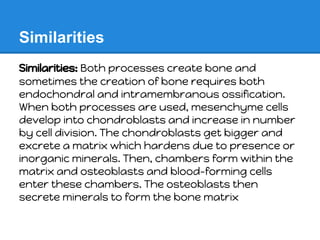
  • 10. Similarities Similarities: Both processes create bone and sometimes the creation of bone requires both endochondral and intramembranous ossification. When both processes are used, mesenchyme cells develop into chondroblasts and increase in number by cell division. The chondroblasts get bigger and excrete a matrix which hardens due to presence or inorganic minerals. Then, chambers form within the matrix and osteoblasts and blood-forming cells enter these chambers. The osteoblasts then secrete minerals to form the bone matrix
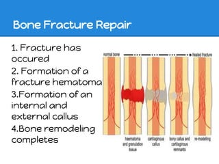
  • 11. Bone Fracture Repair 1. Fracture has occured 2. Formation of a fracture hematoma 3.Formation of an internal and external callus 4.Bone remodeling completes
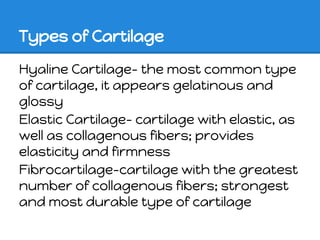
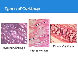
  • 12. Types of Cartilage Hyaline Cartilage- the most common type of cartilage, it appears gelatinous and glossy Elastic Cartilage- cartilage with elastic, as well as collagenous fibers; provides elasticity and firmness Fibrocartilage-cartilage with the greatest number of collagenous fibers; strongest and most durable type of cartilage
  • 13. Types of Cartilage Elastic Cartilage Hyaline Cartilage Fibrocartilage