Staining Steps3509
Download as ppt, pdf0 likes1,539 views

The document outlines the steps for DAKO IHC staining which includes deparaffinization, antigen retrieval, blocking endogenous peroxidase, primary and secondary antibody incubation, DAB development, hematoxylin counterstaining, and coverslipping. It describes the time and temperature for each step and provides troubleshooting tips for issues like negative staining, high background, or weak staining.
1 of 22
Downloaded 56 times

Ad
Recommended
Immunohistochemistry (IHC)Staining Steps
Immunohistochemistry (IHC)Staining StepsLawrence Richards
Ěý
The document outlines the steps for DAKO IHC staining which includes deparaffinization and rehydration of the sample, antigen retrieval, blocking of endogenous peroxidase, incubation with primary and secondary antibodies, development with DAB, counterstaining with hematoxylin, and coverslipping. The process takes approximately 1-2 hours and involves rinsing between steps in various buffers and solvents.Gel extraction protocol using SPRI
Gel extraction protocol using SPRIATPowr
Ěý
This document provides a gel extraction protocol using magnetic beads to purify DNA fragments from an agarose gel. The main steps include: 1) adding a binding solution to the gel slice and incubating to elute DNA fragments, 2) adding magnetic beads and isopropanol to bind DNA to the beads, 3) washing beads to remove contaminants, 4) drying beads and eluting purified DNA from the beads. The protocol aims to maximize DNA yield from the gel using various concentrations of isopropanol in the binding and washing steps.SANDWICH ELISA- BRIEF DESCRIPTION AND PROTOCOL
SANDWICH ELISA- BRIEF DESCRIPTION AND PROTOCOLAnnuja Anandaradje
Ěý
This document provides a protocol for performing a sandwich ELISA to detect MIP-1α. The protocol involves coating a microplate with capture antibody overnight, blocking uncoated sites, adding MIP-1α standards and samples, detecting with a detection antibody and streptavidin HRP conjugate, developing color with substrate, and measuring optical density to quantify MIP-1α levels.Meeting021910
Meeting021910Sally Newton
Ěý
The document summarizes the results of a lab meeting that studied bacterial iron transport. It discusses binding of the siderophore enterobactin at different temperatures, the role of various receptor proteins, and how inhibiting energy production and fluorescently labeling cells affected binding and uptake. It also presents results comparing wild type and mutant strains on their ability to transport iron when exposed to different concentrations of enterobactin and a competing iron source over time.Ziehl neelsen staining
Ziehl neelsen staining Ovya Pugalenthi Aruna
Ěý
The document focuses on Ziehl-Neelsen staining, a differential staining technique used to identify acid-fast organisms like Mycobacterium tuberculosis and M. leprae due to their lipid-rich cell walls. It details the staining procedure, principles, importance, potential errors, and variations in the method, including a modified approach for rapid detection. Additionally, it emphasizes the clinical significance of identifying acid-fast bacilli in diagnosing tuberculosis.Z n staining
Z n stainingmicroarunkumar
Ěý
The document discusses the Ziehl-Neelsen staining technique, which is used to identify acid-fast bacteria such as Mycobacterium, Actinomycetes, and Nocardia. The staining method uses carbol fuchsin as the primary stain, along with heat to aid penetration. Acid-alcohol is used as a decolorizer, followed by methylene blue as the counterstain. Mycobacterium and other acid-fast bacteria will retain the pink carbol fuchsin stain due to their thick, waxy cell walls containing mycolic acid. This allows them to be differentiated from other non-acid fast bacteria that will take on the blue methylene blue counterstainImmunofluorescence Antibody Validation Report for Anti-AMPKα1 Antibody (STJ91...
Immunofluorescence Antibody Validation Report for Anti-AMPKα1 Antibody (STJ91...St John's Laboratory Ltd
Ěý
The antibody validation report (number 91580) details the immunofluorescence analysis procedure using an anti-AMPKα1 polyclonal antibody in rat lung tissue. Key steps include primary and secondary antibody incubations, DAPI counter-staining, and slide mounting, followed by visualization with a Nikon inverted fluorescence microscope. The report outlines specific dilutions, incubation times, and the immunofluorescence protocol, including tissue processing and antigen retrieval methods.Modified Ziehl-Neelsen Stain
Modified Ziehl-Neelsen Stain9925752690
Ěý
This document discusses various staining methods for acid-fast bacteria, including the Ziehl-Neelsen stain and modifications such as Kinyoun's stain, cold stains like Gabbett's method, and stains for tissue sections or spores. Key methods mentioned are Kinyoun's stain, which does not require heating; Gabbett's cold stain which is a two-step method for discolorization and counterstaining; and stains for tissue sections like Fite-Faraco, Fite, and Wade Fite stains which use special treatments to preserve the delicate cell walls of bacteria like M. leprae.MICROANGIOPATHIC HEMOLYTIC ANEMIA
MICROANGIOPATHIC HEMOLYTIC ANEMIAbrijendra72u
Ěý
Plasma exchange therapy is recommended instead of fresh frozen plasma for treating a patient with low platelet count, elevated prothrombin time and PTT, high D-dimer levels, and low fibrinogen levels based on the test results provided. The test results indicate deficiencies in platelets and factors involved in coagulation, suggesting a diagnosis of disseminated intravascular coagulation that plasma exchange therapy may help treat instead of fresh frozen plasma.Immunohistochemistry Guide for şÝşÝߣ Mounted Paraffin Sections
Immunohistochemistry Guide for şÝşÝߣ Mounted Paraffin SectionsElabscience
Ěý
1. The document provides a 13-step immunohistochemistry protocol for slide-mounted paraffin sections. It details the steps for deparaffinizing, antigen retrieval, inactivation of endogenous peroxidase, primary and secondary antibody incubation, signal detection using DAB, hematoxylin counterstaining, dehydration, mounting, and imaging of samples.
2. Key steps include deparaffinizing tissue sections using xylene and graded alcohols, optional antigen retrieval by heating sections in citric acid buffer, blocking endogenous peroxidase with hydrogen peroxide, incubating with primary and secondary antibodies, developing signal using DAB substrate, counterstaining with hematoxylin, and dehydrating[1-5-15] Noggin siRNA Alk Phos Pilot V3
[1-5-15] Noggin siRNA Alk Phos Pilot V3Jay-Paul Choudhary
Ěý
This document outlines the steps for a pilot study examining the effects of noggin siRNA and alkaline phosphatase. It describes plating fibrinogen in wells to create a scaffold, seeding MC3T3 cells on the scaffold with or without BMP-2 treatment, and finally measuring alkaline phosphatase levels after 5 days using a colorimetric assay. The goal is to test the effects of BMP-2 and fibrinogen scaffolds on osteoblast differentiation as measured by alkaline phosphatase levels.Immunohistochemistry Antibody Validation Report for Anti-Phospho-JNK1/2/3 (T1...
Immunohistochemistry Antibody Validation Report for Anti-Phospho-JNK1/2/3 (T1...St John's Laboratory Ltd
Ěý
The document discusses the immunohistochemical analysis protocols for paraffin-embedded human tissues including uterus, colon, liver, and their cancer variants, focusing on the use of jnk1/2/3 (phospho thr183/y185) polyclonal antibody. It details the steps involved in tissue processing, antigen retrieval, antibody incubation, and visualization methods. The procedures emphasize precise dilutions, incubation times, and various staining techniques to assess positive results indicated by a yellow-brown color.Special stains useful in Microbiology laboratory
Special stains useful in Microbiology laboratory9925752690
Ěý
This document provides information on various special stains used to identify microorganisms and cellular structures under the microscope. It discusses stains used for flagella, metachromatic granules, spirochetes, Chlamydia, rickettsia, and fungi. Specific stains covered include Wright's Giemsa, Gram, acid-fast, silver, toluidine blue, calcofluor white, acridine orange, auramine phenol, and lactophenol cotton blue. Procedures for each stain are provided along with what structures or organisms they help identify.Immunofluorescence Antibody Validation Report for Anti-Tubulin β Antibody (ST...
Immunofluorescence Antibody Validation Report for Anti-Tubulin β Antibody (ST...St John's Laboratory Ltd
Ěý
The document presents antibody validation reports 96145-a and 96145-b for the anti-tubulin β antibody, detailing immunofluorescence applications on human uterus and rat kidney tissues, respectively. The protocols include specific dilutions, incubation times, tissue processing steps, and visualization techniques used during the analysis. Both reports emphasize proper handling and staining procedures to ensure accurate imaging using a Nikon inverted fluorescence microscope.Ziehl neelsen staining
Ziehl neelsen stainingDr.Dinesh Jain
Ěý
The document summarizes the Ziehl-Neelsen staining technique used to identify acid-fast bacteria like Mycobacterium tuberculosis. The technique involves staining a smear with carbol fuchsin, heating to allow the stain to penetrate the waxy cell walls, washing with acid to decolorize non-acid fast cells, and counterstaining with methylene blue. Acid fast bacteria will appear bright pink against a blue background under the microscope. The document also describes the Kinyoun cold staining method and provides grading scales used to report acid fast bacilli observations. Gram stain
Gram stain 9925752690
Ěý
This document provides information about Gram staining, including the mechanism, preparation of stains and modifications. Gram staining involves applying crystal violet, iodine, decolorizer like ethanol or acetone, and safranin in sequence. Bacteria that retain the crystal violet-iodine complex appear purple and are Gram positive, while those that lose the complex and take up the safranin counterstain appear pink and are Gram negative. The thickness of the peptidoglycan layer determines this difference. Various modifications to the standard Gram stain procedure are also described.Gram stain
Gram stainAbhishek Singh
Ěý
The document describes the Gram staining procedure, which is used to classify bacteria as either gram-positive or gram-negative. The procedure was developed by Hans Christian Gram in 1884 and involves staining bacteria samples with crystal violet dye followed by iodine to form a complex within the cell. Gram-positive bacteria retain the stain due to their thicker peptidoglycan layer, while gram-negative bacteria wash out the stain more easily due to their thinner peptidoglycan layer. The document also discusses several modifications of the Gram staining technique.Immunofluorescence Antibody Validation Report for Anti-p300 Antibody (STJ97645)
Immunofluorescence Antibody Validation Report for Anti-p300 Antibody (STJ97645)St John's Laboratory Ltd
Ěý
This antibody validation report examines the anti-p300 polyclonal antibody in rat kidney tissue using immunofluorescence. The antibody was diluted at 1:200 and incubated overnight at 4°C, then a Cy3-labeled secondary antibody was applied at 1:300 for 50 minutes at room temperature. Images show the target protein in red and cell nuclei counterstained with DAPI in blue. The report also details the immunofluorescence protocol used, including tissue processing, antigen retrieval, blocking, primary and secondary antibody incubations, mounting, and visualization under a fluorescence microscope.Itb indonesia igem2015_dna_plasmid_isolation
Itb indonesia igem2015_dna_plasmid_isolationSaira Fatima
Ěý
This document describes the alkaline lysis method for isolating DNA plasmids from bacterial cells. It involves three alkaline lysis solutions - Solution I for cell resuspension, Solution II for cell lysis and Solution III for neutralization. Bacterial cells are harvested by centrifugation, lysed with alkaline lysis Solution II, then neutralized with Solution III before plasmid DNA is recovered by ethanol precipitation and dissolved in TE buffer. Detailed recipes and procedures are provided for the three alkaline lysis solutions.Aram english
Aram englishMlala
Ěý
This document lists different rooms in a home including the bedroom, dining room, bathroom, and kitchen. It begins and ends with "The End" suggesting this may be describing a home tour or walk through.Acid fast staining
Acid fast stainingDeborahAR1
Ěý
The document discusses the acid fast staining technique, originally developed by Ziehl and modified by Neelsen, which is used to identify microorganisms with thick waxy walls, specifically members of the genera Mycobacterium, Nocardia, and Cryptosporidium. It details the materials required, the procedure for the staining process, and the expected observations in terms of color differentiation between acid-fast and non-acid-fast bacteria. The staining method employs carbol fuchsin as the primary stain, acid alcohol for decolorization, and methylene blue as a counterstain.Direct microscopic examination
Direct microscopic examinationSt. Xavier's college, maitighar,Kathmandu
Ěý
KOH mount preparation involves mixing specimens like skin or nails with 10-20% potassium hydroxide solution to clear tissues and cellular debris, allowing visualization of fungal hyphae and spores under a microscope. Papanicolaou stain uses acidic and basic dyes to stain cell nuclei blue and cytoplasm colors, aiding identification of candida species. India ink preparation detects encapsulated yeasts like Cryptococcus neoformans in cerebrospinal fluid by staining the background ink black and leaving the capsule clear.Ziehl Neelsen staining
Ziehl Neelsen stainingDr Saqib Hasan
Ěý
This document provides information about Ziehl-Neelsen (ZN) staining for acid-fast bacteria. It describes the principle, reagents, procedure, interpretation and quality control for ZN staining. The principle is that acid-fast bacteria contain mycolic acid in their cell wall, allowing the primary stain to bind strongly and resist decolorization by acid. Reagents include carbol fuchsin, sulfuric acid, and methylene blue. The procedure involves staining, heating, decolorizing, counterstaining and examining under a microscope. Positive results show pink rods on a blue background. Modifications to the sulfuric acid concentration are also discussed.Analysis of pollutants
Analysis of pollutantsShivaji Burungale
Ěý
This document describes several methods for analyzing levels of pollutants like lead in samples. It discusses gravimetric analysis to determine lead chromate levels through precipitation and weighing. It also describes titration methods using EDTA to chelate lead and determine the endpoint potentiometrically. Finally, it discusses using spectrophotometry to measure lead-dithizone complexes and amperometric titration to determine chromium levels in a sample.Staining
StainingMuhammad Iqbal
Ěý
Staining is a microscopy technique that enhances contrast, aiding in the observation of bacteria, with simple and differential staining methods available. Differential staining, notably zn-staining, provides more information about cell types, particularly for mycobacterium tuberculosis. The process involves multiple steps, including dye application, heating, and rinsing, which ultimately allows for the identification of acid-fast and non-acid-fast cells.Isolation of nanocellulose
Isolation of nanocelluloseAhsan Aronno
Ěý
1. Microcrystalline cellulose was mixed with sulfuric acid and hydrolyzed at 45°C for 120 minutes to isolate nanocellulose.
2. The hydrolyzed mixture was washed repeatedly with distilled water until the pH reached 7, then stored at 4°C.
3. Nanocellulose was then mixed with polyvinyl alcohol solution and dispersed via ultrasonication to prepare nanocomposite films of varying nanocellulose content between 2-10 wt%, which were cast and dried into films approximately 150 ÎĽm thick.Pas
PasRashmi Naren Nimbal
Ěý
1. The PAS stain uses periodic acid and Schiff's reagent to demonstrate glycogen, basement membranes, and mucus substances in tissues like skin, liver, and muscle.
2. Periodic acid oxidizes carbohydrates, and the resultant aldehydes react with Schiff's reagent to produce a magenta color.
3. The procedure involves oxidizing tissue sections with periodic acid, treating with Schiff's reagent to develop a magenta color for carbohydrates, and counterstaining with hematoxylin.PARASITES STAINING METHODS
PARASITES STAINING METHODSsharmilak16
Ěý
This document describes staining techniques used to identify microorganisms like microsporidia and Plasmodium species. It explains the Calcofluor stain method used to identify microsporidia by staining the chitin in their endospore walls fluorescent blue-white. The Field's stain method used to identify Plasmodium species in blood films is also outlined, noting the staining characteristics of the parasite at different life stages. Lastly, the document discusses using Lugol's iodine to stain protozoan cysts and ova in faecal wet mounts, making their internal structures more visible.AN OVERVIEW OF IMMUNOHISTOCHEMISTRY(IHC)
AN OVERVIEW OF IMMUNOHISTOCHEMISTRY(IHC)Biswajit Das
Ěý
The document provides a comprehensive overview of immunohistochemistry (IHC), detailing its methodologies for identifying specific cellular epitopes in tissues, particularly for cancer diagnosis. It explains how IHC works, including the roles of antibodies and antigens, and outlines the essential steps in IHC procedures, from slide preparation to staining and mounting. Additionally, it addresses common troubleshooting tips for optimizing IHC results and includes specifics on reagents and equipment necessary for the process.Immunohistochemistry (IHC) Protocol
Immunohistochemistry (IHC) Protocol James Waita
Ěý
The document outlines recommended protocols for immunohistochemistry (IHC) experiments, emphasizing the importance of tissue preparation and the need for standardization to ensure reproducibility. It provides detailed steps for both paraffin and frozen section methods, including fixation, inactivation, antigen retrieval, blocking, antibody incubation, and staining. Additional notes on optimizations, such as avoiding tissue drying and managing autofluorescence in staining, are also mentioned.More Related Content
What's hot (20)
MICROANGIOPATHIC HEMOLYTIC ANEMIA
MICROANGIOPATHIC HEMOLYTIC ANEMIAbrijendra72u
Ěý
Plasma exchange therapy is recommended instead of fresh frozen plasma for treating a patient with low platelet count, elevated prothrombin time and PTT, high D-dimer levels, and low fibrinogen levels based on the test results provided. The test results indicate deficiencies in platelets and factors involved in coagulation, suggesting a diagnosis of disseminated intravascular coagulation that plasma exchange therapy may help treat instead of fresh frozen plasma.Immunohistochemistry Guide for şÝşÝߣ Mounted Paraffin Sections
Immunohistochemistry Guide for şÝşÝߣ Mounted Paraffin SectionsElabscience
Ěý
1. The document provides a 13-step immunohistochemistry protocol for slide-mounted paraffin sections. It details the steps for deparaffinizing, antigen retrieval, inactivation of endogenous peroxidase, primary and secondary antibody incubation, signal detection using DAB, hematoxylin counterstaining, dehydration, mounting, and imaging of samples.
2. Key steps include deparaffinizing tissue sections using xylene and graded alcohols, optional antigen retrieval by heating sections in citric acid buffer, blocking endogenous peroxidase with hydrogen peroxide, incubating with primary and secondary antibodies, developing signal using DAB substrate, counterstaining with hematoxylin, and dehydrating[1-5-15] Noggin siRNA Alk Phos Pilot V3
[1-5-15] Noggin siRNA Alk Phos Pilot V3Jay-Paul Choudhary
Ěý
This document outlines the steps for a pilot study examining the effects of noggin siRNA and alkaline phosphatase. It describes plating fibrinogen in wells to create a scaffold, seeding MC3T3 cells on the scaffold with or without BMP-2 treatment, and finally measuring alkaline phosphatase levels after 5 days using a colorimetric assay. The goal is to test the effects of BMP-2 and fibrinogen scaffolds on osteoblast differentiation as measured by alkaline phosphatase levels.Immunohistochemistry Antibody Validation Report for Anti-Phospho-JNK1/2/3 (T1...
Immunohistochemistry Antibody Validation Report for Anti-Phospho-JNK1/2/3 (T1...St John's Laboratory Ltd
Ěý
The document discusses the immunohistochemical analysis protocols for paraffin-embedded human tissues including uterus, colon, liver, and their cancer variants, focusing on the use of jnk1/2/3 (phospho thr183/y185) polyclonal antibody. It details the steps involved in tissue processing, antigen retrieval, antibody incubation, and visualization methods. The procedures emphasize precise dilutions, incubation times, and various staining techniques to assess positive results indicated by a yellow-brown color.Special stains useful in Microbiology laboratory
Special stains useful in Microbiology laboratory9925752690
Ěý
This document provides information on various special stains used to identify microorganisms and cellular structures under the microscope. It discusses stains used for flagella, metachromatic granules, spirochetes, Chlamydia, rickettsia, and fungi. Specific stains covered include Wright's Giemsa, Gram, acid-fast, silver, toluidine blue, calcofluor white, acridine orange, auramine phenol, and lactophenol cotton blue. Procedures for each stain are provided along with what structures or organisms they help identify.Immunofluorescence Antibody Validation Report for Anti-Tubulin β Antibody (ST...
Immunofluorescence Antibody Validation Report for Anti-Tubulin β Antibody (ST...St John's Laboratory Ltd
Ěý
The document presents antibody validation reports 96145-a and 96145-b for the anti-tubulin β antibody, detailing immunofluorescence applications on human uterus and rat kidney tissues, respectively. The protocols include specific dilutions, incubation times, tissue processing steps, and visualization techniques used during the analysis. Both reports emphasize proper handling and staining procedures to ensure accurate imaging using a Nikon inverted fluorescence microscope.Ziehl neelsen staining
Ziehl neelsen stainingDr.Dinesh Jain
Ěý
The document summarizes the Ziehl-Neelsen staining technique used to identify acid-fast bacteria like Mycobacterium tuberculosis. The technique involves staining a smear with carbol fuchsin, heating to allow the stain to penetrate the waxy cell walls, washing with acid to decolorize non-acid fast cells, and counterstaining with methylene blue. Acid fast bacteria will appear bright pink against a blue background under the microscope. The document also describes the Kinyoun cold staining method and provides grading scales used to report acid fast bacilli observations. Gram stain
Gram stain 9925752690
Ěý
This document provides information about Gram staining, including the mechanism, preparation of stains and modifications. Gram staining involves applying crystal violet, iodine, decolorizer like ethanol or acetone, and safranin in sequence. Bacteria that retain the crystal violet-iodine complex appear purple and are Gram positive, while those that lose the complex and take up the safranin counterstain appear pink and are Gram negative. The thickness of the peptidoglycan layer determines this difference. Various modifications to the standard Gram stain procedure are also described.Gram stain
Gram stainAbhishek Singh
Ěý
The document describes the Gram staining procedure, which is used to classify bacteria as either gram-positive or gram-negative. The procedure was developed by Hans Christian Gram in 1884 and involves staining bacteria samples with crystal violet dye followed by iodine to form a complex within the cell. Gram-positive bacteria retain the stain due to their thicker peptidoglycan layer, while gram-negative bacteria wash out the stain more easily due to their thinner peptidoglycan layer. The document also discusses several modifications of the Gram staining technique.Immunofluorescence Antibody Validation Report for Anti-p300 Antibody (STJ97645)
Immunofluorescence Antibody Validation Report for Anti-p300 Antibody (STJ97645)St John's Laboratory Ltd
Ěý
This antibody validation report examines the anti-p300 polyclonal antibody in rat kidney tissue using immunofluorescence. The antibody was diluted at 1:200 and incubated overnight at 4°C, then a Cy3-labeled secondary antibody was applied at 1:300 for 50 minutes at room temperature. Images show the target protein in red and cell nuclei counterstained with DAPI in blue. The report also details the immunofluorescence protocol used, including tissue processing, antigen retrieval, blocking, primary and secondary antibody incubations, mounting, and visualization under a fluorescence microscope.Itb indonesia igem2015_dna_plasmid_isolation
Itb indonesia igem2015_dna_plasmid_isolationSaira Fatima
Ěý
This document describes the alkaline lysis method for isolating DNA plasmids from bacterial cells. It involves three alkaline lysis solutions - Solution I for cell resuspension, Solution II for cell lysis and Solution III for neutralization. Bacterial cells are harvested by centrifugation, lysed with alkaline lysis Solution II, then neutralized with Solution III before plasmid DNA is recovered by ethanol precipitation and dissolved in TE buffer. Detailed recipes and procedures are provided for the three alkaline lysis solutions.Aram english
Aram englishMlala
Ěý
This document lists different rooms in a home including the bedroom, dining room, bathroom, and kitchen. It begins and ends with "The End" suggesting this may be describing a home tour or walk through.Acid fast staining
Acid fast stainingDeborahAR1
Ěý
The document discusses the acid fast staining technique, originally developed by Ziehl and modified by Neelsen, which is used to identify microorganisms with thick waxy walls, specifically members of the genera Mycobacterium, Nocardia, and Cryptosporidium. It details the materials required, the procedure for the staining process, and the expected observations in terms of color differentiation between acid-fast and non-acid-fast bacteria. The staining method employs carbol fuchsin as the primary stain, acid alcohol for decolorization, and methylene blue as a counterstain.Direct microscopic examination
Direct microscopic examinationSt. Xavier's college, maitighar,Kathmandu
Ěý
KOH mount preparation involves mixing specimens like skin or nails with 10-20% potassium hydroxide solution to clear tissues and cellular debris, allowing visualization of fungal hyphae and spores under a microscope. Papanicolaou stain uses acidic and basic dyes to stain cell nuclei blue and cytoplasm colors, aiding identification of candida species. India ink preparation detects encapsulated yeasts like Cryptococcus neoformans in cerebrospinal fluid by staining the background ink black and leaving the capsule clear.Ziehl Neelsen staining
Ziehl Neelsen stainingDr Saqib Hasan
Ěý
This document provides information about Ziehl-Neelsen (ZN) staining for acid-fast bacteria. It describes the principle, reagents, procedure, interpretation and quality control for ZN staining. The principle is that acid-fast bacteria contain mycolic acid in their cell wall, allowing the primary stain to bind strongly and resist decolorization by acid. Reagents include carbol fuchsin, sulfuric acid, and methylene blue. The procedure involves staining, heating, decolorizing, counterstaining and examining under a microscope. Positive results show pink rods on a blue background. Modifications to the sulfuric acid concentration are also discussed.Analysis of pollutants
Analysis of pollutantsShivaji Burungale
Ěý
This document describes several methods for analyzing levels of pollutants like lead in samples. It discusses gravimetric analysis to determine lead chromate levels through precipitation and weighing. It also describes titration methods using EDTA to chelate lead and determine the endpoint potentiometrically. Finally, it discusses using spectrophotometry to measure lead-dithizone complexes and amperometric titration to determine chromium levels in a sample.Staining
StainingMuhammad Iqbal
Ěý
Staining is a microscopy technique that enhances contrast, aiding in the observation of bacteria, with simple and differential staining methods available. Differential staining, notably zn-staining, provides more information about cell types, particularly for mycobacterium tuberculosis. The process involves multiple steps, including dye application, heating, and rinsing, which ultimately allows for the identification of acid-fast and non-acid-fast cells.Isolation of nanocellulose
Isolation of nanocelluloseAhsan Aronno
Ěý
1. Microcrystalline cellulose was mixed with sulfuric acid and hydrolyzed at 45°C for 120 minutes to isolate nanocellulose.
2. The hydrolyzed mixture was washed repeatedly with distilled water until the pH reached 7, then stored at 4°C.
3. Nanocellulose was then mixed with polyvinyl alcohol solution and dispersed via ultrasonication to prepare nanocomposite films of varying nanocellulose content between 2-10 wt%, which were cast and dried into films approximately 150 ÎĽm thick.Pas
PasRashmi Naren Nimbal
Ěý
1. The PAS stain uses periodic acid and Schiff's reagent to demonstrate glycogen, basement membranes, and mucus substances in tissues like skin, liver, and muscle.
2. Periodic acid oxidizes carbohydrates, and the resultant aldehydes react with Schiff's reagent to produce a magenta color.
3. The procedure involves oxidizing tissue sections with periodic acid, treating with Schiff's reagent to develop a magenta color for carbohydrates, and counterstaining with hematoxylin.PARASITES STAINING METHODS
PARASITES STAINING METHODSsharmilak16
Ěý
This document describes staining techniques used to identify microorganisms like microsporidia and Plasmodium species. It explains the Calcofluor stain method used to identify microsporidia by staining the chitin in their endospore walls fluorescent blue-white. The Field's stain method used to identify Plasmodium species in blood films is also outlined, noting the staining characteristics of the parasite at different life stages. Lastly, the document discusses using Lugol's iodine to stain protozoan cysts and ova in faecal wet mounts, making their internal structures more visible.Immunohistochemistry Antibody Validation Report for Anti-Phospho-JNK1/2/3 (T1...
Immunohistochemistry Antibody Validation Report for Anti-Phospho-JNK1/2/3 (T1...St John's Laboratory Ltd
Ěý
Immunofluorescence Antibody Validation Report for Anti-Tubulin β Antibody (ST...
Immunofluorescence Antibody Validation Report for Anti-Tubulin β Antibody (ST...St John's Laboratory Ltd
Ěý
Immunofluorescence Antibody Validation Report for Anti-p300 Antibody (STJ97645)
Immunofluorescence Antibody Validation Report for Anti-p300 Antibody (STJ97645)St John's Laboratory Ltd
Ěý
Similar to Staining Steps3509 (20)
AN OVERVIEW OF IMMUNOHISTOCHEMISTRY(IHC)
AN OVERVIEW OF IMMUNOHISTOCHEMISTRY(IHC)Biswajit Das
Ěý
The document provides a comprehensive overview of immunohistochemistry (IHC), detailing its methodologies for identifying specific cellular epitopes in tissues, particularly for cancer diagnosis. It explains how IHC works, including the roles of antibodies and antigens, and outlines the essential steps in IHC procedures, from slide preparation to staining and mounting. Additionally, it addresses common troubleshooting tips for optimizing IHC results and includes specifics on reagents and equipment necessary for the process.Immunohistochemistry (IHC) Protocol
Immunohistochemistry (IHC) Protocol James Waita
Ěý
The document outlines recommended protocols for immunohistochemistry (IHC) experiments, emphasizing the importance of tissue preparation and the need for standardization to ensure reproducibility. It provides detailed steps for both paraffin and frozen section methods, including fixation, inactivation, antigen retrieval, blocking, antibody incubation, and staining. Additional notes on optimizations, such as avoiding tissue drying and managing autofluorescence in staining, are also mentioned.Immunohistochemistry (IHC) Protocol
Immunohistochemistry (IHC) ProtocolBoster Biological Technology
Ěý
The document outlines detailed protocols for immunohistochemistry (IHC) tissue preparation, including techniques for paraffin-embedded and frozen sections. It emphasizes the importance of optimizing procedures for different sample types, including steps for antigen retrieval, antibody incubation, and staining. Various precautions and recommendations are provided to ensure reproducibility and accuracy in results.IHC interpretation- and its quality control .pptx
IHC interpretation- and its quality control .pptxDeepshikhaSinghmar
Ěý
Immunohistochemistry (IHC) is a technique for detecting antigens in tissue using antigen-antibody interactions, with key developments made since the 1940s. It involves the use of labeled antibodies, with specific methods for signal amplification, including direct and indirect techniques. Quality control, troubleshooting, and applications of IHC are critical for accurate interpretation, particularly in diagnosing malignancies and characterizing tissue samples.IMMUNOHISTOCHEMISTRY TECHNIQUES-converted.pptx
IMMUNOHISTOCHEMISTRY TECHNIQUES-converted.pptxaamanimutakani
Ěý
The document presents an overview of immunohistochemistry, a technique used to identify cellular or tissue constituents through antigen-antibody interactions. It outlines the principles, methodologies, types of antibodies, labeling techniques, and applications in research and pathology, highlighting the necessity of proper procedure and antibody selection. Additionally, it discusses the limitations, advantages of automation in the staining process, and the importance of understanding antigen location for proper interpretation.IHC protocols.pptx
IHC protocols.pptxMANJULRAJPUT1
Ěý
The immunohistochemistry protocol involves sectioning tissue samples onto slides, deparaffinizing the slides using xylene and ethanol, performing antigen retrieval in a retrieval buffer in a decloaking chamber, blocking peroxidase activity and washing in TBS buffer, incubating with primary and secondary antibodies, staining with hematoxylin and DAB chromogen, dehydrating and mounting the slides for microscopic examination.Immunohistochemistry
Immunohistochemistry Appy Akshay Agarwal
Ěý
This document provides information on immunohistochemistry (IHC), including:
1. IHC is used to detect antigens in tissues through antigen-antibody recognition at the light microscopic level. It applies immunologic principles and techniques to study cells and tissues.
2. The basic principle of IHC is a sharp visualization of target components in cells and tissues based on a satisfactory signal-to-noise ratio.
3. The main steps of IHC are tissue processing, antigen retrieval, primary/secondary antibody incubation, detection, counterstaining, and mounting. Proper controls and interpretation of results are also discussed.Intoduction to IHC using Benchmark XT.pptx
Intoduction to IHC using Benchmark XT.pptxElizabethMarquez56
Ěý
This document provides information on immunohistochemistry (IHC), including:
- IHC is a method to identify specific antigens within tissue sections using antigen-specific antibodies.
- Key steps include preparing the sample, retrieving the antigen, blocking background, detecting the target, and visualizing the sample under a microscope.
- It discusses common IHC reagents like DAB kits, primary and secondary antibodies, and buffers, as well as the basic procedure for running IHC on the BenchMark XT automated slide stainer.Immunohistochemistry description of the Affinity method
Immunohistochemistry description of the Affinity method HadeelAlboaklah
Ěý
1. The document provides tips for operating a fluorescence microscope, including allowing the mercury lamp to stabilize before use, wearing protective eyewear, and observing samples immediately after staining to prevent photobleaching.
2. It describes various methods for immunohistochemistry including counterstaining nuclei with DAPI or propidium iodide, storing stained samples at 4°C for up to a week, and using the avidin-biotin peroxidase complex or labeled streptavidin binding methods to amplify antigen signals.
3. The document lists common chromogens like DAB for use with HRP, counterstains such as hematoxylin and methyl green, and mounting media such as neutral gumImmunohistochemistry description of the fluorescence mehodes and enzymetic m...
Immunohistochemistry description of the fluorescence mehodes and enzymetic m...HadeelAlboaklah
Ěý
This document defines immunohistochemistry and describes techniques for identifying cellular or tissue constituents using antigen-antibody interactions. It discusses antigens, antibodies, antibody-antigen binding, and two main methods - immunofluorescence and enzymatic. The immunofluorescence method uses fluorescent dyes to label antibodies and allow detection under a fluorescence microscope. The enzymatic method uses enzyme-labeled antibodies and reaction with a substrate to yield a colored product detectable by light microscope.Er Pr
Er PrZahoor Ahmed
Ěý
1. ER/PR testing through immunohistochemistry is used to evaluate hormone receptor status in breast cancer and predict response to hormone therapies.
2. The immunohistochemistry procedure involves antigen retrieval, blocking, primary and secondary antibody staining, and chromogen detection of estrogen and progesterone receptors in tumor cells.
3. Several factors can affect immunohistochemistry results including tissue fixation, processing, antibody selection, and use of appropriate controls. Quality control measures are important for optimizing and validating ER/PR testing.Medical laboratory technology-pathology_practical_record_RGUHS_II-Bsc-mlt-PAR...
Medical laboratory technology-pathology_practical_record_RGUHS_II-Bsc-mlt-PAR...KRIPA RAGHUNATHAN
Ěý
The document outlines the Pathology Practical course for the Bachelor of Medical Laboratory Technology, detailing procedures for various staining techniques and laboratory tests. Key topics include paraffin section cutting, special staining methods (like H&E, PAS, and Masson's Trichrome), and hematological assessments such as blood counts and clotting time determinations. It emphasizes proper laboratory practices, such as diagram presentation and reagent preparation, while also explaining the principles and protocols for each staining method.9_Dr Poonam Panjwani Basic principles of IHC.pptx
9_Dr Poonam Panjwani Basic principles of IHC.pptxaditisikarwar2
Ěý
This document discusses immunohistochemistry (IHC), which uses antigen-antibody interactions to identify cellular or tissue constituents to diagnose cancer type and prognosis. IHC utilizes antibodies that bind specifically to antigens on cells. Antibodies are immunoglobulins produced by plasma cells in response to antigens. Antigen-antibody binding is detected using chromogens like DAB, which produce a colored precipitate for visualization under a microscope. The document outlines the IHC process including tissue fixation, antigen retrieval, blocking, primary/secondary antibody incubation, and controls to validate results and avoid artifacts. Automation has improved IHC reproducibility and throughput. IHC is useful for disease diagnosis, prognosis, predicting therapy response, and quantifying markersHEMATOXYLIN AND EOSIN (H & E) Staining.pptx
HEMATOXYLIN AND EOSIN (H & E) Staining.pptxsandeep singh
Ěý
This document provides information on Hematoxylin and Eosin (H&E) staining, which is a common staining technique used in pathology. It describes how H&E staining works by using hematoxylin dye to stain cell nuclei blue and eosin dye to stain other structures pink. The document outlines the standard procedure for H&E staining of paraffin embedded tissue sections and frozen sections. It also discusses considerations for staining, potential problems that can occur, and how to remedy staining issues.principle of Immunohistochemistry and its use in diagnostics
principle of Immunohistochemistry and its use in diagnosticsEkta Jajodia
Ěý
Immunohistochemistry (IHC) localizes antigens in tissues based on antigen-antibody recognition. The principle is visualizing target compounds in tissues with high signal-to-noise ratio. IHC was developed in the 1960s using enzyme labels instead of fluorescent labels to visualize targets under a light microscope. Key steps in IHC include antigen retrieval to unmask antigens, blocking endogenous enzymes, primary antibody incubation, secondary antibody or polymer incubation, and signal development with chromogens. IHC is commonly performed on formalin-fixed paraffin-embedded sections and can identify cell types and localization of proteins to characterize tissues.staining (1).ppt
staining (1).pptAdomatiOresto
Ěý
This document describes tissue staining techniques used in biology. It discusses how basic dyes like hematoxylin stain basophilic structures blue while acidic dyes like eosin stain acidophilic structures pink. The basic steps in staining and mounting tissue sections are also outlined, including deparaffinization, rehydration, staining, dehydration, clearing, and mounting. Specific protocols for hematoxylin and eosin staining are provided.immunohistochemical double staining on formalin fixed paraffin embedded tissue
immunohistochemical double staining on formalin fixed paraffin embedded tissuefeby551064
Ěý
Double stainingimmunohistochemical double staining on formalin fixed paraffin embedded tissue
immunohistochemical double staining on formalin fixed paraffin embedded tissuefeby551064
Ěý
Double stainingImmunohistochemistry in pathology laboratory
Immunohistochemistry in pathology laboratoryAppy Akshay Agarwal
Ěý
Immunohistochemistry (IHC) utilizes labeled antibodies to localize specific antigens in tissue sections through antigen-antibody interactions visualized by markers like fluorescent dyes or enzymes. IHC allows visualization of the distribution and localization of cellular components within tissues. The process involves raising antibodies to target antigens, labeling the antibodies, and applying them to tissue sections using techniques like direct, indirect, or peroxidase anti-peroxidase (PAP) methods. IHC is a sensitive and specific technique that is useful for cancer diagnosis and research applications.Ad
Staining Steps3509
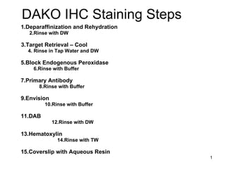
- 1. DAKO IHC Staining Steps 1.Deparaffinization and Rehydration 2.Rinse with DW 3.Target Retrieval – Cool 4. Rinse in Tap Water and DW 5.Block Endogenous Peroxidase 6.Rinse with Buffer 7.Primary Antibody 8.Rinse with Buffer 9.Envision 10.Rinse with Buffer 11.DAB 12.Rinse with DW 13.Hematoxylin 14.Rinse with TW 15.Coverslip with Aqueous Resin
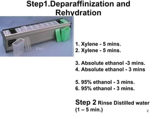
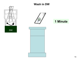
- 2. Step1.Deparaffinization and Rehydration 1. Xylene - 5 mins. 2. Xylene - 5 mins. 3. Absolute ethanol -3 mins. 4. Absolute ethanol - 3 mins 5. 95% ethanol - 3 mins. 6. 95% ethanol - 3 mins. Step 2 Rinse Distilled water (1 – 5 min.)
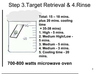
- 3. Step 3.Target Retrieval & 4.Rinse Total: 15 – 18 mins. plus 20 mins. cooling time = 35-38 mins) 1. High - 5 mins. 2. Medium High/Low - 5 mins. 3. Medium - 5 mins. 4. Medium - 3 mins. 5. Cooling time - 20 mins. 700-800 watts microwave oven
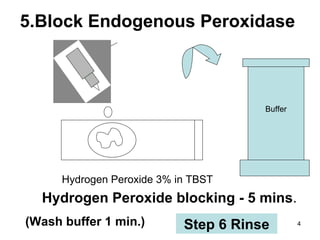
- 4. 5.Block Endogenous Peroxidase Hydrogen Peroxide 3% in TBST Hydrogen Peroxide blocking - 5 mins . (Wash buffer 1 min.) Buffer Step 6 Rinse
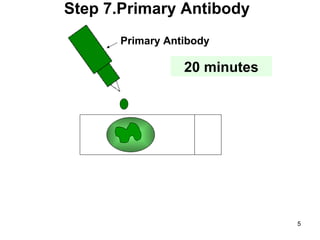
- 5. Primary Antibody Step 7.Primary Antibody 20 minutes
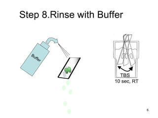
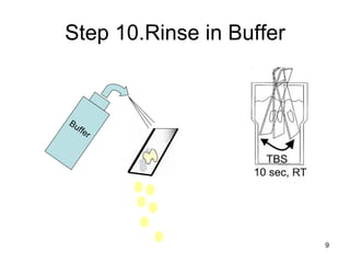
- 6. Step 8.Rinse with Buffer Buffer
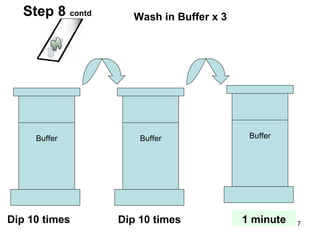
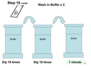
- 7. Buffer Buffer Buffer Wash in Buffer x 3 Dip 10 times Dip 10 times 1 minute Step 8 contd
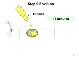
- 8. Envision Step 9.Envision 15 minutes
- 9. Step 10.Rinse in Buffer Buffer
- 10. Buffer Buffer Buffer Wash in Buffer x 3 Dip 10 times Dip 10 times 1 minute Step 10 contd
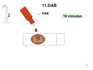
- 11. DAB 11.DAB 10 minutes
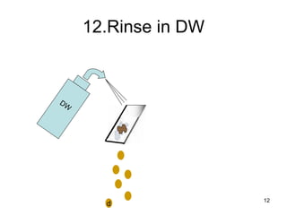
- 12. 12.Rinse in DW DW d
- 13. Wash in DW 1 Minute DW
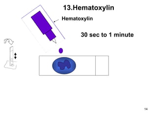
- 14. Hematoxylin 13.Hematoxylin 30 sec to 1 minute
- 15. 14.Rinse in Tap Water
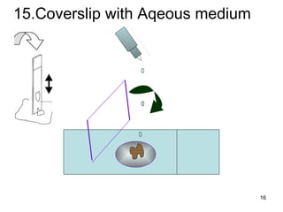
- 16. 15.Coverslip with Aqeous medium
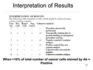
- 17. Interpretation of Results When >10% of total number of cancer cells stained by Ab = Positive
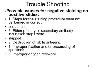
- 18. Trouble Shooting • Possible causes for negative staining on positive slides: 1. Steps for the staining procedure were not performed in correct sequence. 2. Either primary or secondary antibody incubation steps were skipped. 3. Destruction of labile antigens. 4. Improper fixation and/or processing of specimen. 5. Improper antigen recovery.
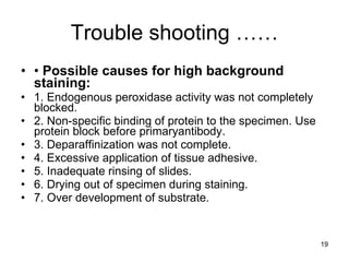
- 19. Trouble shooting …… • Possible causes for high background staining: 1. Endogenous peroxidase activity was not completely blocked. 2. Non-specific binding of protein to the specimen. Use protein block before primaryantibody. 3. Deparaffinization was not complete. 4. Excessive application of tissue adhesive. 5. Inadequate rinsing of slides. 6. Drying out of specimen during staining. 7. Over development of substrate.
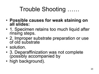
- 20. Trouble Shooting …… Possible causes for weak staining on all slides: 1. Specimen retains too much liquid after rinsing steps. 2. Improper substrate preparation or use of old substrate solution. 3. Deparaffinization was not complete (possibly accompanied by high background).
- 21. Ěý
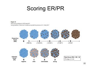
- 22. Scoring ER/PR