Stone disease evaluation in Pathology laboratory: Current prospective.
- 1. Metabolic and Renal Stone Analysis : Current prospective DDrr.. SSaannjjeeeevv MMeehhttaa MMDD Uro Lab. 1
- 2. Stone disease evaluation in Laboratory ŌĆó Series of tests to identify underlying causes. 1.Identify treatable metabolic abnormality 2.Identify underlying medical disease that predisposes to stone formation. 3.Outline a treatment plan ; focal Uro Lab. 2
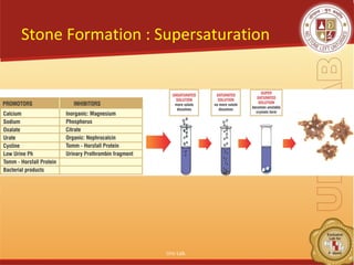
- 3. Why Do Kidney Stones Form? ŌĆó Reasons: Gnetic/dietary/Environmental ŌĆó Urine is supersaturated ŌĆó 2009: Supersaturation can be ŌĆ£fixŌĆØ ŌĆó Therefore, our job is to figure out what is causing the urinary Supersaturation and How best to fix it! Uro Lab. 3
- 4. Stone Formation : Supersaturation ŌĆó k Uro Lab. 4
- 5. Actually up to 65 different chemical compounds are found in urinary calculi. Uro Lab. 5
- 6. Metabolic evaluation ŌĆó Blood and Urinary Tests ; routine ŌĆó Stone Analysis ŌĆó 24 hrs Urinary Metabolic Profile with SUPERSATURTION. New advances in Stone analysis, Blood and Urinary Chemical analysis can find out 90-95% cause.* * Preminger G.M., ŌĆ£Guidelines for medical management of urolithiasisŌĆØ, bussin. meet us kid and urol dis,2005 Uro Lab. 6
- 7. Evaluation : First time stone with low risk ŌĆó Blood screen; Low K and HCO3, High Chloride - RTA High Uric acid - gouty diathesis High Calcium - Pri. Hyperparathyroidism Low Phosphorus ŌĆō Renal phosphate leak. ŌĆó Stone analysis ; all cases Uro Lab. 7
- 8. Evaluation: First time stone ŌĆó Urine Urinalysis : Routine pH > 7.5 - infection lithiasis pH < 5.5 - Uric acid lithiasis Sediments for crystalluria Urine culture : Urea-splitting organisms ŌĆō infection lithiasis. Screening / quantitative Cystine Uro Lab. 8
- 9. 24 hrs Urine metabolic profile ; Extensive Uro Lab. 9
- 10. 24 hrs extensive Metabolic evaluation: indications ŌĆó Stone recurrence ŌĆó Motivated patients wants to investigate. ŌĆó Select one-time formers: - Solitary Kidney - Renal insufficiency. - Residual stone burden. ŌĆó All children Uro Lab. 10
- 11. Extensive Metabolic Evaluation 24 hrs Urine collections. Stone risk factors : Volume Calcium , Calcium to creatinine index. Oxalate . ?Primary hyperoxaluria Citrate Uric acid. Uro Lab. 11
- 12. Extensive Metabolic Evaluation ŌĆó Dietary risk factors: Sodium, Potassium Magnesium Urinary analytes : phosporus, sulfate, Urea Marker for accuracy : Creatinine. Repeat 24 hrs Urine collection 4-6 weeks post intervantion. Uro Lab. 12
- 13. 24 hrs Urine metabolic profile graph Uro Lab. 13
- 14. Follow-up in same patient Uro Lab. 14
- 15. Stone analysis Uro Lab. 15
- 16. Renal Calculus Analysis ŌĆó Essential step in the examination and initial treatment of Urolithiasis. ŌĆó Composition yields fundamental information of pathogenesis of disease like ; - Metabolic abnormality. - Presence of infection. - Possible artifacts. - Drug metabolism. Uro Lab. 16
- 17. Integrated analysis: Techniques ŌĆó Optical Crystallogrphy ŌĆó Chemical Microscopy. ŌĆó Polarizing Microscopy. ŌĆó Infrared spectroscopy. ŌĆó X-ray diffraction. ŌĆó Electron Microscopy ŌĆó Fluorescence and chromatography. ŌĆó Final , semi quantitative, modified estimate from above results. * herringlab.com Uro Lab. 17
- 18. Significance of Stone analysis ŌĆó Exact composition gives important clue as to how Stone formed. ŌĆó Information may not available from any other type of work-up. ŌĆó Identify factors leading to clinical events. ŌĆó Identify Risk factors. Uro Lab. 18
- 19. Significance of Stone analysis Three categories : 1.Composition and hardness of Renal Stones. 2.Composition and its predictive value. 3.Composition and related metabolic abnormalities. Uro Lab. 19
- 20. Hardness Factor of Stone Calcium Oxalate Dihydrate 1.0 Calcium Oxalate Monohydrate 1.3 Hydroxy-peptite 1.1 Brushite 2.2 Uric Acid/ Urate 1.0 Cystine 2.4 Carbonate Apatite 1.3 Struvite 1.0 Mixed Stone 1.0 * Ringden I, Scand J Urol Nephrol.2007;41(4):316-23 Uro Lab. 20
- 21. Clinical Significance: Hardness pattern in Stone. ŌĆó Useful in describing consistency in individual. ŌĆó Formulation of treatment strategies. - Number of re-treatments. - Number of Shock waves. ŌĆó Energy index (KV x number of shock waves). ŌĆó PMID:17763224( PubMed-indexed for MEDLINE) Uro Lab. 21
- 22. Calcium stoneŌĆ”. ŌĆó Pure Calcium oxalate: More Acid urine, Low urine volume, high oxalate excretion. ŌĆó Mixed Stone formers ; High Calcium, pH and Stone formation rate. High Calcium excretion. * Schroeppel j Smith et all ; J Am Soc Nephrol 1997;8:568A Uro Lab. 22
- 23. CalciumŌĆ”.. ŌĆó Calcium Oxalate Monohydrate : Hypomagnesuria, acid urine, low volume More hard then dihydrate. ŌĆó Calcium Oxalate Dihydrate : - hypercalciuria. High Urine pH and hypocitraturia. Uro Lab. 23
- 24. Calcium Stone with ŌĆ” Carbonate apatite : may indicate Renal Tubular Acidosis (RTA). - Increases with amount of apatite. ( 5 ŌĆō 39%). Brushite Stones : Consider Renal tubular Acidosis (RTA). Uro Lab. 24
- 25. Struvite Stones ŌĆó Mixed Stone, infection. Mostly Proteus Ammonium Urate ŌĆó Elders : associated with infection. ŌĆó Children : May also form as result of hyperuricosuria, but NO urinary tract infection. ŌĆó Rare in pure form (0.03%) Uro Lab. 25
- 26. Uric Acid ŌĆó Hyperuricemia, hyperuricosuria. ŌĆó Low Urine Ph. < 6.2 ŌĆó Causes: - Gout. - Myeloproliferative processes associated with pathological increased purine metabolism. - Chemotherapy and Radiotherapy. Uro Lab. 26
- 27. Rare Cystine : ŌĆó Cysteinuria. Acidic ŌĆó Autosomal recessive disorder. Xenthene: ŌĆó Xanthinuria. ŌĆó Absence of Xanthinooxidase. ŌĆó Genetic autosomal hereditary recessive enzyme disorder. Uro Lab. 27
- 28. Conclusion. ŌĆó Combined with Optical Crystallography, appropriate Blood & 24 Urine metabolic work-up with super-saturation, it can find out the cause of stone formation in nearly all cases ŌĆó Supersaturation Index is GOLD Standard to know exactly patho-physiological basis of Stone formation to guide proper treatment. Uro Lab. 28
- 29. Thank You ! Uro Lab. 29


