


Synoptophore
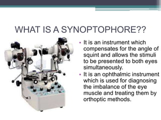
- 2. WHAT IS A SYNOPTOPHORE?? • It is an instrument which compensates for the angle of squint and allows the stimuli to be presented to both eyes simultaneously. • It is an ophthalmic instrument which is used for diagnosing the imbalance of the eye muscle and treating them by orthoptic methods.
- 3. WHAT IS IT USED FOR?? • It is used to investigate the potential for binocular function in the presence of a manifest squint. • Specifically used in children (from 3 years of age). • Also used to detect suppression and Abnormal retinal correspondance.
- 4. HOW DOES IT WORK?? • Consists of two cylindrical tubes with a mirrored right angled bend • A +6.50 D lens in each eyepiece which optically sets the testing distance to about 6 metres. • Pictures are inserted in a slide carrier situated at the outer end of each tube. • Two tubes are supported on columns which enable the pictures to be moved in relation to each other. • It can measure horizontal, vertical and torsional misalignments simultaneously.
- 6. ESTIMATION OF GRADES OF BINOCULAR VISION • Grade 1- (SIMULTANEOUS PERCEPTION) tested by two dissimilar but not mutually antagonistic pictures, such as a bird and a cage • Grade 2- (FUSION) Ability of the two eyes to produce a composite picture from two similar pictures, each of which is incomplete in one small different detail. • Grade 3- (stereopsis) Ability to obtain an impression of depth by the superimposition of two pictures of the same object which have been taken from slightly different angles.
- 8. DETECTION OF NORMAL/ABNORMAL ARC • Done by determining the subjective and objective angles of squint. • In normal retinal correspondance, these two angles are equal. • In ARC, objective angle is greater than the subjective angle and the difference between the two is called ANGLE OF ANOMALY.
- 9. THANK YOU!!!