Technique of bursectomy
Download as pptx, pdf1 like214 views
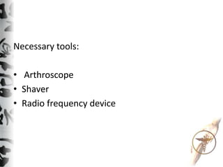
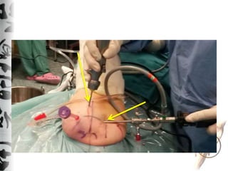
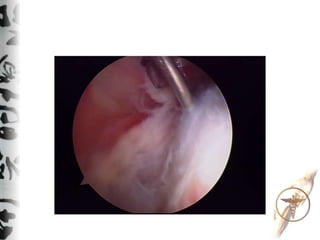
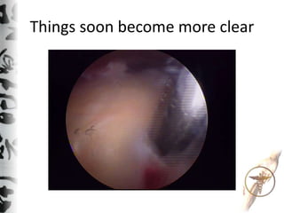
Technique of Bursectomy involves using an arthroscope, shaver, and radio frequency device through two portals, posterior and lateral, to perform bursectomy. Initially the view is blurred but things soon become more clear as the procedure progresses.
1 of 7
Download to read offline

Ad
Recommended
Rotator cuff 2008 final
Rotator cuff 2008 finalShoulder Library
╠²
This document discusses rotator cuff tears, including their indications, treatment options, and results. It provides an overview of rotator cuff anatomy and function. It describes the various types and classifications of rotator cuff tears and discusses the history and evolution of rotator cuff repair techniques. Treatment options are discussed depending on factors like the patient's age, tear size and chronicity. Expected results are outlined based on the pre-operative tissue quality and repair achieved.Bone defects thessal2010
Bone defects thessal2010Shoulder Library
╠²
This document discusses treatment options for bone defects in the glenoid and humeral head that can cause recurrent shoulder instability. It finds that humeral head (Hill-Sachs) defects occur in 65-93% of cases depending on the number of dislocations, while glenoid defects occur in 5-56% of cases. Treatment depends on the size and engagement of the defects. For large Hill-Sachs lesions, options include bone grafting, arthroplasty, or the remplissage procedure. For significant glenoid bone loss over 20-30%, options include soft tissue repair plus bone grafting or procedures like Bristow or Latarjet to add bone to the glenoid. The document advocatesRc repair philosophy and technique microhand 2014
Rc repair philosophy and technique microhand 2014Shoulder Library
╠²
This document summarizes the philosophy and techniques for arthroscopic treatment of rotator cuff tears. It discusses restoring the balance between the functional demands on the shoulder and the capacity of the rotator cuff by lowering demands, increasing cuff capacity, or repairing tears. Various tear patterns such as partial, full thickness, crescent, U-shaped, and massive contracted tears are described. Surgical techniques including debridement, acromioplasty, margin convergence, and interval slides are outlined. Good results are reported for small and medium tears, and massive tears with less fatty infiltration, while irreparable tears may require tendon transfers or arthroplasty.Impingement syndromes
Impingement syndromesShoulder Library
╠²
This document discusses various types of shoulder impingement syndromes. It begins with a brief history, noting that while Meyer first described the process in 1931, Neer classified and named shoulder impingement syndrome in 1972. It then describes current classifications, including primary and secondary subacromial impingement, coracohumeral impingement, glenoid (internal) impingement, ASI (AnteroSuperior Impingement), and PSGI (PosteroSuperior Glenoid Impingement). The bulk of the document focuses on describing these various impingement syndromes in more detail, including causes, presentations, treatments, and relevant anatomy. It provides an overview of imaging and clinical exams forŽĆŽüŽēŽä╬┐ ╬Ą╬Š╬¼Žü╬ĖŽü╬Ę╬╝╬▒
ŽĆŽüŽēŽä╬┐ ╬Ą╬Š╬¼Žü╬ĖŽü╬Ę╬╝╬▒Shoulder Library
╠²
This document discusses the treatment of first-time shoulder dislocations. It finds that arthroscopic stabilization has lower recurrence rates compared to conservative treatment, especially for young athletes. Arthroscopy allows visualization and repair of common lesions like Bankart tears and Hill-Sachs defects. Studies show arthroscopic stabilization reduces recurrence to 16% versus 47% for conservative care. Arthroscopy provides excellent outcomes with minimal pain and quick return to previous activity levels. It is the recommended approach for young, active patients to prevent future dislocations and allow continued athletic participation.╬╗╬╣╬▓╬▒╬┤╬Ą╬╣╬¼ 2012
╬╗╬╣╬▓╬▒╬┤╬Ą╬╣╬¼ 2012Shoulder Library
╠²
This document provides an overview of shoulder arthroscopy presented by Manos Antonogiannakis. It discusses shoulder anatomy, patient positioning, basic arthroscopic tools, and common arthroscopic procedures such as rotator cuff repair, biceps tenodesis/tenotomy, calcifying tendonitis repair, frozen shoulder treatment, and anterior instability procedures like Bankart repair. The presentation emphasizes proper portal placement, hemostasis, and having the necessary instrumentation prepared before beginning the arthroscopic procedure.Massive rot cuf
Massive rot cufShoulder Library
╠²
Massive rotator cuff tears present unique challenges for repair. The document discusses techniques for arthroscopic repair based on tear pattern, including releases to improve mobility. For crescent tears, a double row fixation is recommended. L-shaped and U-shaped tears are repaired with side-to-side sutures converging the margin. Massive contracted immobile tears may require interval slides. Outcomes are generally good, though strength deficits can remain. Proper patient selection considering fatty degeneration and mobility is important for success.Traumatic glenohumeral instability final
Traumatic glenohumeral instability finalShoulder Library
╠²
The document summarizes key points from a seminar on shoulder instability and scapular dyskinesis. It discusses the anatomy of the shoulder and factors contributing to instability, including traumatic injuries and muscular dysfunction. It presents classifications for different instability profiles and evaluates treatments based on the size of glenoid and humeral bone defects. The concepts of engaging versus non-engaging Hill-Sachs lesions are introduced, and the evolving model of on-track versus off-track lesions is described, which considers both glenoid track width and size of the Hill-Sachs interval. Remplissage is highlighted as an arthroscopic technique for engaging Hill-Sachs lesions.Shoulder arthroscopy general
Shoulder arthroscopy generalShoulder Library
╠²
Shoulder arthroscopy has evolved significantly since its origins in the 1930s. Diagnostic arthroscopy allowed better visualization of shoulder anatomy and pathology. Therapeutic arthroscopy now enables repair of most shoulder injuries including rotator cuff tears, instability, SLAP lesions, and arthritis. Advancements include stronger suture configurations, graft materials, and techniques to mobilize massive contracted tears. Arthroscopy provides less invasive alternatives to open surgery for treating shoulder conditions with equal or better outcomes including lower morbidity, faster recovery, and earlier return to activity. Further developments may include arthroscopic repair of bone defects and use of tendon substitutes.Evag rot cuf
Evag rot cufShoulder Library
╠²
This document discusses arthroscopic rotator cuff repair. It begins with an overview of rotator cuff disease and the history of rotator cuff repair surgery. The document then outlines the goals and techniques of arthroscopic rotator cuff repair surgery, including preparation, visualization, tear evaluation and closure methods. Post-operative rehabilitation is also addressed. The document concludes with a discussion of studies showing high success rates for arthroscopic repair and comparisons of advantages and disadvantages to open surgical techniques.╬▒Žü╬ĖŽü╬┐Žā╬║ŽīŽĆ╬ĘŽā╬Ę ŽÄ╬╝╬┐Žģ ╬╝ŽäŽć ŽĆ╬▒Žü╬▒╬║╬┐╬╗╬┐ŽŹ╬Ė╬ĘŽā╬Ę
╬▒Žü╬ĖŽü╬┐Žā╬║ŽīŽĆ╬ĘŽā╬Ę ŽÄ╬╝╬┐Žģ ╬╝ŽäŽć ŽĆ╬▒Žü╬▒╬║╬┐╬╗╬┐ŽŹ╬Ė╬ĘŽā╬ĘShoulder Library
╠²
This document discusses shoulder arthroscopy and postoperative monitoring. It begins by outlining the range of motion and planes of movement in the shoulder joint, which make it prone to injuries. The history of shoulder arthroscopy is then reviewed, from the first cadaver procedure in 1931 to advances like arthroscopic rotator cuff repair in the 1980s. Diagnostic arthroscopy techniques are covered, including anesthesia, positioning, room setup, and basic tools. Therapeutic arthroscopy is then outlined for treating various shoulder issues like rotator cuff tears, instability, SLAP lesions, frozen shoulder, AC joint problems, and arthritis. Success rates for arthroscopic shoulder stabilization are reported to be over 90% in several studies.╬Ą╬Š╬Ą╬╗╬»╬Š╬Ą╬╣Žé ŽāŽä╬Ę╬Į ╬▒Žü╬ĖŽü╬┐Žā╬║╬┐ŽĆ╬╣╬║╬« Žć╬Ą╬╣Žü╬┐ŽģŽü╬│╬╣╬║╬« Žä╬ĘŽé ╬▒╬║ŽüŽē╬╝╬╣╬┐╬║╬╗╬Ą╬╣╬┤╬╣╬║╬«Žé
╬Ą╬Š╬Ą╬╗╬»╬Š╬Ą╬╣Žé ŽāŽä╬Ę╬Į ╬▒Žü╬ĖŽü╬┐Žā╬║╬┐ŽĆ╬╣╬║╬« Žć╬Ą╬╣Žü╬┐ŽģŽü╬│╬╣╬║╬« Žä╬ĘŽé ╬▒╬║ŽüŽē╬╝╬╣╬┐╬║╬╗╬Ą╬╣╬┤╬╣╬║╬«ŽéShoulder Library
╠²
This document discusses advances in arthroscopy of the acromioclavicular (AC) joint. It describes the anatomy of the AC joint and common pathologies like osteolysis, arthritis, cysts, and dislocations that can be treated arthroscopically. Techniques for arthroscopic distal clavicle excision, AC joint reconstruction for dislocations using grafts or ligament transfers are presented. The author's preference is for open reduction and fixation for AC dislocations due to more precise reduction and residual scarring's effect on stability. Several case examples are provided.╬║╬▒╬╗╬▒╬╝╬¼Žä╬▒ 2016 ╬▒Žü╬ĖŽü╬»Žä╬╣╬┤╬▒ ŽÄ╬╝╬┐Žģ
╬║╬▒╬╗╬▒╬╝╬¼Žä╬▒ 2016 ╬▒Žü╬ĖŽü╬»Žä╬╣╬┤╬▒ ŽÄ╬╝╬┐ŽģShoulder Library
╠²
This document discusses shoulder arthritis and treatment options. It begins by describing the anatomy of the shoulder joint and causes of shoulder pain such as osteoarthritis, rotator cuff tears, and fractures. Symptoms of shoulder arthritis include pain, reduced range of motion, grinding, and stiffness. Diagnosis involves physical exams, imaging like x-rays and MRIs, and blood tests. Non-surgical treatments include medications, injections, physical therapy and lifestyle modifications. Surgical options include joint preserving procedures like arthroscopy and synovectomy for early arthritis or shoulder replacements like hemiarthroplasty, total shoulder replacement, and reverse total shoulder replacement for more severe arthritis. Outcomes of shoulder replacements are generally good with implant survival rates ofPortals navigation
Portals navigationShoulder Library
╠²
This document provides an overview of shoulder arthroscopy procedures including patient positioning, room setup, skin marking of bony landmarks, creation of portals, and navigation of the glenohumeral joint and subacromial space. It discusses the lateral decubitus position as the preferred approach and describes skin marking for the basic posterior, anterior superior, anterior inferior, and posterior lateral portals. Key steps are outlined including insertion of the arthroscope through the posterior portal for initial visualization of the glenohumeral joint.Traumatic shoulder dislocation 2017 kat
Traumatic shoulder dislocation 2017 katShoulder Library
╠²
This document discusses the management of traumatic anterior shoulder dislocations. It begins by describing the shoulder's anatomy and how its mobility makes it prone to instability. It then reviews the history and clinical examination findings that help determine appropriate treatment. Arthroscopic findings from studies of acute and chronic dislocations are presented, showing common lesions like Bankart tears. Treatment options are explored, including arthroscopic stabilization which can address all lesions with minimal morbidity. Arthroscopy allows accurate diagnosis and repair of injuries while facilitating early rehabilitation. The conclusion is that arthroscopy is now often the treatment of choice for traumatic shoulder dislocations.Posterior instability
Posterior instabilityShoulder Library
╠²
This document discusses posterior shoulder instability. It begins by outlining different types of posterior instability such as locked dislocation, traumatic unidirectional instability, and atraumatic instability. It emphasizes the importance of obtaining a thorough history and clinical examination to determine the mechanism of injury, position of apprehension, and ability to demonstrate instability. Imaging can help identify labral tears or bone abnormalities like glenoid dysplasia. Arthroscopic surgical techniques are recommended for treating traumatic unidirectional instability and involve repairing the labrum through posterior and accessory portals. Post-operative rehabilitation is outlined. Careful examination is key to properly diagnosing and treating posterior shoulder instability.Massive rct salonica 2106
Massive rct salonica 2106Shoulder Library
╠²
1) Massive rotator cuff tears lower the functional capacity of the shoulder and can cause clinical symptoms when functional demands exceed capacity.
2) Treatment aims to restore equilibrium between demands and capacity by lowering demands, increasing capacity, repairing the cuff, or using tendon transfers or reverse shoulder arthroplasty.
3) Complete release of the torn tendons from surrounding structures is required to properly assess reparability before attempting repair without tension or considering alternative treatments.Double row athlitiatriko 2008
Double row athlitiatriko 2008Shoulder Library
╠²
This document discusses the history and techniques of double row arthroscopic rotator cuff repair. It provides details on the anatomy and function of the rotator cuff. Tears can range from partial to full thickness and increase in prevalence with age. Double row repair techniques aim to restore the footprint and increase fixation points compared to single row repair. While it provides stronger repair, double row is more time consuming and technically challenging. Factors like tear size and chronicity impact repair outcomes.Mdi physiotherapists - nikos
Mdi physiotherapists - nikosShoulder Library
╠²
This document discusses multi-directional shoulder instability (MDI). MDI is characterized by instability in at least two directions, usually anterior, posterior, or inferior. It is commonly caused by repetitive overhead motion stretching the shoulder capsule beyond its limits. Clinically, MDI presents with vague symptoms, laxity and translation in multiple directions on examination. Treatment involves strengthening dynamic stabilizers through physical therapy, with surgery such as arthroscopic capsular plication considered if conservative measures fail. Outcomes are generally good, though some residual pain and instability may remain long term.Mri in corellation to surgery
Mri in corellation to surgeryShoulder Library
╠²
The document summarizes a study on arthroscopic remplissage for recurrent anterior shoulder instability. 48 patients underwent remplissage in addition to Bankart repair, with a mean follow-up of 37 months. The failure rate was 6.3%, and 93.7% were satisfied without restrictions. Scores on the ASES, Rowe, and Oxford scales all significantly improved post-operatively without loss of range of motion. The study concludes remplissage enhances Bankart repair for managing instability, with good results and no effect on shoulder movement.Fixation techniques in rot cuff repair
Fixation techniques in rot cuff repairShoulder Library
╠²
This document discusses techniques for repairing rotator cuff tears arthroscopically. It begins by describing the classification of partial versus full thickness tears and massive tears involving two or more tendons. For massive contracted immobile tears, interval slides can be performed through the anterior and posterior intervals to regain mobility. Repair techniques depend on the tear pattern, such as side-to-side sutures for U-shaped tears or interval slides for massive immobile tears. Results of arthroscopic repair for massive tears show 84-94% excellent or good results according to several studies. The key is to match the repair technique to the tear morphology and mobility to minimize strain on the repair.Installation problems
Installation problemsShoulder Library
╠²
1. The document outlines potential problems that may occur with arthroscopic tower equipment and provides protocols for addressing each issue.
2. It describes the key components and circuits of the arthroscopic tower system, including the power supply, image, water, shaver, and coagulation circuits.
3. Standard protocols are proposed for responding to different types of malfunctions, such as identifying the faulty circuit, diagnosing the specific issue, and either fixing or replacing the problematic component. Maintaining calm and clearing identifying the root cause are emphasized.New developments in shoulder arthroscopy
New developments in shoulder arthroscopyShoulder Library
╠²
This document discusses new developments in arthroscopic shoulder surgery. It describes various shoulder pathologies that can be addressed with arthroscopic surgery, including painful conditions like rotator cuff tears, biceps tendinosis, and frozen shoulder. It also discusses unstable shoulder conditions like anterior glenohumeral instability. The document provides details on surgical techniques for treating various partial and full-thickness rotator cuff tears arthroscopically. It also describes options for managing massive, contracted rotator cuff tears as well as glenohumeral instability with significant bone loss, such as the arthroscopic Latarjet procedure.Shoulder sports related injuries
Shoulder sports related injuriesShoulder Library
╠²
1. Shoulder injuries are common in sports and can be acute or chronic. They range from mild sprains to traumatic dislocations and are often painful and mobility-restricting.
2. MRI and CT scans are important imaging modalities to diagnose shoulder injuries and assess soft tissue damage, bone defects, and other pathology like tumors or fractures. MR arthrography provides high accuracy for labral tears.
3. Common acute injuries include dislocations, rotator cuff tears, and injuries to the biceps tendon. Chronic overload can also cause tendinopathy and impingement. The size and chronicity of rotator cuff tears affects prognosis.Instability and bone loss. pptx
Instability and bone loss. pptxShoulder Library
╠²
This document discusses shoulder dislocation with bone defects. It begins with an introduction to the speaker, Dr. Manos Antonogiannakis, and the conference he is presenting at. The document then covers various topics related to shoulder instability and bone defects, including the types of instability, static and dynamic stabilizers of the shoulder, classifications of glenoid and humeral bone defects, techniques for addressing bone defects like the Latarjet procedure and remplissage, and results from studies on these techniques. It provides images and diagrams to illustrate the concepts and surgical approaches.Evolution of tsa1
Evolution of tsa1Shoulder Library
╠²
The evolution of shoulder arthroplasty has progressed through several generations of prosthesis designs from the late 19th century to present day. Early designs in the 1890s-1950s aimed to replicate the native anatomy but had high failure rates due to issues like wear, loosening, and infection. Modular designs in the 1980s improved positioning and sizing but still did not fully restore anatomy. Current third generation prostheses from the 1990s onward are anatomically designed with variable sizes and offsets to more closely mimic the native joint mechanics and center of rotation. Reverse total shoulder arthroplasty, developed in the 1970s-1990s, has also improved through lateralized and inferiorly tilted component designs to maximize deltoid function for patients with rotator cMultidirectional shoulder instability
Multidirectional shoulder instabilityShoulder Library
╠²
This document discusses multi-directional shoulder instability (MDI). MDI is characterized by subluxations or dislocations in at least two directions, usually anteriorly, posteriorly, or inferiorly. It is commonly seen in overhead athletes and is associated with capsular laxity. Clinical examination reveals laxity and translation in multiple directions. Treatment involves strengthening dynamic stabilizers through physical therapy initially, with surgery such as arthroscopic capsular plication considered if conservative measures fail. Post-operative rehabilitation is important for successful outcomes. Long-term, over half of untreated MDI patients experience pain and instability.How to do a sd & a dc resection
How to do a sd & a dc resectionShoulder Library
╠²
The document describes the steps for performing a subacromial decompression and distal clavicle resection arthroscopically. It discusses positioning the patient laterally, creating portals for accessing the subacromial space, performing a bursectomy and acromioplasty by resecting the anterior and lateral edges of the acromion, and resecting the distal 6-8 mm of the clavicle parallel to the AC joint from multiple portals. Post-operative checks include verifying 8-10 mm of space in the acromioclavicular joint with shoulder abduction and external rotation.INTERPRETATION OF LABORATORY INVESTIGATIONS.pptx
INTERPRETATION OF LABORATORY INVESTIGATIONS.pptxEliLawluvi
╠²
THE DOCUMENT SUMMARIZES THE KEY COMPONENTS OF INTERPRETING FULL BLOOD CUNTMore Related Content
More from Shoulder Library (20)
Shoulder arthroscopy general
Shoulder arthroscopy generalShoulder Library
╠²
Shoulder arthroscopy has evolved significantly since its origins in the 1930s. Diagnostic arthroscopy allowed better visualization of shoulder anatomy and pathology. Therapeutic arthroscopy now enables repair of most shoulder injuries including rotator cuff tears, instability, SLAP lesions, and arthritis. Advancements include stronger suture configurations, graft materials, and techniques to mobilize massive contracted tears. Arthroscopy provides less invasive alternatives to open surgery for treating shoulder conditions with equal or better outcomes including lower morbidity, faster recovery, and earlier return to activity. Further developments may include arthroscopic repair of bone defects and use of tendon substitutes.Evag rot cuf
Evag rot cufShoulder Library
╠²
This document discusses arthroscopic rotator cuff repair. It begins with an overview of rotator cuff disease and the history of rotator cuff repair surgery. The document then outlines the goals and techniques of arthroscopic rotator cuff repair surgery, including preparation, visualization, tear evaluation and closure methods. Post-operative rehabilitation is also addressed. The document concludes with a discussion of studies showing high success rates for arthroscopic repair and comparisons of advantages and disadvantages to open surgical techniques.╬▒Žü╬ĖŽü╬┐Žā╬║ŽīŽĆ╬ĘŽā╬Ę ŽÄ╬╝╬┐Žģ ╬╝ŽäŽć ŽĆ╬▒Žü╬▒╬║╬┐╬╗╬┐ŽŹ╬Ė╬ĘŽā╬Ę
╬▒Žü╬ĖŽü╬┐Žā╬║ŽīŽĆ╬ĘŽā╬Ę ŽÄ╬╝╬┐Žģ ╬╝ŽäŽć ŽĆ╬▒Žü╬▒╬║╬┐╬╗╬┐ŽŹ╬Ė╬ĘŽā╬ĘShoulder Library
╠²
This document discusses shoulder arthroscopy and postoperative monitoring. It begins by outlining the range of motion and planes of movement in the shoulder joint, which make it prone to injuries. The history of shoulder arthroscopy is then reviewed, from the first cadaver procedure in 1931 to advances like arthroscopic rotator cuff repair in the 1980s. Diagnostic arthroscopy techniques are covered, including anesthesia, positioning, room setup, and basic tools. Therapeutic arthroscopy is then outlined for treating various shoulder issues like rotator cuff tears, instability, SLAP lesions, frozen shoulder, AC joint problems, and arthritis. Success rates for arthroscopic shoulder stabilization are reported to be over 90% in several studies.╬Ą╬Š╬Ą╬╗╬»╬Š╬Ą╬╣Žé ŽāŽä╬Ę╬Į ╬▒Žü╬ĖŽü╬┐Žā╬║╬┐ŽĆ╬╣╬║╬« Žć╬Ą╬╣Žü╬┐ŽģŽü╬│╬╣╬║╬« Žä╬ĘŽé ╬▒╬║ŽüŽē╬╝╬╣╬┐╬║╬╗╬Ą╬╣╬┤╬╣╬║╬«Žé
╬Ą╬Š╬Ą╬╗╬»╬Š╬Ą╬╣Žé ŽāŽä╬Ę╬Į ╬▒Žü╬ĖŽü╬┐Žā╬║╬┐ŽĆ╬╣╬║╬« Žć╬Ą╬╣Žü╬┐ŽģŽü╬│╬╣╬║╬« Žä╬ĘŽé ╬▒╬║ŽüŽē╬╝╬╣╬┐╬║╬╗╬Ą╬╣╬┤╬╣╬║╬«ŽéShoulder Library
╠²
This document discusses advances in arthroscopy of the acromioclavicular (AC) joint. It describes the anatomy of the AC joint and common pathologies like osteolysis, arthritis, cysts, and dislocations that can be treated arthroscopically. Techniques for arthroscopic distal clavicle excision, AC joint reconstruction for dislocations using grafts or ligament transfers are presented. The author's preference is for open reduction and fixation for AC dislocations due to more precise reduction and residual scarring's effect on stability. Several case examples are provided.╬║╬▒╬╗╬▒╬╝╬¼Žä╬▒ 2016 ╬▒Žü╬ĖŽü╬»Žä╬╣╬┤╬▒ ŽÄ╬╝╬┐Žģ
╬║╬▒╬╗╬▒╬╝╬¼Žä╬▒ 2016 ╬▒Žü╬ĖŽü╬»Žä╬╣╬┤╬▒ ŽÄ╬╝╬┐ŽģShoulder Library
╠²
This document discusses shoulder arthritis and treatment options. It begins by describing the anatomy of the shoulder joint and causes of shoulder pain such as osteoarthritis, rotator cuff tears, and fractures. Symptoms of shoulder arthritis include pain, reduced range of motion, grinding, and stiffness. Diagnosis involves physical exams, imaging like x-rays and MRIs, and blood tests. Non-surgical treatments include medications, injections, physical therapy and lifestyle modifications. Surgical options include joint preserving procedures like arthroscopy and synovectomy for early arthritis or shoulder replacements like hemiarthroplasty, total shoulder replacement, and reverse total shoulder replacement for more severe arthritis. Outcomes of shoulder replacements are generally good with implant survival rates ofPortals navigation
Portals navigationShoulder Library
╠²
This document provides an overview of shoulder arthroscopy procedures including patient positioning, room setup, skin marking of bony landmarks, creation of portals, and navigation of the glenohumeral joint and subacromial space. It discusses the lateral decubitus position as the preferred approach and describes skin marking for the basic posterior, anterior superior, anterior inferior, and posterior lateral portals. Key steps are outlined including insertion of the arthroscope through the posterior portal for initial visualization of the glenohumeral joint.Traumatic shoulder dislocation 2017 kat
Traumatic shoulder dislocation 2017 katShoulder Library
╠²
This document discusses the management of traumatic anterior shoulder dislocations. It begins by describing the shoulder's anatomy and how its mobility makes it prone to instability. It then reviews the history and clinical examination findings that help determine appropriate treatment. Arthroscopic findings from studies of acute and chronic dislocations are presented, showing common lesions like Bankart tears. Treatment options are explored, including arthroscopic stabilization which can address all lesions with minimal morbidity. Arthroscopy allows accurate diagnosis and repair of injuries while facilitating early rehabilitation. The conclusion is that arthroscopy is now often the treatment of choice for traumatic shoulder dislocations.Posterior instability
Posterior instabilityShoulder Library
╠²
This document discusses posterior shoulder instability. It begins by outlining different types of posterior instability such as locked dislocation, traumatic unidirectional instability, and atraumatic instability. It emphasizes the importance of obtaining a thorough history and clinical examination to determine the mechanism of injury, position of apprehension, and ability to demonstrate instability. Imaging can help identify labral tears or bone abnormalities like glenoid dysplasia. Arthroscopic surgical techniques are recommended for treating traumatic unidirectional instability and involve repairing the labrum through posterior and accessory portals. Post-operative rehabilitation is outlined. Careful examination is key to properly diagnosing and treating posterior shoulder instability.Massive rct salonica 2106
Massive rct salonica 2106Shoulder Library
╠²
1) Massive rotator cuff tears lower the functional capacity of the shoulder and can cause clinical symptoms when functional demands exceed capacity.
2) Treatment aims to restore equilibrium between demands and capacity by lowering demands, increasing capacity, repairing the cuff, or using tendon transfers or reverse shoulder arthroplasty.
3) Complete release of the torn tendons from surrounding structures is required to properly assess reparability before attempting repair without tension or considering alternative treatments.Double row athlitiatriko 2008
Double row athlitiatriko 2008Shoulder Library
╠²
This document discusses the history and techniques of double row arthroscopic rotator cuff repair. It provides details on the anatomy and function of the rotator cuff. Tears can range from partial to full thickness and increase in prevalence with age. Double row repair techniques aim to restore the footprint and increase fixation points compared to single row repair. While it provides stronger repair, double row is more time consuming and technically challenging. Factors like tear size and chronicity impact repair outcomes.Mdi physiotherapists - nikos
Mdi physiotherapists - nikosShoulder Library
╠²
This document discusses multi-directional shoulder instability (MDI). MDI is characterized by instability in at least two directions, usually anterior, posterior, or inferior. It is commonly caused by repetitive overhead motion stretching the shoulder capsule beyond its limits. Clinically, MDI presents with vague symptoms, laxity and translation in multiple directions on examination. Treatment involves strengthening dynamic stabilizers through physical therapy, with surgery such as arthroscopic capsular plication considered if conservative measures fail. Outcomes are generally good, though some residual pain and instability may remain long term.Mri in corellation to surgery
Mri in corellation to surgeryShoulder Library
╠²
The document summarizes a study on arthroscopic remplissage for recurrent anterior shoulder instability. 48 patients underwent remplissage in addition to Bankart repair, with a mean follow-up of 37 months. The failure rate was 6.3%, and 93.7% were satisfied without restrictions. Scores on the ASES, Rowe, and Oxford scales all significantly improved post-operatively without loss of range of motion. The study concludes remplissage enhances Bankart repair for managing instability, with good results and no effect on shoulder movement.Fixation techniques in rot cuff repair
Fixation techniques in rot cuff repairShoulder Library
╠²
This document discusses techniques for repairing rotator cuff tears arthroscopically. It begins by describing the classification of partial versus full thickness tears and massive tears involving two or more tendons. For massive contracted immobile tears, interval slides can be performed through the anterior and posterior intervals to regain mobility. Repair techniques depend on the tear pattern, such as side-to-side sutures for U-shaped tears or interval slides for massive immobile tears. Results of arthroscopic repair for massive tears show 84-94% excellent or good results according to several studies. The key is to match the repair technique to the tear morphology and mobility to minimize strain on the repair.Installation problems
Installation problemsShoulder Library
╠²
1. The document outlines potential problems that may occur with arthroscopic tower equipment and provides protocols for addressing each issue.
2. It describes the key components and circuits of the arthroscopic tower system, including the power supply, image, water, shaver, and coagulation circuits.
3. Standard protocols are proposed for responding to different types of malfunctions, such as identifying the faulty circuit, diagnosing the specific issue, and either fixing or replacing the problematic component. Maintaining calm and clearing identifying the root cause are emphasized.New developments in shoulder arthroscopy
New developments in shoulder arthroscopyShoulder Library
╠²
This document discusses new developments in arthroscopic shoulder surgery. It describes various shoulder pathologies that can be addressed with arthroscopic surgery, including painful conditions like rotator cuff tears, biceps tendinosis, and frozen shoulder. It also discusses unstable shoulder conditions like anterior glenohumeral instability. The document provides details on surgical techniques for treating various partial and full-thickness rotator cuff tears arthroscopically. It also describes options for managing massive, contracted rotator cuff tears as well as glenohumeral instability with significant bone loss, such as the arthroscopic Latarjet procedure.Shoulder sports related injuries
Shoulder sports related injuriesShoulder Library
╠²
1. Shoulder injuries are common in sports and can be acute or chronic. They range from mild sprains to traumatic dislocations and are often painful and mobility-restricting.
2. MRI and CT scans are important imaging modalities to diagnose shoulder injuries and assess soft tissue damage, bone defects, and other pathology like tumors or fractures. MR arthrography provides high accuracy for labral tears.
3. Common acute injuries include dislocations, rotator cuff tears, and injuries to the biceps tendon. Chronic overload can also cause tendinopathy and impingement. The size and chronicity of rotator cuff tears affects prognosis.Instability and bone loss. pptx
Instability and bone loss. pptxShoulder Library
╠²
This document discusses shoulder dislocation with bone defects. It begins with an introduction to the speaker, Dr. Manos Antonogiannakis, and the conference he is presenting at. The document then covers various topics related to shoulder instability and bone defects, including the types of instability, static and dynamic stabilizers of the shoulder, classifications of glenoid and humeral bone defects, techniques for addressing bone defects like the Latarjet procedure and remplissage, and results from studies on these techniques. It provides images and diagrams to illustrate the concepts and surgical approaches.Evolution of tsa1
Evolution of tsa1Shoulder Library
╠²
The evolution of shoulder arthroplasty has progressed through several generations of prosthesis designs from the late 19th century to present day. Early designs in the 1890s-1950s aimed to replicate the native anatomy but had high failure rates due to issues like wear, loosening, and infection. Modular designs in the 1980s improved positioning and sizing but still did not fully restore anatomy. Current third generation prostheses from the 1990s onward are anatomically designed with variable sizes and offsets to more closely mimic the native joint mechanics and center of rotation. Reverse total shoulder arthroplasty, developed in the 1970s-1990s, has also improved through lateralized and inferiorly tilted component designs to maximize deltoid function for patients with rotator cMultidirectional shoulder instability
Multidirectional shoulder instabilityShoulder Library
╠²
This document discusses multi-directional shoulder instability (MDI). MDI is characterized by subluxations or dislocations in at least two directions, usually anteriorly, posteriorly, or inferiorly. It is commonly seen in overhead athletes and is associated with capsular laxity. Clinical examination reveals laxity and translation in multiple directions. Treatment involves strengthening dynamic stabilizers through physical therapy initially, with surgery such as arthroscopic capsular plication considered if conservative measures fail. Post-operative rehabilitation is important for successful outcomes. Long-term, over half of untreated MDI patients experience pain and instability.How to do a sd & a dc resection
How to do a sd & a dc resectionShoulder Library
╠²
The document describes the steps for performing a subacromial decompression and distal clavicle resection arthroscopically. It discusses positioning the patient laterally, creating portals for accessing the subacromial space, performing a bursectomy and acromioplasty by resecting the anterior and lateral edges of the acromion, and resecting the distal 6-8 mm of the clavicle parallel to the AC joint from multiple portals. Post-operative checks include verifying 8-10 mm of space in the acromioclavicular joint with shoulder abduction and external rotation.╬Ą╬Š╬Ą╬╗╬»╬Š╬Ą╬╣Žé ŽāŽä╬Ę╬Į ╬▒Žü╬ĖŽü╬┐Žā╬║╬┐ŽĆ╬╣╬║╬« Žć╬Ą╬╣Žü╬┐ŽģŽü╬│╬╣╬║╬« Žä╬ĘŽé ╬▒╬║ŽüŽē╬╝╬╣╬┐╬║╬╗╬Ą╬╣╬┤╬╣╬║╬«Žé
╬Ą╬Š╬Ą╬╗╬»╬Š╬Ą╬╣Žé ŽāŽä╬Ę╬Į ╬▒Žü╬ĖŽü╬┐Žā╬║╬┐ŽĆ╬╣╬║╬« Žć╬Ą╬╣Žü╬┐ŽģŽü╬│╬╣╬║╬« Žä╬ĘŽé ╬▒╬║ŽüŽē╬╝╬╣╬┐╬║╬╗╬Ą╬╣╬┤╬╣╬║╬«ŽéShoulder Library
╠²
Recently uploaded (20)
INTERPRETATION OF LABORATORY INVESTIGATIONS.pptx
INTERPRETATION OF LABORATORY INVESTIGATIONS.pptxEliLawluvi
╠²
THE DOCUMENT SUMMARIZES THE KEY COMPONENTS OF INTERPRETING FULL BLOOD CUNTAD-SAFE: An Initiative to Build Understanding of ARIA and Skills Needed to Gu...
AD-SAFE: An Initiative to Build Understanding of ARIA and Skills Needed to Gu...PVI, PeerView Institute for Medical Education
╠²
Chair and Presenter, Ana M. Franceschi, MD, PhD, and Petrice M. Cogswell, MD, PhD, discuss AlzheimerŌĆÖs disease in this CME/MOC/NCPD/CPE/AAPA/IPCE activity titled ŌĆ£AD-SAFE: An Initiative to Build Understanding of ARIA and Skills Needed to Guide Treatment Decisions and Support Rapid Recognition and Response in Radiology.ŌĆØ For the full presentation, downloadable Practice Aids, and complete CME/MOC/NCPD/CPE/AAPA/IPCE information, and to apply for credit, please visit us at https://bit.ly/42nd09H. CME/MOC/NCPD/CPE/AAPA/IPCE credit will be available until May 26, 2026.Tuberculosis Nepal 2025 National Plan.pptx
Tuberculosis Nepal 2025 National Plan.pptxDr. Anu Marhatta
╠²
The presentation contains preventive therapy, the DOTS program, and the national program of Nepal. This material is intended for educational purposes only.COUGH AND THIER DRUGS AND MODE OF ACTION.pptx
COUGH AND THIER DRUGS AND MODE OF ACTION.pptxPackialakshmiP
╠²
COUGH AND THIER DRUGS AND MODE OF ACTIONAnti-Infective Agents: Classification, Uses & Formulations Detailed Guide to...
Anti-Infective Agents: Classification, Uses & Formulations Detailed Guide to...Sajini
╠²
This presentation covers various classes of anti-infective agents including antifungal, antiviral, antitubercular, antimalarial, urinary tract anti-infectives, and sulphonamides. It provides classification, properties, storage conditions, uses, brand names, and pharmaceutical formulations of each drug. Ideal for pharmacy, nursing, and medical students preparing for pharmacology and mediciinferential statistics Part - 1 i.e Parametric tests
inferential statistics Part - 1 i.e Parametric testsBabitha Devu
╠²
This part deals with the Inferential Statistics.
Basic Terminologies.
Parametric Test.
T-test
paired & unpaired T -test
Z testDrugs Acting on the Autonomic Nervous System ŌĆō Classification, Properties & Uses
Drugs Acting on the Autonomic Nervous System ŌĆō Classification, Properties & UsesSajini
╠²
This presentation offers a comprehensive overview of drugs acting on the autonomic nervous system (ANS), including sympathomimetic, sympatholytic, parasympathomimetic, and anticholinergic agents. It explains the classification, mechanism of action, properties, formulation, brand names, and clinical uses of key drugs like adrenaline, dopamine, propranolol, atropine, and pilocarpine. Ideal for students and professionals in pharmacy, medicine, and healthcare.Updates_in Head__Neck TNM staging- 9th edition.pptx
Updates_in Head__Neck TNM staging- 9th edition.pptxDr. Maroti Wadewale
╠²
The Union for International Cancer Control (UICC) and the American Joint Committee on Cancer (AJCC) collaborate closely to produce the globally recognized TNM Classification of Malignant Tumours. Therefore, when discussing the "9th edition of UICC head and neck staging," it's largely in alignment with the updates introduced by the AJCC's Version 9. The UICC's TNM Core Committee finalized the 9th edition of the TNM Classification, with publication anticipated in August 2025.
The key updates for head and neck cancers in the 9th edition (or Version 9) reflect an ongoing effort to improve prognostic accuracy and align staging with contemporary clinical understanding and treatment outcomes. Here are the significant changes, particularly those relevant to head and neck:
* Emphasis on Personalized Care and Prognostic Refinements: The 9th edition reflects a greater focus on personalized care, incorporating refinements that aim to better predict patient outcomes.
* Revised Criteria for Specific Head and Neck Cancers:
* Nasopharyngeal Carcinoma (NPC): This site has seen substantial revisions, as highlighted in the previous response on AJCC updates. Key changes include:
* More precise definition of T3 disease (unequivocal evidence of bone involvement).
* Introduction of advanced radiologic extranodal extension (ENE) as an N3 criterion. This acknowledges the prognostic impact of ENE seen on imaging.
* Subclassification of M1 disease into M1a (3 or fewer metastatic lesions) and M1b (more than 3 lesions) to better stratify prognosis in metastatic settings.
* Redefined Stage Groups for NPC, with T1-2N0-1 now often falling into Stage I, and Stage IV being exclusively for metastatic disease, further subdivided by the M1a/M1b categories.
* Salivary Gland Cancers: Revised criteria based on updated imaging and anatomical features are being incorporated.
* HPV-Related Oropharyngeal Cancers: New staging is introduced for HPV-associated oropharyngeal cancers to better reflect their distinct biological behavior and prognosis, which is generally more favorable than HPV-negative oropharyngeal cancers. This often involves specific considerations for nodal burden.
* Integration of Imaging and Anatomical Features: The updates are grounded in recent evidence, incorporating insights from advanced imaging techniques and a deeper understanding of anatomical spread.
* International Collaboration: These updates are the result of collaborative efforts between the AJCC and UICC, involving input from cancer registries, clinical outcomes data, and disease-specific experts worldwide. The goal is to provide a unified and globally applicable staging system.
* Dynamic Update Process: Similar to the AJCC's shift from "Editions" to "Versions" for specific sites, the UICC is also exploring more flexible ways to share future TNM updates.
In essence, the 9th edition of the UICC staging system for head and neck cancers, particularly in areas like NPC and HPVMastering the Review Article: Structure, Strategy & Success
Mastering the Review Article: Structure, Strategy & SuccessRajendra Dev Bhatt
╠²
A scoping search identified various types of review articles. For this training, most common types were selected, highlighting their key features, strengths, weaknesses, and uses.Navigating the Open Enrollment Period for Medicare Supplement Insurance in Sa...
Navigating the Open Enrollment Period for Medicare Supplement Insurance in Sa...dfwdirectinsurance
╠²
Understanding Medicare options can feel like navigating a maze, especially during the open enrollment period. For residents of Sarasota approaching age 65 or already enrolled in Medicare, this window offers a vital opportunity to secure additional health coverage that helps reduce out-of-pocket costs. If you're planning to enhance your Medicare plan, it's essential to understand the rules, deadlines, and options that apply to Medicare supplement insurance in Sarasota.Aspirin powder or Acetyl salicylic acid powder.docx
Aspirin powder or Acetyl salicylic acid powder.docxkopalsharma85
╠²
pharmacy exercise on aspirin powderUpdate on Anesthesia for Pediatric Ophthalmic Surgery.pptx
Update on Anesthesia for Pediatric Ophthalmic Surgery.pptxDr.Umang Sharma
╠²
Based on practices on my hospital and 2021 bja articleViddha karma in Ayurveda-Dr Mahesh Kumar.pdf
Viddha karma in Ayurveda-Dr Mahesh Kumar.pdfCBPACS, Khera Dabar, Najafgarh New Delhi- 73
╠²
Ayurveda have description of various treatment modalities. Viddhakarma is ayurvedic treatment method described in ancient ayurveda literature. Its actually a Vedhana karma.
Application of Viddha karma in clinical practice is now popular.Growth hormone by Dr Kondam AmbareeshaGoud
Growth hormone by Dr Kondam AmbareeshaGoudDr K Ambareesha Goud PhD
╠²
Growth hormone (GH) secretion from anterior pituitary is regulated by the hypothalamus and the mediators of GH actions. Major regulatory factors include GH releasing hormone (GHRH), somatostatin (SRIF), GH releasing peptide (ghrerin) and insulin-like growth factor (IGF-I).Ratricharya according to ayurveda along with day and night pattern in various...
Ratricharya according to ayurveda along with day and night pattern in various...DR DHARMENDRA BINJHWAR
╠²
This slide are more importents for ayurveda students and teachers because i have mentioned in this slide night time routine in ayurveda the ancient science of India along with day and night pattern in various counteries within in one ppt. thanks for watching i will be greatful for your suggestion and feedback... please like share and suppourtTheories of Learning and Teaching.......
Theories of Learning and Teaching.......Avantika Gupta
╠²
Definition of Teaching:
ŌĆ£Teaching is a form of interpersonal influence aim at changing the behavior potential of another personŌĆØ.
Nature and Characteristics of Teaching:
’āś Teaching is giving information.
’āś Teaching is causing to learn.
’āś Teaching is a matter of helping the child to respond to his environment in an effective manner.
’āś Teaching is helping a child to adjust himself to his environment.
’āś Teaching is stimulation and encouragement.
’āś Teaching is guidance.
’āś Teaching is Training the emotions of the child.
’āś Teaching is a mean of preparation.
’āś Teaching is an art and science.
General Theory of Teaching:
The General Theory of Teaching, also known as the "Theory of Teaching" or "General Theory of Instruction," is a comprehensive framework that outlines the fundamental principles and processes of teaching.
Benefits of the General Theory of Teaching:
ŌĆó Improved Student Learning
ŌĆó Increased Teacher Efficacy
ŌĆó Enhanced Teacher Professional Development
ŌĆó Better Student Engagement
CONCLUSION
ŌĆó Teaching theory is prescriptive.
ŌĆó Teachers and pupils are the major variables of teaching theory.
ŌĆó It is narrow and specific.
ŌĆó It is based upon learning theory, learning conditions and learning components.
ŌĆó While learning theory are formulated by conducting experiments on animals teaching theory is developed by dealing with human subjects in normal situations.
ŌĆó It is concerned with effective learning and development of pupils.
Learning is a complex and multifaceted process that involves the acquisition, processing, and retention of knowledge, skills, and attitudes.
The relatively permanent change in a person's knowledge or behavior due to experience.
Characteristics of Learning:
ŌĆó Learning is Growth.
ŌĆó Learning is adjustment.
ŌĆó Learning is purposeful.
ŌĆó Learning is intelligent.
ŌĆó Learning is active.
ŌĆó Learning is both individual and social.
ŌĆó Learning is the product of the environment.
ŌĆó Learning is experience.
Learning Theory: Learning theory refers to the body of knowledge that explains how people learn and acquire new knowledge, skills, and attitudes.
Types of Teaching Theory:
1.Formal Theory of Teaching
ŌĆó Meutic Theory of teaching.
ŌĆó Communication theory of teaching.
ŌĆó Moulding theory of teaching.
ŌĆó The mutual enquiry theory.
2.Descriptive theory of teaching
ŌĆó Theories of instruction
ŌĆó Prescriptive theory of teaching
3.Normative theory of teaching
ŌĆó Cognitive theory of teaching
ŌĆó Theory of teacher behaviour
ŌĆó Psychological theory of teaching
ŌĆó General theory of teaching
FORMAL THEORY OF TEACHING
Formal theory of teaching is also known as philosophical theory. The theory which is based upon certain logic, certain metaphysical, epistemological assumptions and propositions is known as formal theory of teaching.
1.Meutic Theory of Teaching:
ŌĆó This theory conceives that teaching process helps to recollect or unfold that knowledge with questioning techniques.
ŌĆó The SocraticŌĆÖs method is an essential for this theory.
ŌĆó The heredity plays an importAD-SAFE: An Initiative to Build Understanding of ARIA and Skills Needed to Gu...
AD-SAFE: An Initiative to Build Understanding of ARIA and Skills Needed to Gu...PVI, PeerView Institute for Medical Education
╠²
Navigating the Open Enrollment Period for Medicare Supplement Insurance in Sa...
Navigating the Open Enrollment Period for Medicare Supplement Insurance in Sa...dfwdirectinsurance
╠²
Ratricharya according to ayurveda along with day and night pattern in various...
Ratricharya according to ayurveda along with day and night pattern in various...DR DHARMENDRA BINJHWAR
╠²
Ad
Technique of bursectomy
- 1. Technique of Bursectomy Center for Shoulder Arthroscopy IASO General Hospital Athens, Greece Head: Manos Antonogiannakis Tasos Deligeorgis
- 2. Necessary tools: ŌĆó Arthroscope ŌĆó Shaver ŌĆó Radio frequency device
- 3. 2 portals necessary ŌĆó Posterior ŌĆó Lateral
- 7. Things soon become more clear