The Gross Anatomy of external and Middle ear Middle Ear
•Download as PPTX, PDF•
0 likes•15 views

middle ear has 6 boundaries


The Gross Anatomy of external and Middle ear Middle Ear
- 1. Anatomy of Middle ear Dr. Suresh Managutti
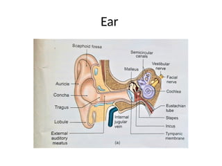
- 2. Ear
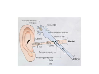
- 3. Middle ear (Tympanic cavity) • Location- petrous part of temporal bone • Filled with air • Lined by mucous membrane • Full adult size at birth • Communications- in front with lateral wall of nasopharynx – auditory tube • Behind with mastoid antrum through aditus to antrum
- 4. Pistol
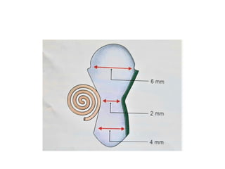
- 5. Measurements • Vertical diameter- 15 mm • AP diameter- 15 mm • TD- 6mm at roof, 2 mm middle, 4mm at floor
- 6. Subdivisions of middle ear • Epitympanum (head of malleus, body & short process of incus) • Mesotympanum (handle of malleus, long process of incus and stapes) • Hypotympanum
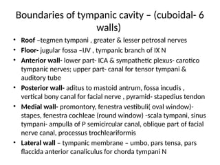
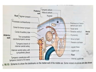
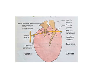
- 8. Boundaries of tympanic cavity – (cuboidal- 6 walls) • Roof –tegmen tympani , greater & lesser petrosal nerves • Floor- jugular fossa –IJV , tympanic branch of IX N • Anterior wall- lower part- ICA & sympathetic plexus- carotico tympanic nerves; upper part- canal for tensor tympani & auditory tube • Posterior wall- aditus to mastoid antrum, fossa incudis , vertical bony canal for facial nerve , pyramid- stapedius tendon • Medial wall- promontory, fenestra vestibuli( oval window)- stapes, fenestra cochleae (round window) -scala tympani, sinus tympani- ampulla of P semicircular canal, oblique part of facial nerve canal, processus trochleariformis • Lateral wall – tympanic membrane – umbo, pars tensa, pars flaccida anterior canaliculus for chorda tympani N
- 13. Lateral wall
- 18. Ear ossicles
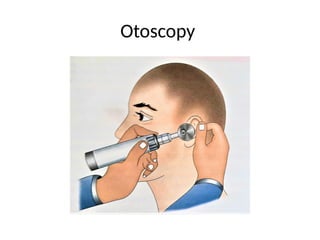
- 20. Otoscopy
- 21. Syringing
- 23. Applied anatomy • Otitis media- ASOM, CSOM- complications • Otosclerosis • Hyperacusis • Tympanic membrane perforation • Myringotomy, Myringoplasty, Tympanoplasty • Facial nerve & chorda tympani injury • Eustachian catarrh