Salivarygland neoplasm by numan h.k.d.e.t dental clg1DrMohammad Uddin
油
This document provides information on salivary gland neoplasms:
- It describes the anatomy and locations of the major salivary glands.
- The most common benign tumor is pleomorphic adenoma, while mucoepidermoid carcinoma is the most common malignant tumor.
- Other tumors discussed include adenolymphoma, oncocytoma, and adenoid cystic carcinoma.
- Surgical excision is the primary treatment for salivary gland tumors. Care must be taken during parotid surgery to preserve the facial nerve.
Salivarygland neoplasm by numan(h.k.d.e.t.dental clg)DrMohammad Uddin
油
This document provides information about salivary gland neoplasms. It discusses the anatomy of major and minor salivary glands. It describes different tumors that can occur in the parotid gland, submandibular gland and minor salivary glands. The most common benign tumor is pleomorphic adenoma, while mucoepidermoid carcinoma is the most common malignant tumor. Different tumors are classified as epithelial or connective tissue tumors. Clinical features, pathology, treatment and prognosis are outlined for various tumors including pleomorphic adenoma, adenolymphoma, oncocytoma and malignant tumors.
1. The three major salivary glands are the parotid, submandibular, and sublingual glands. The parotid gland is the largest.
2. Pleomorphic adenoma is the most common benign salivary gland tumor, accounting for 80% of parotid tumors.
3. Mucoepidermoid carcinoma is the most common malignant salivary gland tumor. It typically presents as a slow growing swelling in the parotid or submandibular region.
4. Surgery is the primary treatment for salivary gland tumors. For malignant tumors, surgery may be combined with radiation or chemotherapy.
The document summarizes the anatomy and tumors of the parotid gland. It describes the location and lobes of the parotid gland, its blood supply and innervation. It then discusses the most common tumor types including pleomorphic adenoma, Warthin's tumor, mucoepidermoid carcinoma, adenoid cystic carcinoma and metastatic carcinomas. It provides details on the histology, characteristics and treatment considerations for each tumor type.
The document discusses the anatomy and tumours of the parotid gland. It describes the location and lobes of the parotid gland, its blood supply and innervation. It then discusses the various types of tumours that can occur in the parotid gland and other salivary glands, including pleomorphic adenoma, Warthin's tumour, mucoepidermoid carcinoma, adenoid cystic carcinoma, and metastatic carcinomas. It provides details on the histology, presentation and characteristics of these tumour types.
This document discusses neoplasms (tumors) of the salivary glands. It begins by describing the major and minor salivary glands. The most common benign tumors are pleomorphic adenoma, Warthin's tumor, and oncocytoma. The most common malignant tumors are mucoepidermoid carcinoma and adenoid cystic carcinoma. Factors like size and location of the gland affect likelihood of malignancy. Surgery is the main treatment and complications can include facial nerve paralysis, fluid collections, and Frey's syndrome.
Salivary gland tumors can be benign or malignant. The majority are benign and arise most commonly in the parotid gland. Histologically, tumors are best classified based on their patterns. Treatment depends on the type and severity of the tumor, with more aggressive surgery and radiation used for malignant tumors. Prognosis varies significantly depending on the specific tumor type.
imaging of scrotum [Repaired] [Repaired].pptxdypradio
油
The scrotum contains the testes and epididymides. On ultrasound, the normal anatomy includes the oval testes with homogeneous echotexture and color flow. Potential pathological findings include infections like epididymitis, tumors such as seminomas which appear hypoechoic and well-defined, and traumatic injuries or torsion which may demonstrate absent flow. Malignancies require evaluation for metastases while infections require treatment with antibiotics. Imaging guides diagnosis and management of scrotal pathologies.
This document provides an overview of salivary gland tumors including their anatomy, epidemiology, classification, and management. It discusses the most common benign tumors like pleomorphic adenoma and Warthin's tumor as well as malignant tumors such as mucoepidermoid carcinoma and adenoid cystic carcinoma. The document outlines the clinical presentation, investigations, treatment and prognosis of various salivary gland tumors. It emphasizes complete surgical excision of benign tumors and importance of postoperative radiation for malignant tumors.
The parotid gland is located below and in front of the ear. It has two lobes and is drained by Stenson's duct which opens into the mouth. The gland has a capsule and structures like arteries pass through it. It is supplied by parasympathetic and sympathetic nerves. Common tumors include pleomorphic adenoma and Warthin's tumor. Mucoepidermoid carcinoma and adenoid cystic carcinoma are malignant tumors that can occur. Surgical excision is the main treatment for tumors but radiotherapy may also be used for malignant ones. Complications after parotidectomy include facial nerve injury and salivary fistula.
The parotid gland is located below and in front of the ear. It has two lobes and is drained by Stenson's duct into the mouth. The gland receives nerve supply from the facial nerve and parasympathetic fibers from the glossopharyngeal nerve. Common tumors of the parotid include pleomorphic adenoma, Warthin's tumor and mucoepidermoid carcinoma. Surgical excision is the main treatment for parotid tumors and complications can include facial nerve injury, hemorrhage or infection.
This document discusses salivary gland tumors. It begins by providing a brief history of salivary gland surgery. It then describes the different types of salivary glands and their anatomy. The major salivary glands discussed are the parotid, submandibular, and sublingual glands. It also discusses minor salivary glands. The document then covers the classification, etiology, clinical features, treatment and prognosis of different benign and malignant salivary gland tumors including pleomorphic adenoma, Warthin's tumor, oncocytoma, ductal papillomas and monomorphic adenomas.
This document discusses pleomorphic adenoma, the most common benign tumor of the salivary glands. It typically presents as a slow-growing, painless mass in the parotid gland. Other key details include:
- It represents about 60% of parotid tumors and occurs most often in females ages 30-60.
- On imaging, it appears as a well-defined mass that is hypoechoic on ultrasound and enhances with contrast on CT/MRI.
- The facial nerve must be carefully preserved and dissected during surgery to remove these tumors from the parotid gland in order to prevent facial paralysis.
Testicular tumors are rare.
1 2 % of all malignant tumors.
Most common malignancy in men in the 15 to 35 year age group.
Benign lesions represent a greater percentage of cases in children than in adults.
Most curable solid neoplasm
Oral squamous cell carcinoma is a malignant tumor that may occur anywhere within the oral cavity. It is locally invasive, infrequently metastasizes to ipsilateral regional lymph nodes, and rarely spreads to distant sites. Risk increases dramatically when alcohol use exceeds 6 oz of distilled liquor, 15 oz of wine, or 36 oz of beer/day. The combination of heavy smoking and alcohol abuse is estimated to raise the risk 100-fold in women and 38-fold in men.
Purpose:
The purpose of this webinar is to help participants learn how to prevent oral squamous cell carcinoma.
The document summarizes the anatomy, blood supply, innervation, and common tumors of the major and minor salivary glands. The major salivary glands are the parotid, submandibular, and sublingual glands. The parotid gland is the largest and is located deep to the ear. The submandibular gland is below the mandible. The sublingual gland is beneath the tongue. Common benign tumors include pleomorphic adenoma and Warthin's tumor, while common malignant tumors include mucoepidermoid carcinoma and adenoid cystic carcinoma. Surgery is the primary treatment for salivary gland tumors.
This document provides information about neck masses, including the major structures in the neck, lymph nodes, and the differential diagnosis. It discusses the major structures that can be palpated in the neck, such as the thyroid gland and lymph nodes. The differential diagnosis is divided into congenital, inflammatory, and neoplastic categories. Common congenital masses discussed include thyroglossal duct cysts, cystic hygromas, ectopic thyroid, plunging ranula, branchial cleft cyst, and dermoid cysts. Inflammatory masses are usually self-limiting.
1) Salivary gland tumors are mostly benign (80%), with the majority originating in the parotid glands.
2) Pleomorphic adenoma is the most common benign tumor, representing 80% of parotid gland tumors.
3) Mucoepidermoid carcinoma is the most common malignant salivary gland tumor in both adults and children, typically presenting as a slow-growing mass in the parotid gland or palate.
Childhood mumps, certain bacterial infections (for example, of the tonsils or teeth), and other diseases that are typically more common among adults (such as AIDS, Sj旦gren syndrome, diabetes mellitus, sarcoidosis, and bulimia) often cause swelling of the major salivary glands.
This document provides information on diseases of the salivary glands. It discusses the anatomy of the major and minor salivary glands. It then covers specific diseases including mumps, sialolithiasis, Sjogren's syndrome, and various neoplasms of the salivary glands such as pleomorphic adenoma, Warthin's tumor, mucoepidermoid carcinoma, and adenoid cystic carcinoma. For each condition, it discusses clinical features, investigations, and treatment options.
This document discusses tumors of the larynx, including benign and malignant tumors. It provides details on various benign tumors such as papillomas, paragangliomas, schwannomas, and hemangiomas. It then focuses on malignant tumors, specifically squamous cell carcinoma which is the most common. Details are given on risk factors, pathology, staging, symptoms, workup and various treatment options for laryngeal cancer such as cordectomy, laryngectomy, and chemoradiotherapy.
This document discusses the structure and pathology of the oral cavity and esophagus. It begins by describing the general four-layer structure of the esophagus which includes the mucosa, submucosa, muscularis and serosa layers. It then provides more details on the histology and functions of each layer. The document goes on to describe common oral pathologies like aphthous ulcers, oral candidiasis, leukoplakia and erythroplakia. It also discusses salivary gland structures, diseases like Sjogren's syndrome, mucoceles, ranulas, sialolithiasis and various benign and malignant salivary gland tumors.
Surgical management of colorectal cancer.pptxHamSayshi1
油
Surgical treatment of Colorectal Cancer Current Treatment Guidelines 2024...A reveiw of literature
palliative management of CRC and Mechanical bowel preparation in case of CRC alongwith Treatment guidleines of grade 4 CRC in presence of metastasis
Solubilization in Pharmaceutical Sciences: Concepts, Mechanisms & Enhancement...KHUSHAL CHAVAN
油
This presentation provides an in-depth understanding of solubilization and its critical role in pharmaceutical formulations. It covers:
Definition & Mechanisms of Solubilization
Role of surfactants, micelles, and bile salts in drug solubility
Factors affecting solubilization (pH, polarity, particle size, temperature, etc.)
Methods to enhance drug solubility (Buffers, Co-solvents, Surfactants, Complexation, Solid Dispersions)
Advanced approaches (Polymorphism, Salt Formation, Co-crystallization, Prodrugs)
This resource is valuable for pharmaceutical scientists, formulation experts, regulatory professionals, and students interested in improving drug solubility and bioavailability.
Chair and Presenters Sara A. Hurvitz, MD, FACP, Carey K. Anders, MD, FASCO, and Vyshak Venur, MD, discuss metastatic HER2-positive breast cancer in this CME/NCPD/CPE/AAPA/IPCE activity titled Fine-Tuning the Selection and Sequencing of HER2-Targeting Therapies in HER2-Positive MBC With and Without CNS Metastases: Expert Guidance on How to Individualize Therapy Based on Latest Evidence, Disease Features, Treatment Characteristics, and Patient Needs and Preferences. For the full presentation, downloadable Practice Aids, and complete CME/NCPD/CPE/AAPA/IPCE information, and to apply for credit, please visit us at https://bit.ly/4f8sUs7. CME/NCPD/CPE/AAPA/IPCE credit will be available until March 2, 2026.
Flag Screening in Physiotherapy Examination.pptxBALAJI SOMA
油
Flag screening is a crucial part of physiotherapy assessment that helps in identifying medical, psychological, occupational, and social barriers to recovery. Recognizing these flags ensures that physiotherapists make informed decisions, provide holistic care, and refer patients appropriately when necessary. By integrating flag screening into practice, physiotherapists can optimize patient outcomes and prevent chronicity of conditions.
Creatines Untold Story and How 30-Year-Old Lessons Can Shape the FutureSteve Jennings
油
Creatine burst into the public consciousness in 1992 when an investigative reporter inside the Olympic Village in Barcelona caught wind of British athletes using a product called Ergomax C150. This led to an explosion of interest in and questions about the ingredient after high-profile British athletes won multiple gold medals.
I developed Ergomax C150, working closely with the late and great Dr. Roger Harris (1944 2024), and Prof. Erik Hultman (1925 2011), the pioneering scientists behind the landmark studies of creatine and athletic performance in the early 1990s.
Thirty years on, these are the slides I used at the Sports & Active Nutrition Summit 2025 to share the story, the lessons from that time, and how and why creatine will play a pivotal role in tomorrows high-growth active nutrition and healthspan categories.
Cardiac Arrhythmia definition, classification, normal sinus rhythm, characteristics , types and management with medical ,surgical & nursing, health education and nursing diagnosis for paramedical students.
At Macafem, we provide 100% natural support for women navigating menopause. For over 20 years, we've helped women manage symptoms, and in 2024, we're proud to share their heartfelt experiences.
The course covers the steps undertaken from tissue collection, reception, fixation,
sectioning, tissue processing and staining. It covers all the general and special
techniques in histo/cytology laboratory. This course will provide the student with the
basic knowledge of the theory and practical aspect in the diagnosis of tumour cells
and non-malignant conditions in body tissues and for cytology focusing on
gynaecological and non-gynaecological samples.
Stability of Dosage Forms as per ICH GuidelinesKHUSHAL CHAVAN
油
This presentation covers the stability testing of pharmaceutical dosage forms according to ICH guidelines (Q1A-Q1F). It explains the definition of stability, various testing protocols, storage conditions, and evaluation criteria required for regulatory submissions. Key topics include stress testing, container closure systems, stability commitment, and photostability testing. The guidelines ensure that pharmaceutical products maintain their identity, purity, strength, and efficacy throughout their shelf life. This resource is valuable for pharmaceutical professionals, researchers, and regulatory experts.
2. History:
1.Patient Information:
Name: Muhammad Iqbal Masih
Age: 55 year
Gender: Male
Marital status: Married
Religion: Christian
Known Hypertensive and X-ray operator by profession comes to the Out-patient department with
following complaints:
Painful swelling in the left cheek-----6 months
3. History:
2.History of presenting illness:
Mr. Iqbal was in his usual state of health 6 months back when he first noticed a swelling in left parotid region
initially about the size of pea & gradually increase in size to a size of lemon. The swelling is painful. The pain is
intermittent, dull in character worsened by eating, and occasionally radiates to the left ear. He denies any history
of fever, weight loss, or facial weakness. He has no history of dry mouth, altered taste sensations & dysphonia.
He has no other similar swelling elsewhere in the body.
3.Past Medical History: Hypertension (controlled with medication), No history of diabetes or other significant
medical conditions.
4.Past Surgical History: He had history of cholecystectomy few years back.
5.Personal History: Smoker & Occasional alcohol consumption.
6.Family History: No family history of head & neck cancer, salivary gland tumors or other malignancies.
4. Clinical Examination:
General Examination:
Patient of average build physique & height is conscious, cooperative, and well-oriented to time, place, and
person. Vital signs are within normal limits. No signs of pallor and cyanosis and clubbing etc. Rest of general
physical examination is unremarkable.
Head and Neck Examination:
A firm, non-tender, mobile swelling is palpable in the left parotid region just below the left ear lobule, swelling is
rubbery in consistency with negative translumination.The overlying skin appears normal and have mild
erythema. There is no local rise of temperature. The swelling measures approximately 2x2 cm in size.
Intraoral Examination: No abnormalities detected.
Neck Examination: No cervical lymphadenopathy detected.
Nerve Examination: Facial and trigeminal nerve examination unremarkable.
5. Differential diagnosis:
Parotid gland tumors
1. Warthins Tumor
2. Pleomorphic adenoma
Metastasis tumor from skin, oral cavity etc.
Lipoma
7. Ultrasound of swelling:
Ultrasound shows a well-defined homogeneous hypoechoic lesion 2x2 cm in superficial
lobe of left parotid gland.
The lesion shows multiple cystic spaces with internal septations.
No evidence of invasion into adjacent structures
There is no intralesional flow signal.
The rest of the gland shows normal echo pattern without dilated ducts and stone.
The right submandibular gland and superficial lobes of both parotid glands show normal
echo pattern, size without focal lesion/ dilated ducts.
The thyroid gland shows normal echo pattern without focal lesion.
There is no cervical lymphadenopathy.
8. CT-SCAN:
Axial scans shows the presence of an ovoid-shape, well-defined,
homogeneous lesion of 2x2cm, with rapid enhancement located in the left
parotid gland.
No invasion of the surrounding structures was noted.
No displacement of the surrounding structured noted.
Rest of the scan appears normal.
11. Introduction:
Tumors of the salivary glands are:
Most heterogeneous group of tumors.
Greatest diversity of morphologic features.
uncommon.
The majority of these neoplasms are benign 80%
and only 20% are malignant.
The various types of salivary gland tumors are best distinguished by their histologic patterns.
12. Anatomy:
3 major salivary glands:
1. The parotid glands
2. The submandibular glands
3. The sublingual glands
Other locations: lateral margin of tongue, palate, lips, buccal mucosa.
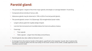
13. Parotid gland:
The parotid gland - largest of the three major glands and weighs on average between 14 and 30 g.
Composed almost entirely of serous cells.
Sebaceous glands may be observed in 10% to 42% of normal parotid glands .
The parotid glands contain 3 to 32(average: 20) intraglandular lymph nodes.
Largest salivary gland & roughly wedge shaped.
Lies b/w Sternomastoid and mandible below the External auditory meatus.
Coverings :
True capsule
False capsule a layer from the deep cervical fascia.
Parotid glands duct also called as Stensons duct
It emerges from the anterior border of the gland, superficial to the masseter muscle, then it pierces the
buccinator muscle & opens into the oral cavity on the inner surface of the cheek, usually opposite to the
maxillary second molar.
14. Lobes of Parotid gland:
Parotid divided into:
1. Superficial-------80%
2. Deep lobes-------20%
by the facial nerve.
Fasciovenous plane of Patey.
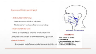
16. Structures within the parotid gland
1. External carotid artery :
Gives terminal branches in the gland
Maxillary artery and superficial temporal artery.
2. Retromandibular vein :
Formed by union of sup. Temporal and maxillary vein
joins post. Auricular vein to form the external jugular vein.
3.The facial nerve:
Enters upper part of posteromedial border and divides into:
17. Nerve supply of parotid gland:
Parasympathetic fibres :- Secretomotor
Preganglionic fibres arise from the inferior salivatory nucleus
Pass through glossopharyngeal nerve
Relay in ottic ganglion
Postganglionic fibres reach the gland through auriculotemporal nerve
Sympathetic fibres :- Vasomotor
Sensory nerves :- Auriculotemporal nerve
19. Rule of 80s:
80% of parotid tumors are benign.
80% of parotid tumors are Pleomorphic adenomas.
80% of salivary gland Pleomorphic adenomas occur in the parotid .
80% of parotid Pleomorphic adenomas occur in the superficial lobe.
80% of untreated Pleomorphic adenomas remain benign.
20. Epidemiology:
Uncommon neoplasms.
2%-3%of all head and neck neoplasms.
Most salivary gland tumors originate in the parotid glands (64%-80%), malignancy (15%- 32%).
7-11% occur in the submandibular glands, malignancy (37% - 45%).
less than 1% in the sublingual glands,malignancy (70%-90%),
9%-23% in the minor glands.
Benign tumors account for 63% to 78% of all salivary gland neoplasms.
21. Etiology:
Viruses- EBV, CMV, Polyoma virus
Ionizing radiation.
Increased occupational risks- asbestos, nickel compounds or silica dust.
Employment in the woodworking, rubber industries & beauty saloons.
Lifestyle- Warthins tumors showed a strong association with cigarette smoking.
Endogenous hormones.
24. Pleomorphic adenoma:
It is also known as Mixed salivary tumor
It is the most common benign tumor of salivary glands 80 %
Characterized by neoplastic proliferation of parenchymatous glandular cells along with myoepithelial
components
60-70%- Parotid glands ,40-60%- Submandibular glands, 40-70%- Minor salivary glands Seldomly- Sublingual
glands. Age: 30-50 years Sex: female> male 3:1 4:1
1. In Parotid- Presents in the superior lobe as a mass over the angle of the mandible, below and infront of
the ear.
2. Painless, slow growing, firm mass, initially small in size and begins to increase in size.
3. Recurrent tumor- multinodular, fixed on palpation.
4. Palate intraorally common site.
25. Warthins tumor:
Warthins tumour, also known as adenolymphoma or cystadenoma lymphomatosum, is a benign tumour.
They are the second most common benign salivary gland tumours (515%) Mainly seen in older men, after
the 5th
& 6th
decade of life.
They have been associated with cigarette smoking as well as radiation exposure.
They are almost exclusively seen in the parotid gland, especially in the inferior pole, and are rarely seen in the
peri-parotid nodes.
They can occur synchronously or metachronously in the same or bilateral glands. (10-15% Bilateral)
They are also known to occur with other salivary gland neoplasms such as pleomorphic adenoma and
salivary duct carcinoma.
Clinically, they present as painless, small slow-growing soft cystic fluctuant swellings. Facial palsy is rare.
Malignant transformation is extremely rare. Recurrences are very rare and may be due to multifocal tumours.
26. Mucoepidermoid tumor:
Commonest type of malignant salivary tumor in adults
Commonest malignant tumor of parotid in childhood
Common in middle age (35-65 years of age)
Female predilection Blue / Red in color
Parotid is the most common site of tumor
2nd common is palate minor salivary gland
Radiation etiological factor
t(11;19)(q21;p13) chromosome translocation resulting in a MECT1-MAML2 fusion gene
Presents as painless, slow-growing mass that is firm or hard.
Grossly Un encapsulated mass with cystic spaces
Facial nerve involvement in late stages
27. ADENOID CYSTIC CARCINOMA
Slow growing, aggressive neoplasm.
2nd
MC malignant tumor.
Common malignant tumor- submandibular, sublingual & minor salivary ,
2/3rd
occurs in minor salivary glands.
Clinical Features: 1) MC seen in females 5th
-6th
decade. Local recurrence common (30-50%). 2) Parotid,
submaxillary, palate & tongue - MC involved. 3) Early local pain (surface ulceration), FN palsy, local invasion &
fixation to deeper structure. LN metastasis 10%-30%.
4) Tendency to spread through perineural spaces (20%-30%)
Commonly involved nerves- Facial nerve, mandibular & maxillary nerve Pathway for invasion of the skull base.
More frequent- advanced, recurrent & high grade tumors.
28. Acinic cell carcinoma
3rd
most comman malignant Ca. of parotid gland.
Low malignancy. M:F=3:2, More common in males
mainly in middle ages (44yrs)
Tumor may be multifocal or B/L.
Clinically Painless lump, Encapsulated & lobulated. Chiefly occurs Parotid (80%)
Most common intraoral site Lips & buccal mucosa
Local recurrence & distal metastasis. Has the best survival rate of any salivary cancer.
Excision of a facial nerve is not justified unless it is involved.
29. Squamous cell carcinoma:
Primary salivary gland SCC is very rare(<1%)
Parotid (80%), submandibular gland(20%)
Age : 60 to 65years, M:F= 2:1.
History of previous radiotherapy.
30. Grade of tumor:
Low grade:
Well-differentiated cells with little cellular atypia
High proportion of mucous cells
Prominent cyst formation
Intermediate grade: intermediate features
High grade:
Poorly differentiated with cellular pleomorphism
High proportion of squamous cells
Solid with few if any cysts
31. Malignancy should be suspected when:
Rapid growth
Facial nerve palsy
Painful
Skin infiltration
Get fixed to massester muscle Trismus
Feels stony hard
Presence of lymph nodes in neck
34. Main investigations: ( USG,FNAC & CT Scan )
FNAC:-
1) Accuracy95-98%
2) Diff benign from malignant
disease.
3) The key to successful FNAC is
immediate
evaluation of the specimen for
adequacy.
Ultrasound:
1) Ideal tool for the initial assessment of
superficially located tumors of the parotid and
submandibular gland Distinguish intrinsic from
extrinsic neoplasm.
2) USG f/o malignant tumors include
ill-defined margins,
heterogeneous architecture,
subcutaneous invasion,
& the presence of LN metastases.
35. CT & MRI:
1) Effective modalities for imaging the size, the local, and the regional extension of the
primary tumor and the neck metastasis & to differentiate intra from extra glandular mass.
2) CT IOC for subtle cortical involvement & bone destruction.
3) MRI IOC for bone marrow invasion.
4) MRIIOC for detecting perineural spread.
5) Contrast-enhanced MRI IOC for intracranial invasion
Disadvantage Of MRI :-
1) Less sensitive in cystic lesions.
2) Inability to detect calcification.
36. Investigations:
1 )Plain X ray
2) X ray chest To R/O secondaries.
3) OPG To R/O mandibular involvement.
4) Open biopsy Rarely used due to risk of recurrence & FN damage Useful HP guidance for use of
palliative CTRT, poor surgical candidate, obvious malignancy.
5) Sialography:-
a) C/I:-Acute infection, Iodine allergy, Multiple myeloma.
b) Limitation:- Mass < 2mm, Deep lobe pathology.
6) Radiosialography Tc99 To detect mass lesion & parenchyma function No use in ductal system study.
7) Colour doppler sonography Non invasive Evaluates vascular anatomy.
8) PET Differentiate benign from malignant lesions.
38. Indications of facial nerve sacrifice:
Preoperative weakness / paralysis of nerve
Intraoperative evidence of gross invasion
Tumors transgressing through facial nerve from superficial to deep lobe
Nerve stump is checked for frozen section for negative margins, if positive,
mastoidectomy & nerve dissection is required
39. Indications of PORT:
1) High-grade tumor
2) Deep lobe cancers
3) All T3 and T4 cancers
4) Recurrent disease
5) Documented LN metastasis
6) Extraparotid extension
7) Gross/microscopic residual disease
8) Tumor involving or close to the facial
nerve.
1) Clinically cervical Lympadenopathies
(15%).
2) Parotid tumor bigger than 4cm Occult
metastasis risk >20%.
3) High grade malignancy Occult
metastasis risk >25%.
Indications of neck dissection:
43. Chemotherapy useful in pallation
& in inoperable cases.
Combination regimen have not
proven better results
2 groups
Epidermoid like tumor
i.e.
Sq. cell CA
Mucoepidermoid Ca.
Adeno Ca like tumors i.e.
Adenoid cystic Ca,
Acinic cell Ca,
Ca. ex polymorphic Ca
Methotrexate
Cisplatin
Adriamycin
Cisplatin
5-flurouracil
COMPLICATIONS
OF PAROTID
SURGERY