Unit 3 sence organs ear and nose(1)
- 1. UNIT 3: SENSE ORGANS ŌĆō NOSE AND EARS ŌĆó Chemical senses ŌĆóSense of hearing and balance Campbell et.al, 2010 ŌĆō CHAPTER 50
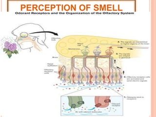
- 2. 1. SENSE OF SMELL ’éóReceptor cells of smell are OLFACTORY CELLS ’éóOlfactory cells are located within olfactory epithelium high in the roof of the nasal cavity.
- 4. PERCEPTION OF SMELL ’éóThe gas molecules in the air dissolves in the mucus of the nasal cavity. ’éóIt stimulates the microvilli of olfactory cells where it bonds to the odorant receptors ’éóThis cause an impulse to be send from olfactory cell through the sensory nerve fibers, to the olfactory bulb and then to the temporal lobe of the cerebrum. ’éóSmell is integrated and perceived.
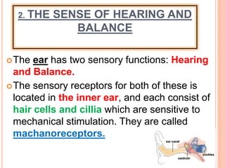
- 6. 2. THE SENSE OF HEARING AND BALANCE ’éóThe ear has two sensory functions: Hearing and Balance. ’éóThe sensory receptors for both of these is located in the inner ear, and each consist of hair cells and cillia which are sensitive to mechanical stimulation. They are called machanoreceptors.
- 8. FUNCTIONS OF DIFFERENT PARTS OF THE EAR ’āśOUTER EAR ’āśMIDDLE EAR ’āśINNER EAR
- 9. THE OUTER EAR ’éóPinna ŌĆō Concentrate sound waves in the direction of the external auditory canal. ’éóExternal Auditory canal ’éóŌĆō Transport sound waves from the pinna to the tympanic membrane. ’éó- Contain fine hairs and cerumin glands that secrete cerumin (earwax) to help guard the ear against foreign material and insects. (smell)
- 10. THE OUTER EAR ’éóTympanic membrane ’éóŌĆō A thin membrane that covers the opening between the inner- and middle ear. ’éó- Converts soundwaves into vibrations. (starts to vibrate)
- 11. MIDDLE EAR ’éó3 Bony ossicles e.g.: (start to vibrate): - Malleus ŌĆō transmit vibration to incus - Incus ŌĆō transmit vibrations to stapes - Stapes ŌĆō transmit vibrations to oval window (fenestra ovalis) ’éóOval window ŌĆō start to vibrate and cause waves in liquid (perilymph) in cochlea. ’éóEustachian tube ŌĆō Equalize the pressure between the atmosphere and the inside of the ear. (Connected with the pharynx).
- 12. MIDDLE EAR
- 13. INNER EAR ’éóCochlea: - Snail shaped canal. - Divided in 3 canals separated by membranes 1. Vestibular canal (scale vestibuli) ŌĆō top canal, filled with perilymph. Receives vibration from oval window, form waves in perilymph, causes Reissner membrane to form waves.
- 14. 2. Cochlear canal (Scala media) ŌĆō middle canal, filled with endolymph. ’āśForm waves in endolymph, that causes Basilar membrane to wave up and down. ’āśContains the receptor cells for hearing: Organ of Corti - which pushes the stereocilia against the tectorial membrane, causes an impulse which is send through the cochlear nerves to the temporal lobe of the brain for integration.
- 15. 3. Tympanic canal (Scala tympani)ŌĆō bottom canal, filled with perilymph. Form waves which are carried to the round window (fenestra rotunda). ’éóRound Window: absorb excess sound waves to prevent echoing in the ear.
- 17. 3. INNER EAR: SEMI CIRCULAR CANALS ’éóContain machanorecepters (cristae) ŌĆō detect rotational or angular movement of the head. ’éóCristae- located in the ampulla (enlarged base of semi circular canals)in the endolymph found in the semi circular canals. ’éó- Consist of hair cells, supporting cells, stereocillia imbedded in a gelatin capsule called cupula, and nerve fibers.
- 18. INNER EAR: SEMI CIRCULAR CANALS ’éóMovement of the head ’éócauses the endolymph to move around in the ampulla, ’éóthe cupula moves, ’éóbending the stereocilia, ’éócausing an impulse send through the vestibular nerve ’éóto the cerebellum of the brain for integration.
- 19. CRISTAE
- 20. 4. INNER EAR: UTRICULUS AND SACCULUS ’éóEnlarged area below the semi circular canals. ’éóContain mechanoreceptors (macula) ŌĆō that detects straight line movement of the head in any direction ŌĆō gravitational equilibrium. ’éóMacula: ’é¦ Consist of hair cells with stereocilia embedded in a gelatin membrane called otolithic membrane with otoliths (crystals) ontop, supporting cells and vestibular nerves.
- 21. 4. INNER EAR: UTRICULUS AND SACCULUS ’éóIf a person stops suddenly, ’éóthe endolymph in the utriculus and sacculus move around, ’éó the otolithic membrane moves, ’éóbending the stereocilia, ’éówhich sends an impulse through the vestibular nerves ’éóto the cerebellum of the brain to maintain balance.
- 22. MACULA FOUND IN SACCULUS AND UTRICULUS
- 26. Fig. 50-8 Hair cell bundle from a bullfrog; the longest cilia shown are about 8 ┬Ąm (SEM). Auditory canal Eustachian tubePinna Tympanic membrane Oval window Round window Stapes Cochlea Tectorial membrane Incus Malleus Semicircular canals Auditory nerve to brain Skull bone Outer ear Middle ear Inner ear Cochlear duct Vestibular canal Bone Tympanic canal Auditory nerve Organ of Corti To auditory nerve Axons of sensory neurons Basilar membrane Hair cells
- 27. SENSE OF BALANCE
- 28. Fig. 50-11 Vestibular nerve Semicircular canals Saccule Utricle Body movement Hairs Cupula Flow of fluid Axons Hair cells Vestibule


