


More Related Content
What's hot (20)
Similar to Viral conjunctivitis (20)


Recently uploaded (20)


Viral conjunctivitis
- 1. Dr M.Abdullah Younas PGR Ophthalmology DHQ Teaching Hospital Gujranwala,Pakistan
- 2. Sources of study:- ď‚— Kanski(90%) ď‚— Oxford handbook of ophthalmology(4%) ď‚— Wills eye manual(4%) ď‚— Harpers(2%)
- 3. Introduction:- ď‚— Common External Ocular infection. ď‚— In 90% cases,Adenovirus is the causative agent. ď‚— May be Sporadic,or occur in epidemics.
- 4. Causative Agents:- ď‚— Adenovirus conjunctivitis(>90% cases). ď‚— Herpes simplex keratoconjunctivitis. ď‚— Herpes zoster conjunctivitis. ď‚— Picorna viruses(Enterovirus and coxsackie virus). ď‚— Poxvirus conjunctivitis. ď‚— Myxovirus conjunctivitis. ď‚— Parammyxovirus conjunctivitis. ď‚— ARBOR virus conjunctivitis.
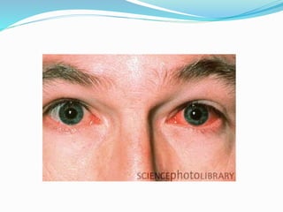
- 5. Symptoms:- ď‚— Watering ď‚— Redness ď‚— Irritation(Radak). ď‚— Itching. ď‚— Photophobia(When Cornea is involved).
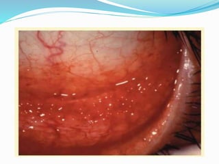
- 6. Signs(Anterior to posterior):- ď‚— Eyelids : edema,Ranging from mild to Severe. ď‚— Lymphadenopathy: Common.Tender Pre-auricular nodes. ď‚— Conjunctiva: Hyperemia,Follicles.May be Papillae(Particularly superior tarsal conjunctiva). ď‚— Severe Inflammation: may be associated with conj.Hamorrhages, chemosis, membranes(Rare) and pseudomembranes.Sometimes conj Scarring.
- 7. Signs(Cont’d):-  Keratitis(Adenoviral): Epithelial microcysts in the early stage. punctate epithelial keratitis:Usually occur in 7-10 days of onset of symptoms.Resolving in 2 weeks. Anterior Stromal infiltrates/SEI:may persist for months or years. Anterior uveitis: Usually mild.
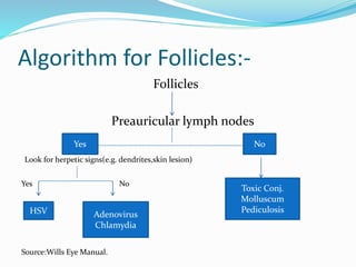
- 8. Algorithm for Follicles:- Follicles Preauricular lymph nodes Look for herpetic signs(e.g. dendrites,skin lesion) Yes No Source:Wills Eye Manual. Yes No HSV Adenovirus Chlamydia Toxic Conj. Molluscum Pediculosis
- 14. Adenoviral Conjunctivitis ď‚— Non-enveloped, double stranded DNA viruses, which replicate within the nucleus of host cells. General reservoir is only human.
- 15. Type of adenoviral conjunctivitis:- ď‚— Epidemic keratoconjunctivitis (EKC) ď‚— Nonspecific acute follicular conjunctivitis ď‚— Pharyngoconjunctival fever (PCF) ď‚— Chronic /relapsing adenoviral conjunctivitis
- 16. Spread of infection:- ď‚— Facilitated by i)virus can survive on dry surfaces for weeks. ii)Viral shedding may occur for many days before clinical features are apparent. ď‚— Transmission by i)Contact with Respiratory or ocular secretions. ii)Via Contaminated Fomites such as Towels. iii)Route of transmission is usually Eye-Hands-Eyes. In Clinical setting,Eye-Instruments-Eye.
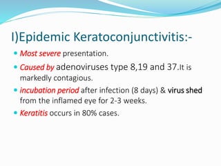
- 17. I)Epidemic Keratoconjunctivitis:- ď‚— Most severe presentation. ď‚— Caused by adenoviruses type 8,19 and 37.It is markedly contagious. ď‚— incubation period after infection (8 days) & virus shed from the inflamed eye for 2-3 weeks. ď‚— Keratitis occurs in 80% cases.
- 18. II)Non-specific acute follicular Conj. ď‚— Most common form of acute follicular conjunctivitis ď‚— Caused by adenovirus serotypes 1 to 11 & 19 ď‚— Milder form of acute follicular conjunctivitis. ď‚— Unilateral symptoms, Other eye involved 1-2 days later, but less severely. ď‚— Patient may have systemic symptoms such as sore throat or common cold.
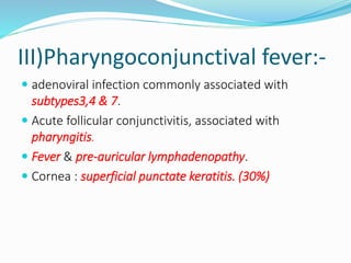
- 19. III)Pharyngoconjunctival fever:- ď‚— adenoviral infection commonly associated with subtypes3,4 & 7. ď‚— Acute follicular conjunctivitis, associated with pharyngitis. ď‚— Fever & pre-auricular lymphadenopathy. ď‚— Cornea : superficial punctate keratitis. (30%)
- 20. IV)Chronic/relapsing adenoviral conj. ď‚— Rare ď‚— Gives a clinical picture of chronic non-specific follicles/papillas. ď‚— Can persist over years, but eventually self limiting.
- 21. Herpes simplex Virus:- ď‚— Causes Follicular conjunctivitis particularly in primary disease. ď‚— Usually unilateral. ď‚— Often Associated skin lesions. ď‚— Minute,Micro dendrites may be mistaken for punctate epithelial keratitis,But Corneal sensation is reduced in HSV (Source:Harper).
- 22. Acute hemorrhagic conjunctivitis:- ď‚— Usually occurs in tropical areas. ď‚— Caused by Enterovirus and coxsackie virus(Picorna virus family). ď‚— Rapid onset,resolves within 1-2 weeks.
- 23. Molluscum Contagiosum:- ď‚— Caused by dsDNA pox virus. ď‚— Peak incidence of getting the virus is 2-4years. ď‚— Typically,Virus causes a skin lesion. ď‚— When skin lesion is on the lash line area of eyelid,it causes viral shedding and follicular conjunctivitis. ď‚— Examine eyelash line carefully when Chronic,unilateral eye irritation and mild discharge is present.
- 25. Systemic viral infections Causing Conjunctivitis:- ď‚— Measles , mumps , Varicella ,HIV etc.
- 26. Investigations:- ď‚— Giemsa stain. ď‚— PCR ď‚— Viral culture. ď‚— Immunochromatography. ď‚— Serology. ď‚— For other causes in non-resolving cases.
- 27. TREATMENT Adenoviral conjunctivitis:- ď‚— Supportive treatment for amelioration of symptoms is the only treatment required and includes: I)Artificial tears 4x/d.Preferably preservative free. II)Topical Anti Histamines and vasoconstrictors. III)Cold Compresses IV)Discontinuation of contact lens wear.
- 28. (Cont’d) V)Removal of membranes/pseudomembranes. VI)Topical antibiotics. VII)Povidone-Iodine:kills free adenoviruses. VIII)Topical Steroids:For severe Membranous or Pseudo-membranous conjunctivitis and SEIs.
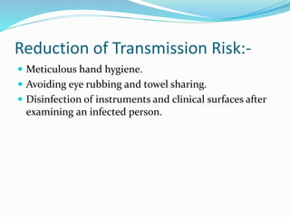
- 29. Reduction of Transmission Risk:- ď‚— Meticulous hand hygiene. ď‚— Avoiding eye rubbing and towel sharing. ď‚— Disinfection of instruments and clinical surfaces after examining an infected person.
- 30. Acute Haemorrhagic Conjunctivitis Treatment:- ď‚— Prophylactic measures similar to EKC. ď‚— Supportive measures same as Adenoviral. ď‚— Usually the disease has a self-limiting course of 7 days.
- 31. Molluscum treatment:- ď‚— Usually the lesion is self-limiting in immunocompetent patient. ď‚— Removal is needed to address secondary Conjunctivitis or for Cosmetic reasons. ď‚— Expression by making a nick in the skin by a needle is usually effective.
- 32. Herpes Simplex Treatment:- ď‚— Usually self limiting. ď‚— Topical antiviral drugs control the infection effectively and prevent recurrences. ď‚— Supportive measures are similar with Adenoviral.