This document provides a summary and illustrated glossary of the standardized lexicon for reporting degenerative lumbar disk pathology. It begins by explaining the need for standardized terminology in radiology reports. It then defines and provides diagrams to illustrate various normal and pathological disk findings, including congenital variants, bulges, herniations, annular tears, and degenerative changes. Classification schemes and diagnostic algorithms are presented to clearly define and differentiate common lumbar disk lesions using standardized terms. The goal is to establish familiarity and consistency with this standardized lexicon for radiology residents and physicians in training and practice.
This study retrospectively analyzed 310 patients treated for zygomatic bone and zygomatic arch fractures over a 10-year period. The majority of patients were young adult males injured in road traffic accidents. Patients were divided into three groups based on fracture location: zygomatic bone only, zygomatic arch only, or both. Most fractures were of the zygomatic bone alone. Treatment modalities included open reduction and internal fixation via miniplates and screws for 90.6% of patients and closed reduction for 9.4%. All patients achieved satisfactory postoperative results without complications, demonstrating the effectiveness of the treatment approaches used.
This document summarizes information about AxioMed Spine Corp and its Freedom cervical and lumbar total disc replacement technologies. It discusses AxioMed's mission, clinical studies that have been conducted in Europe and the US, positive outcomes from those studies compared to other disc replacement technologies and spinal fusion, regulatory approvals in Europe and plans for FDA approval, and AxioMed's intellectual property and manufacturing capabilities. The Freedom discs are designed to restore natural motion and function to diseased spinal segments.
L-WP-1004, Benefits of Viscoelastic DiscJames Kuras
油
The document discusses the benefits of a viscoelastic lumbar total disc replacement (TDR) compared to first generation TDR technologies. It notes that first generation TDRs, which use metal-on-metal or metal-on-polyethylene cores, lack the viscoelastic properties needed to replicate the shock absorbing function of a natural disc. In contrast, the Freedom Lumbar Disc's viscoelastic polymer core allows it to expand radially and axially, approximating the stiffness of a natural disc and providing properties like the restoration of lordosis and natural motion. Long-term studies of first generation TDRs found they placed excessive stress on facets and led to facet degeneration, while the viscoelastic FLD aims
Structural Targets for Prevention of Post Traumatic OAOARSI
油
David Hunter MBBS, PhD, FRACP
Florance and Cope Chair of Rheumatology, Professor of Medicine
University of Sydney and Royal North Shore Hospital
Chair, Institute of Bone and Joint Research
Chair, Musculoskeletal, Sydney Medical Program
Consultant Rheumatologist, North Sydney Orthopedic and Sports Medicine
Regeneration around Furcations- Challenges and Pitfalls- Guest lecture as a part of Dr NTRUHS Zonal CDE programme at MNR Dental College, Sangareddy, India on 21/3/2014 and at Meghna Dental College, Nizamabad, India on 24/7/2014.
This document discusses potential interventions for preventing hip fractures in geriatric patients. It summarizes a new technique called Anisotropy Restoring Femoroplasty (ARF) that aims to restore mechanical properties to the proximal femur through a minimally invasive procedure. ARF involves inserting metallic elements embedded in an isotropic filler like calcium phosphate into the femur via small drill holes. Testing in a porcine model found ARF restored load and strength to levels comparable to healthy bone. The researchers believe ARF may help reduce hip fracture risk in high-risk patients, and could be performed alongside traditional hip fracture repair.
The document summarizes a study conducted at Baystate Medical Center on surgical rib fixation (SRF) in patients with flail chest or severe rib fractures. The study compared outcomes between patients aged 65 and older and those aged 64 and younger who underwent SRF. The results showed that the median number of ventilator days, injury severity score, intensive care unit length of stay, and hospital length of stay were similar between the two age groups, suggesting there should be no age restrictions for SRF. The goals are to improve outcomes for rib fracture patients and establish a national registry through further research.
A Large Intra-Articular Ossicle in the Knee Joint-A Rare Occurrence_Crimson P...CrimsonPublishersAICS
油
This document presents a case report of a rare occurrence of a large intra-articular ossicle in the knee joint of a 14-year-old male. Magnetic resonance imaging found a large ossicle impinging on the anterior cruciate ligament, causing lifting of the ligament anteriorly and laterally. Intra-articular ossicles in the knee are uncommon but can grow to a significant size, as seen in this case, producing mass effect and clinically mimicking an ACL injury. The etiology of intra-articular ossicles is unclear but may be congenital, traumatic, or degenerative in origin.
The document discusses guidelines for deep vein thrombosis (DVT) prophylaxis for orthopedic trauma patients. It notes that many existing guidelines do not adequately address trauma patients, who have higher DVT risks due to immobility from injury. A review found that 77% of patients transferred to the authors' hospitals did not receive pre-transfer DVT prophylaxis, including 67% of hip fracture patients despite being at high risk. The authors developed new DVT prophylaxis guidelines for orthopedic trauma patients to help standardize care and lower DVT risks.
This document summarizes key issues in treating fractures of the distal humerus. It discusses the increasing incidence of these fractures, especially in elderly women. Classification systems and preoperative planning are outlined. Surgical approaches like the olecranon osteotomy are described, along with techniques for fracture stabilization and indications for total elbow arthroplasty in complex fractures of elderly patients.
An adolescent male football player presented with heel pain that had worsened over a year. Initial conservative treatment provided temporary relief but the pain intensified. Imaging revealed an osteoid osteoma, a benign bone tumor, in the calcaneus. Surgical excision of the tumor completely resolved the athlete's pain. Osteoid osteomas are rare in athletes but should be considered for persistent hindfoot pain atypical of common conditions like tendinitis.
This document provides information on pediatric supracondylar fractures of the elbow. It discusses that these fractures are common in children and often present with neurovascular injuries. Nondisplaced or minimally displaced fractures can be treated with immobilization, while displaced fractures typically require closed reduction and pin fixation. Fractures with preoperative neurovascular deficits or those that cannot be adequately reduced closed may require open reduction to address entrapment issues. Open reduction through an anterior incision allows exposure and release of trapped nerves and vessels. Postoperative monitoring is important to identify any developing complications.
This study examined the effect of mechanical environment on healing of critical-sized femoral defects in rats, finding that applying an initial period of loose fixation ("reverse dynamization") followed by rigid fixation led to better healing than rigid fixation alone, as assessed by radiology, histology, and mechanical testing. The reverse dynamization approach counters current clinical practice of immediate rigid fixation for large segmental defects and warrants further optimization and study to determine if it could improve treatment of such injuries in patients.
This document discusses osteoporosis and its imaging features. It begins by defining osteoporosis as a reduction in bone quantity with normal bone quality, seen as increased bone porosity. Radiographic signs include increased radiolucency, cortical thinning, and altered trabecular patterns. Spinal manifestations include vertebral compression fractures and wedge deformities. The pelvis, femur, and sacrum are also commonly involved sites. Quantitative CT and dual-energy X-ray absorptiometry (DXA) are introduced as methods to measure bone mineral density and diagnose osteoporosis based on T-scores. Regional variants and imaging patterns of disuse osteoporosis are also described.
This document provides information on Paget's disease, including:
- It is a chronic bone disorder characterized by abnormal bone remodeling that can cause bone deformities and fractures.
- The cause is unknown but may involve viruses or genetic factors. It most commonly affects older adults and bones like the pelvis and spine.
- Symptoms can include bone pain, stiffness, fractures, and hearing loss. Lab tests show elevated alkaline phosphatase levels. Imaging like x-rays are used for diagnosis.
- The disease involves abnormal bone breakdown and formation seen on imaging as thickened and misshapen bones. While often asymptomatic, treatment with medications may be used for painful symptoms.
This document provides an overview of disc replacement surgery. It describes what the surgery is, who is a candidate for it, how it is performed, and the benefits and risks. Disc replacement surgery involves removing a damaged disc and replacing it with an artificial disc made of metal and plastic materials. It is an alternative to spinal fusion for patients with degenerative disc disease who have not found relief from non-surgical treatments. The surgery aims to reduce pain and preserve motion in the operated spine segment. India is highlighted as a lower-cost location for this procedure compared to other countries like the US.
Retrograde Intramedullary Nail with Femoral Head Allograft for Large Deficit ...skisnfeet
油
The document summarizes a study that evaluated the outcomes of using a retrograde intramedullary nail with femoral head allograft for large defect tibiotalocalcaneal arthrodesis. Eleven patients were included who had this procedure for conditions such as Charcot neuroarthropathy, avascular necrosis, or revision fusion. While complications occurred in six patients, eight patients were considered successes based on clinical and radiographic criteria, such as stability and union. The technique provides a powerful one-stage method to address large bony deficits but also carries risk, as only partial unions were observed in some cases. Overall, it was deemed a useful technique for this difficult patient population.
The anterior cruciate ligament (ACL) is a key ligament in the knee that prevents anterior tibial translation and rotational loads. It frequently tears during high-impact sports. The ACL inserts on the femur and tibia and is composed of two bundles that restrain movement differently based on knee flexion angle. While partial ACL tears may be treated nonsurgically, complete tears typically require surgical reconstruction using a graft to replace the torn ligament. Postoperative rehabilitation focuses initially on regaining range of motion and strength before gradually progressing to sport-specific activities.
This document describes a study examining variations in the anatomy of blood vessels that supply the iliac bone. The researchers dissected 216 iliac regions from cadavers and clinical cases. They observed four main variations in the origin and branching patterns of the deep circumflex iliac artery, which is the primary blood vessel supplying the iliac bone. The relationships between this artery and surrounding anatomical landmarks like the anterior superior iliac spine were also measured. Documenting these variations will help surgeons more safely harvest vascularized bone grafts from the iliac crest while reducing complications.
1) The document describes a case of an intra-articular distal radius fracture with significant deformity and displacement seen on radiographs.
2) Several techniques are discussed for surgical treatment including mobilizing fracture fragments, using intact structures like the ulnar head to help rebuild support, and building the fracture back to the volar locking plate which can help achieve and maintain reduction.
3) Volar locked plating provides adequate stability for early range of motion rehabilitation and typically leads to excellent healing and functional outcomes, though care must be taken to ensure the locking screws are placed just below the subchondral bone to avoid joint penetration.
1) Periprosthetic femur fractures around hip implants are increasingly common as more elderly patients maintain active lifestyles with hip replacements.
2) Evaluation involves plain radiographs and surgery is usually needed except for non-displaced Vancouver type A and some B/C patterns.
3) Surgical treatment follows plate fixation principles to restore length, alignment and rotation without disrupting fracture fragments. Bridge plating is preferred over anatomic reduction. Long locking plates provide stable fixation, especially in osteoporotic bone.
This document describes a new expandable prosthesis called the JR prosthesis that is designed to replace vertebral bodies after corpectomy. The prosthesis has dual cage and plate functions to provide immediate stabilization of the spine. It was tested in cadavers and then used in 14 patients with vertebral tumors who underwent corpectomy. All patients experienced reduced pain and improved neurological function after surgery except those with the most severe pre-operative deficits. The prosthesis provided immediate stabilization of the spine and this was maintained long-term in surviving patients, with no significant complications. This clinical experience represents the first report of an expandable prosthesis with both cage and plate functions in a single device.
Osteoarthritis is a degenerative joint disease that causes joint pain and stiffness. It involves the breakdown of cartilage and underlying bone within a joint. Symptoms include joint pain, tenderness, and stiffness. It commonly affects the hands, feet, spine, hips and knees. Treatment focuses on lifestyle changes like exercise and weight loss, along with pain medications and joint replacement surgery for severe cases. Osteoarthritis is the most common and leading cause of chronic disability in the United States.
Dr.salah.radiology.radiological approach to bone diseasesabas_lb
油
This document provides guidance on evaluating solitary bone lesions based on radiographic findings. Key factors include the patient's age, location and characteristics of the lesion, presence of cortical destruction or a periosteal reaction, and whether the lesion is mono- or polyostotic. Together these factors can help determine if a lesion is likely benign and slow-growing or potentially malignant and aggressive.
1) Paget's disease is a chronic bone disorder characterized by excessive abnormal bone remodeling that commonly affects older adults, with prevalence increasing from 4% in those over 40 to 11% over 80.
2) It typically involves multiple bones (polyostotic) and bones asymmetrically. Common sites are the pelvis, spine, skull, and proximal long bones.
3) Radiographically, Paget's disease presents with thickened bone cortex, accentuated trabecular pattern, and enlarged bone size ("classic triad"). In the pelvis, thickening occurs along the iliopubic and ilioschial lines. In the spine, vertebrae have a "picture frame"
Intradural Disc Herniation, an Infrequent Spine Disease, Case ReportAliceMary13
油
Intradural disc herniation (IDH) is a rare disease that affects the spine, difficult to diagnose clinically and radiologically and easily mistaken with other types of disc herniations.
In this article is presented the case of a patient diagnosed and treated with IDH and reported it to share the diagnostic process as well
as treatment performed, so that we can contribute to the proper
management of these patients.
This document discusses abnormalities in spinal discs and vertebrae. It begins by describing disc degeneration which can include drying, fibrosis, bulging, cracking and bone spurs. Determining if changes are due to aging or pathology is difficult with observation alone. The document then discusses various pathologies in more detail such as herniations, fractures, stenosis, infections and bone alterations. Classifications for grading severity are provided. In summary, the document provides an overview of diverse spinal abnormalities with a focus on precise terminology and imaging appearances.
This document provides an overview of imaging appearances of disk disease and degenerative spondylosis of the lumbar spine. It describes normal lumbar spine anatomy and the appearances and terminology used to describe various pathologies. Degenerative changes that can be seen include intervertebral osteochondrosis, disk desiccation, endplate changes, Schmorl's nodes, spondylosis deformans, annular tears, and herniated disks. The terminology used to classify herniations is also discussed. Other degenerative changes mentioned include apophyseal arthrosis and epidural lipomatosis.
Degenerative spine disease involves three main areas: the intervertebral disc, vertebral bodies/end plates, and posterior elements. Changes to the intervertebral disc include decreased water/proteoglycan content leading to distorted collagen fibers and tears in the annulus fibrosis. Vertebral endplates can show three stages of degeneration. Posterior element changes include facet joint osteoarthritis with osteophytes/hypertrophy, ligamentum flavum hypertrophy/cysts, and spinal canal/foraminal stenosis. Imaging plays an important role in evaluating these degenerative changes and their effects.
X-rays are the oldest and most commonly used form of medical imaging. A radiologist will analyze x-ray images and send a report to the referring physician. Accurate diagnosis often requires two x-ray images at right angles. Structures with high density like bones appear white, while low density structures like soft tissue appear grey. Fractures appear as a dark line where bone continuity is lost. Interpretation requires checking patient details and producing a report detailing observations and diagnosis. The ABCs approach involves evaluating Adequacy, Bones, Cartilage, and Soft Tissues.
Presentation1, radiological imaging of degenerative and inflammatory disease ...Abdellah Nazeer
油
This document discusses radiological imaging findings of degenerative and inflammatory spine diseases. It provides detailed descriptions and images to illustrate various abnormalities that can be seen, including disc degeneration, herniations, fractures, spinal stenosis, and infections. Key findings are organized by specific pathologies such as disc bulges, protrusions, extrusions, sequestrations, migrating fragments, and vertebral bone marrow changes. Imaging features of conditions like osteoarthritis, synovial cysts, ligamentous thickening, and spinal infections are also reviewed. The document emphasizes the importance of accurate terminology in radiological descriptions and clinical diagnosis of spinal abnormalities.
This document discusses the treatment of distal radius fractures through plating. It begins with an overview of distal radius anatomy and the columnar classification system used to guide treatment. It then discusses the pathophysiology of distal radius fractures and associated injuries. Treatment options range from closed reduction to open reduction with plating or other internal fixation. Plating allows for anatomic restoration and stable fixation, enabling early return of wrist function. Factors such as fracture pattern, displacement, and patient needs help determine the appropriate treatment. Complications of plating include tendon issues and potential need for plate removal.
A 66-year-old male presented with non-specific lower back pain. MRI revealed disc degeneration including annular bulging and Schmorl's nodes in the upper lumbar spine. The diagnosis was lumbar spondylosis. Disc degeneration results from mechanical stress and age-related changes and can lead to annular tears, nuclear material changes, and instability. Later stages involve further disc resorption and replacement by fibrocartilage. Nerve fibers in the outer annulus can contribute to discogenic pain.
This document provides an illustrated review of degenerative changes in the spine. It discusses:
1. Degenerative change is a biomechanically related process starting within the intervertebral disc (A-changes) and progressing to involve surrounding structures (B-changes) and eventually more distant areas (C-changes).
2. A-changes include degeneration of the nucleus pulposus seen as reduced signal on MRI. B-changes involve the annulus fibrosus, endplates, and bone marrow and include fissures, herniations, and bone marrow changes.
3. C-changes are advanced changes like facet joint osteoarthritis, ligamentum flavum hypertrophy,
This document discusses degenerative diseases of the spine. It notes that degenerative change is considered a response to mechanical or metabolic injury rather than a disease. Common causes of degeneration include mechanical micro-insults, macro-insults like fractures, and metabolic processes. Imaging can accurately characterize degenerative processes, identify abnormalities, and assist in determining treatment. Degeneration may involve single segments or spread horizontally or vertically to adjacent segments. Specific degenerative changes discussed include disc degeneration, bulges, tears, herniations, endplate and bone marrow changes, facet joint changes, ligamentum flavum thickening, and spinal stenosis.
Neglected fracture neck of femur in young adultsZahid Iqbal
油
A 35-year-old woman presented with left hip pain and limited movement 3 months after falling down stairs during pregnancy and sustaining a left femoral neck fracture. X-rays showed the fracture had not healed. The patient underwent open reduction and internal fixation with a fibular strut graft and dynamic hip screw to repair the neglected fracture. Post-operatively, her hip movements improved and she was discharged on antibiotics with weight-bearing restrictions for 6 weeks.
Diagnostic imaging of bones and joints (1)Vijaya Krishna
油
Plain film radiography is often the initial imaging modality used to evaluate bones and joints. Key factors that determine the radiographic appearance of structures include composition, thickness, and contrast medium usage. Standard views such as anteroposterior, lateral, and oblique are used to demonstrate anatomy while minimizing radiation exposure. Interpretation of radiographs involves assessing alignment, bone density, cartilage spaces, and soft tissues for abnormalities that may indicate conditions such as trauma, degeneration, inflammation, or infection.
This document discusses spondylolisthesis, defined as the anterior or posterior displacement of one vertebra on another. It describes classifications based on anatomy and etiology, radiological grading scales, clinical presentation, diagnostic imaging including X-rays, CT, MRI, and myelography. Treatment options are discussed including non-operative care with bracing and exercise, as well as surgical techniques such as decompression with or without fusion, interbody fusion approaches, reduction methods, instrumentation, and complications. Surgical treatment aims to prevent slip progression, stabilize the segment, correct deformity, relieve pain and reverse neurological deficits.
This document discusses the effects of aging on the spine. It covers how aging impacts the vertebral bodies, intervertebral discs, endplates, facet joints, muscles, ligaments, and the biomechanics of the spinal functional unit. Key changes include a loss of bone mineral density in the vertebrae and endplates, dehydration and stiffening of intervertebral discs, arthritis in the facet joints, and weakening of the muscles and ligaments. These changes alter the load distribution and biomechanics of the spine, increasing risks for conditions like spinal stenosis and fractures. Physiotherapy can help treat age-related spinal symptoms and prevent further deterioration.
Diagnostic Imaging of Bones and Joints.pptRaj Harshwal
油
Plain film radiography is often the initial imaging modality used to evaluate bones and joints. Key factors that determine the radiographic appearance of structures include composition, thickness, and contrast medium usage. Standard views such as anteroposterior, lateral, and oblique are used to demonstrate anatomy while minimizing radiation exposure. Interpretation of radiographs involves assessing alignment, bone density, cartilage spaces, and soft tissues for abnormalities that may indicate conditions such as trauma, degeneration, inflammation, or infection.
Diagnostic Imaging of Bones and Joints.pptGurumurthy B R
油
Plain film radiography is often the initial imaging modality used to evaluate bones and joints. Key factors that determine the radiographic appearance of structures include composition, thickness, and contrast medium usage. Standard views such as anteroposterior, lateral, and oblique are used to demonstrate anatomy while minimizing radiation exposure. Interpretation of radiographs involves assessing alignment, bone density, cartilage spaces, and soft tissues for abnormalities that may indicate conditions such as trauma, degeneration, or disease.
Lumbar disc extrusion clinical relation with sizevinod naneria
油
This document summarizes 15 case studies of patients with lumbar disc extrusion or herniation. The key points are:
1) There is often no correlation between the size of the disc protrusion/extrusion and the amount of pain or neurological symptoms experienced by the patient.
2) Disc herniations can spontaneously reduce in size or even disappear over time without surgery, resulting in improved clinical outcomes for patients.
3) Conservative treatment with non-operative procedures is a well-accepted method for treating lumbar disc issues with radiculopathy and can result in good-to-excellent outcomes for many patients.
Distraction osteogenesis, also called callus distraction, callotasis and osteodistraction, is a process used in orthopedic surgery, podiatric surgery, and oral and maxillofacial surgery to repair skeletal deformities and in reconstructive surgery
How's your disk illustrative glossary of degenerative disk lesions using standardized lexicon
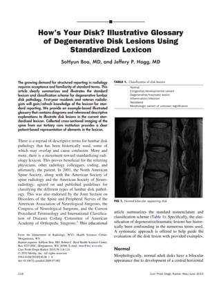
1. Hows Your Disk? Illustrative Glossary
of Degenerative Disk Lesions Using
Standardized Lexicon
SoHyun Boo, MD, and Jeffery P. Hogg, MD
The growing demand for structured reporting in radiology
requires acceptance and familiarity of standard terms. This
article clearly summarizes and illustrates the standard
lexicon and classi鍖cation scheme for degenerative lumbar
disk pathology. First-year residents and veteran radiolo-
gists will gain/refresh knowledge of the lexicon for stan-
dard reporting. We provide an example-based illustrated
glossary that contains diagrams and referenced descriptive
explanations to illustrate disk lesions in the current stan-
dardized lexicon. Collected cross-sectional imaging of the
spine from our tertiary care institution provides a clear
patient-based representation of elements in the lexicon.
There is a myriad of descriptive terms for lumbar disk
pathology that has been historically used, some of
which may overlap and cause confusion. More and
more, there is a movement toward standardizing radi-
ology lexicon. This proves bene鍖cial for the referring
physicians, other radiology colleagues, coding, and
ultimately, the patient. In 2001, the North American
Spine Society, along with the American Society of
spine radiology and the American Society of Neuro-
radiology, agreed on and published guidelines for
classifying the different types of lumbar disk pathol-
ogy. This was also endorsed by the Joint Section on
Disorders of the Spine and Peripheral Nerves of the
American Association of Neurological Surgeons, the
Congress of Neurological Surgeons, and the Current
Procedural Terminology and International Classi鍖ca-
tion of Diseases Coding Committee of American
Academy of Orthopedic Surgeons.1
This educational
article summarizes the standard nomenclature and
classi鍖cation scheme (Table 1). Speci鍖cally, the clas-
si鍖cation of degenerative/traumatic lesions has histor-
ically been confounding in the numerous terms used.
A systematic approach is offered to help guide the
evaluation of the disk lesion with provided examples.
Normal
Morphologically, normal adult disks have a bilocular
appearance due to development of a central horizontal
From the Department of Radiology, WVU Health Sciences Center,
Morgantown, WV.
Reprint requests: SoHyun Boo, MD, Robert C. Byrd Health Sciences Center,
Box 9235 HSC, Morgantown, WV 26506. E-mail: sboo@hsc.wvu.edu.
Curr Probl Diagn Radiol 2010;39:118-124.
息 2010 Mosby, Inc. All rights reserved.
0363-0188/2010/$36.00 路 0
doi:10.1067/j.cpradiol.2009.07.002
FIG 1. Normal bilocular appearing disk.
TABLE 1. Classi鍖cation of disk lesions
Normal
Congenital/developmental variant
Degenerative/traumatic lesion
In鍖ammation/infection
Neoplasia
Morphologic variant of unknown signi鍖cance
118 Curr Probl Diagn Radiol, May/June 2010
2. band of 鍖brous tissue within the nucleus. Normal
does not give regard to the clinical context and does
not include aging, adaptive, developmental, or degen-
erative changes, which may be clinically normal
25%
90属
Normal disk
FIG 2. Schematic of normal disk considered as a 360-degree arc.
FIG 3. Congenital developmental variant of disk which has undergone
morphologic change secondary to scoliosis in this case.
Generalized
disk bulge
25%
90属
FIG 4. Generalized disk bulge. Disk displacement involves 肋50%
(180属) of ring apophyses.
central canal zone
central zone
(right or left)
or
subarticular zone
nucleus pulposusannulus fibrosus
foraminal zone
(pedicle zone)
extra-foraminal zone
(far lateral zone)
FIG 5. Normal disk consists of the nucleus pulposus and surrounding
annulus 鍖brosus. Disk hernias can be further described in its zonal
location.
FIG 6. Annular tears are seen as high intensity nucleus pulposus
existing through areas of separation between the dark annular 鍖bers.
Intravertebral hernias are also seen.
TABLE 2. Subcategories of degenerative/traumatic lesions
Annular tear/鍖ssure
Herniation (extrusion/protrusion)
Degeneration (desiccation, spondylosis deformans,
intervertebral osteochondrosis)
Curr Probl Diagn Radiol, May/June 2010 119
3. 90属
Focal Hernia
25%
90属
Broad-based hernia
25%
FIG 7. Localized displacement of disk can be a focal (禄25%) or broad-based (25-50%) hernia.
90属
Protrusion
25%
or
FIG 8. Protrusion type hernia.
120 Curr Probl Diagn Radiol, May/June 2010
4. (Fig 1).1
The normal disk can be considered as a
360-degree arc divided into 4 quadrants (Fig 2).1
Congenital Developmental Variant
These abnormal disks have undergone morphologic
changes to adapt to abnormal growth of the spine such
as in scoliosis or spondylolisthesis (Fig 3).
There is generalized displacement of disk beyond
the endplates that constitutes a disk bulge. By conven-
tion, bulging disks involve 肋50% (180属) of the ring
apophyses (Fig 4).1
By this standard lexicon, a bulge
is not a hernia.
Degenerative/Traumatic
Degenerative/traumatic change in the disk is a broad
category that again is purely descriptive and identi鍖es
and does not imply any relationship to the clinical
context, pathology, or need for treatment. Several
subcategories of degenerative/traumatic lesions are
identi鍖ed in Table 2.
90属
Extrusion
25%
or
FIG 9. Extrusion type hernias.
Curr Probl Diagn Radiol, May/June 2010 121
5. Generalized
disk bulge
25%
Identify displacement
of disk.
(generalized vs.. localized)
Is
displacement > 50%
(180 degrees)
of the edge of the ring
apophyses?
YES
NO
Is displacement
< 25%?
Are the widest
edges of displaced disk,
in any plane, less than
the distance
between the edges of
the hernia base?
Localized
Hernia
Focal Broad-based
Focal
Extrusion
Focal Protrusion
Are the widest
edges of displaced
disk,
in any plane, greater
than the distance
between the edges of
the hernia base?
Broad-based
Extrusion
Broad-based
Protrusion
ONSEY
YES YESNO NO
90属
Broad-based
hernia 25%
90属
Focal
hernia
25%
Generalized
bulge
90属
FIG 10. Algorithm for analysis of disk displacement.
122 Curr Probl Diagn Radiol, May/June 2010
6. Annular Tears
Annular tears or 鍖ssures describe breaks or separation
between the dark annular 鍖bers that extend concentri-
cally, transversely, or radially, allowing the more high
intensity nucleus to exit through (Figs 5 and 6).2,3
Intravertebral hernia is also seen in Fig 6.
Disk Hernias
By convention, a hernia is de鍖ned as localized dis-
placement of disk material, whether it be nucleus,
cartilage, annular 鍖bers, etc, beyond the intervertebral
disk space. This interspace is de鍖ned by the vertebral
body endplates and ring apophyses, exclusive of os-
teophytes.
Localized displacement of disk can be categorized
as focal or broad-based. It is usually best appreciated
on axial imaging (Fig 7). However, the displacement
of disk may occur in any plane. Visualization in all
planes must occur to properly categorize herniations.
Furthermore, disk herniations can be de鍖ned as
protrusions vs extrusions. These are typically easily
de鍖ned on sagittal images, but again, the de鍖nition
must be applied in any plane. The base of the hernia is
the widest part in a protrusion (Fig 8), whereas the
herniated disk is wider than the base in an extrusion
(Fig 9A-C). We provide an algorithm for analysis of
disk displacement (Fig 10).
Degeneration
Degeneration is a broad description that can include
changes seen with apparent desiccation (Fig 11) or
鍖brosis of disk, disk space narrowing, vacuum gas
phenomena, diffuse generalized bulging disk, endplate
sclerosis, and osteophytosis.1,2
When these changes
occur, they can be subcategorized as spondylosis
FIG 11. Degenerative change from disk desiccation.
FIG 12. Spondylosis deformans.
FIG 13. Intervertebral osteochondrosis.
Curr Probl Diagn Radiol, May/June 2010 123
7. deformans2
(Fig 12), typically seen with aging, and/or
intervertebral osteochondrosis2
(Fig 13), which is a
more pronounced pathologic change within the disk.
REFERENCES
1. Fardon DF, Milette PC. Nomenclature and classi鍖cation of
lumbar disc pathology: Recommendations of the Combined
Task Forces of the North American Spine Society, American
Society of Spine Radiology, and American Society of Neu-
roradiology. Spine 2001;26:E93-E113.
2. Grossman RI, Yousem DM. Neuroradiology: The Requisites,
2nd edition. St Louis, MO: Mosby, 2003.
3. Harnsberger HR, et al. Diagnostic and Surgical Imaging
Anatomy: Brain, Head and Neck, Spine. Salt Lake City, UT:
Amirsys, 2006.
124 Curr Probl Diagn Radiol, May/June 2010