Gluteal region
- 1. GLUTEAL REGION Varun Walia (145)
- 2. Contents: ïī Gluteal region and boundaries ïī Cutaneous innervations ïī Muscles of gluteal region ïī Arteries of gluteal region ïī Nerves of gluteal region ïī Applied Aspect
- 3. Introduction: ïī Transitional area b/w trunk and lower extremity ïī Anatomically it is a part of trunk. Functionally it is a part of lower extremity ïī Gluteal region includes rounded, posterior buttocks and laterally placed hip region
- 4. Boundaries:
- 5. Superficial fascia: ïī Thick, dense, well developed, laden with large quantities of fat (specially in women) that: - Gives the characteristic convexity to the buttock - Forms a thick cushion over the ischial tuberosity ïī It contains cutaneous innervations, vessels and lymphatics
- 7. Cutaneous vessels & lymphatics: ïī Blood supply: -branches of superior and inferior gluteal arteries ïī Lymphatics: - lateral group of superficial inguinal lymph nodes
- 8. Deep Fascia ïī Is continuation of the fascia lata (deep fascia of the thigh) ïī At the lower border of the gluteus maximus, fascia lata splits to enclose the muscle ïī Above the gluteus maximus, the deep fascia continues as one layer covering the gluteus medius & gets attached to iliac crest ïī Laterally the fascia merges with the iliotibial tract Fascia over gluteus medius Tensor fascia lata Gluteal fascia Iliotibial tract
- 9. âĒ Gluteus maximus âĒ Gluteus medius âĒ Gluteus minimus âĒ Tensor fascia lata âĒ Piriformis âĒ Obturator internus âĒ Superior Gemellus âĒ Inferior Gemellus âĒ Quadratus femoris âĒ obturator externus Muscles of the Gluteal Region
- 10. ïī Largest muscle in the body ïī Forms the prominence of buttock ilium S C Gluteus Maximus
- 11. ïī Origin: ïīOuter surface of ilium behind the posterior gluteal line ïīLumbar fascia ïīPosterior surface of sacrum & coccyx ïīSacrotuberous ligament ïī Insertion: ïī Most of the muscle (3/4th) inserted into the iliotibial tract ïīDeeper fibers inserted to the gluteal tuberosity ïī Nerve supply: ïīInferior gluteal nerve (L5, S1, 2)
- 12. Actions: ï Extends & laterally rotates the hip joint ï Extends the knee joint (through iliotibial tract) ï Gives simultaneous stability to the hip and knee joints through the iliotibial tract
- 13. Structures under the cover of gluteus maximus: ïī gluteus Medius & minimus ïī rectus femoris ïī Piriformis ïī obturator internus with two gemelli ïī Quadratus femoris ïī obturator externus ïī Origin of four hamstring from ischial tuberosity ïī Insertion of pubic fibers of ad. magnus Muscles:-
- 14. ïī Superior gluteal vessels ïī inferior gluteal vessels ïī internal pudendal ïī Medial femoral circumflex artery ïī trochanteric & cruciate anastomosis Vessels:-
- 15. ïī Superior gluteal ïī inferior gluteal ïī sciatic ïī Post. cut. Nerve of thigh ïī nerve to quadratus femoris ïī pudendal nerve ïī nerve to obturator internus ïī perforating cutaneous nerves Nerves:-
- 16. ïī Bones & joints- ilium, ischial tuberosity, upper end of femur with greater trochanter, sacrum, coccyx, hip joint &sacroiliac joint ïī Ligaments- sacrotuberous, sacrospinous & ischiofemoral ïī Bursa- trochanteric bursa of glut. maximus, of ischial tuberosity, & bet. glut. max, & vastus lateralis
- 17. Ligaments of gluteal region: 2 ligaments: Sacrospinous, connecting sacrum to ischial spine Sacrotuberous, connecting sacrum to ischial tuberosity They convert the greater & lesser sciatic notches into greater & lesser sciatic foramina Their main function is to: -Stabilize the sacrum -Prevent its posterior rotation at the sacroiliac joint
- 18. ïīOrigin: outer surface of ilium between the middle and posterior gluteal lines ïīInsertion: Lateral surface of greater trochanter ïīNerve supply: Superior gluteal nerve (L4,5, S1) ïīAction: ïīAbducts & medially rotates the thigh ïīSteady pelvis in walking Gluteus Medius
- 19. ïīOrigin: outer surface of ilium ïīInsertion: Anterior surface of greater trochanter ïīNerve supply: Superior gluteal nerve (L4,5, S1) ïīAction: Abducts & medially rotates the thigh Gluteus Minimus
- 20. ïīOrigin: Anterior surface of sacrum between the anterior sacrum foramina ïīInsertion: Apex of greater trochanter ïīNerve supply: direct branch from L5, S1&S2 ïī Action: lateral rotator of femur Piriformis
- 21. ïī Origin: Deep surface of obturator membrane and surrounding bone ïī Insertion: Medial side of greater trochanter above the trochanteric fossa ïī Nerve supply: nerve to obturator internus ïī Action: Lateral rotator of femur Obturator internus
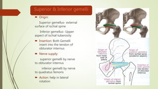
- 22. ïī Origin: Superior gemellus- external surface of ischial spine Inferior gemellus- Upper aspect of ischial tuberosity ïī Insertion: Both Gemelli insert into the tendon of obturator internus ïī Nerve supply: superior gemelli by nerve to obturator internus inferior gemelli by nerve to quadratus femoris ïī Action: help in lateral rotation Superior & Inferior gemelli
- 23. ïīOrigin: Lateral border of ischial tuberosity ïīInsertion: Quadrate tubercle of femur ïīNerve supply: nerve to quadratus femoris ïīAction: lateral rotator of thigh Quadratus femoris
- 24. ïī Origin: Obturator membrane Ramus of pubis Ramus of ischium ïī Insertion: Trochanteric fossa on the medial aspect of greater trochanter ïī Nerve supply: Obturator nerve ïī Action: Lateral rotator of femur Obturator externus
- 25. Arteries ïī Inferior gluteal artery âĒ Originates from the ant. Trunk of the internal iliac artery âĒ Supplies adjacent muscles and desends through the gluteal region into the posterior thigh ïī Superior gluteal artery âĒ Originates form the post. Trunk of the internal iliac artery âĒ Divides into a superficial and a deep branch ïī Internal pudendal artery ïī Various anastomosis
- 26. Arterial anastomosis of the gluteal region ïī Cruciate anastomosis: âĒ Present in the lower part of the gluteal region. âĒ Arteries taking part in anastomosis are inferior gluteal artery, first perforating artery and the lateral & medial Circumflex Femoral arteries. ïī Trochanteric artery: âĒ Seen in relation to greater trochanter. âĒ Arteries taking part in anastomosis are superior gluteal artery and the medial & lateral circumflex femoral arteries.
- 27. Nerves in gluteal region
- 28. ïą Structures passing through greater sciatic foramen ïī Piriformis ïī Structures passing above the piriformis are: superior gluteal nerve and superior gluteal vessels ïī Structures passing below the piriformis are: Inferior gluteal nerve and vessels, sciatic nerve, posterior cutaneous nerve of thigh, nerve to quadratus femoris, pudendal nerve, internal pudendal vessels, nerve to obturator internus ïą Structures passing through lesser sciatic foramen ïī Pudendal nerve, Internal pudendal vessels, nerve to obturator internus, tendon of obturator internus
- 29. Applied ïī I/m injection is given in superolateral quadrant of gluteal region to avoid injury to nerves
- 30. ïī When gluteus maximus is paralysed, the patient cannot stand up from sitting posture without support. ïī When the gluteus Medius & minimus are paralysed, patient sways on the paralysed side while walking. This is known as lurching gait. When bilateral, the gait is called as waddling gait. ïī Trendelenburg sign: Normally when the body weight is supported on one limb, the glutei of the supported side raise the opposite (unsupported) side of the pelvis. However if abductor mechanism is defective, the unsupported side of the pelvis drops and this is known as positive trendelenburg sign.


