Normal anatomical variants
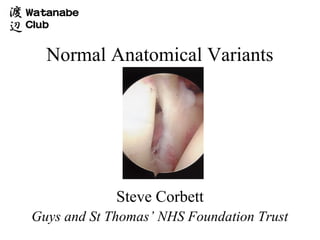
- 1. Normal Anatomical Variants Steve Corbett Guys and St Thomas’ NHS Foundation Trust
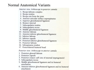
- 5. Normal Anatomical Variants • Superior labrum / Biceps • Biceps • Anterosuperior Quadrant • AS head • AI Quadrant • Superior Cuff / Head • PI Quadrant • Posterior Cuff / Head • PS Quadrant • GHJ Surfaces • Bursa
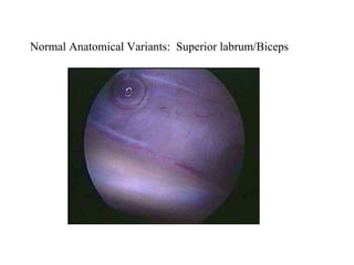
- 6. Normal Anatomical Variants: Superior labrum/Biceps
- 7. Normal Anatomical Variants: Superior labrum/Biceps
- 8. Normal Anatomical Variants: Superior labrum/Biceps • 15% loosely attached meniscal type labrum • 1-5mm width
- 9. Normal Anatomical Variants: Superior labrum/Biceps
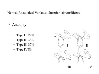
- 11. Normal Anatomical Variants: Superior labrum/Biceps • Anatomy – Type I 22% – Type II 33% – Type III 37% I II – Type IV 8% III IV
- 12. Normal Anatomical Variants: Superior labrum/Biceps • Vincula Biceps • Bifid Biceps – Small strands of – 1 part attached to cable mesentry – 2nd part attached to – Pass from biceps to tubercle surrounding capsule • Complete absence
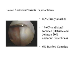
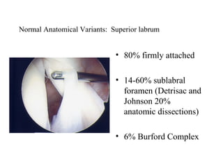
- 13. Normal Anatomical Variants: Superior labrum • 80% firmly attached • 14-60% sublabral foramen (Detrisac and Johnson 20% anatomic dissections) • 6% Burford Complex
- 14. Normal Anatomical Variants: Superior labrum • 80% firmly attached • 14-60% sublabral foramen (Detrisac and Johnson 20% anatomic dissections) • 6% Burford Complex
- 15. Normal Anatomical Variants: Superior labrum • 80% firmly attached • 14-60% sublabral foramen (Detrisac and Johnson 20% anatomic dissections) • 6% Burford Complex
- 16. Normal Anatomical Variants: Superior labrum • 80% firmly attached • 14-60% sublabral foramen (Detrisac and Johnson 20% anatomic dissections) • 6% Burford Complex
- 17. Normal Anatomical Variants: Superior labrum • 6% Burford Complex – Cord like MGHL – No labral tissue ant/sup glenoid – Surfaces smooth
- 18. Normal Anatomical Variants: Superior labrum • 6% Burford Complex – Cord like MGHL – No labral tissue ant/sup glenoid – Surfaces smooth
- 19. Normal Anatomical Variants: Superior labrum • 6% Burford Complex – Cord like MGHL – No labral tissue ant/sup glenoid – Surfaces smooth
- 20. Normal Anatomical Variants: Superior labrum • Divides sup. 2/5 and inf. 3/5. • Variable in depth
- 21. Normal Anatomical Variants: Subscapularis / SGHL • Leading edge may be split or bifid • 3% • SGHL present in nearly 100%, Occassionally frayed
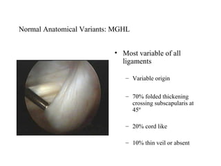
- 22. Normal Anatomical Variants: MGHL • Most variable of all ligaments – Variable origin – 70% folded thickening crossing subscapularis at 45º – 20% cord like – 10% thin veil or absent
- 23. Normal Anatomical Variants: MGHL • Most variable of all ligaments – Variable origin – 70% folded thickening crossing subscapularis at 45º – 20% cord like – 10% thin veil or absent
- 24. Normal Anatomical Variants: MGHL • Most variable of all ligaments – Variable origin – 70% folded thickening crossing subscapularis at 45º – 20% cord like – 10% thin veil or absent
- 26. Normal Anatomical Variants: Anterior Inferior Labrum • 95% smooth attachment • 5% meniscoid – Probe can be inserted but labrum not detached
- 27. Normal Anatomical Variants: Anterior Inferior Labrum • 95% smooth attachment • 5% meniscoid – Probe can be inserted but labrum not detached
- 28. Normal Anatomical Variants: IGHL • aIGHL – Variable attachment to labrum – Distinct superior band not always present (Defined by Turkel et al) – May hypertrophy when MGHL absent
- 29. Normal Anatomical Variants: Inferior capsular recess • Normally smooth • Delicate synovial covering • Small fenestrations • Post. Sup. Band pIGHL not always well visualised (Schwartz et al)
- 30. Normal Anatomical Variants: Bare area • Bare area – 2-3 mm – 2-3 cm – Frequent indentations, deep holes – Size varies with age (De Palma)
- 31. Normal Anatomical Variants: Bare area • Must distinguish from Hill Sachs
- 32. Normal Anatomical Variants: Superior cuff • Layer of capsule and synovium • Rotator cable
- 33. Normal Anatomical Variants: Posterosuperior cuff • May have fenestrations in superficial layers
- 34. Normal Anatomical Variants: Posterior labrum / Capsule • 95% firmly attached • 5% meniscoid, firmly attached at periphery
- 35. Normal Anatomical Variants: Posterior labrum / Capsule • Normal to have a deep cleft in capsule posterior to labrum
- 36. Thank you

