11 lab pet
This document provides an overview of positron emission tomography (PET). PET is a noninvasive imaging technique that uses radioactive tracers to measure metabolic activity in the body. It was developed in the 1970s and was the first method to provide functional information about the brain. PET works by injecting a radioactive tracer tagged to glucose or other compounds, which is detected as it accumulates in tissues. When the radioactive tracer decays, it emits positrons that annihilate with electrons, producing pairs of gamma rays. PET cameras detect these gamma rays simultaneously to determine where the decay occurred and construct 3D images. PET is used to diagnose and monitor conditions like cancer, heart disease, neurological disorders, and seizures.


Recommended
More Related Content
Similar to 11 lab pet (20)
More from Khaleeque Memon (13)
Recently uploaded (20)
11 lab pet
- 1. BIOMEDICAL ENGINEERING SYSTEM Positron Emission Tomography (P.E.T) ENGR KHALEEQUE AHMED Sir syed university of engineering & technology Karachi
- 2. What is PET Ō¢║PET is a noninvasive, diagnostic imaging technique for measuring the metabolic activity of cells in the human body. Ō¢║It was developed in the mid 1970s and it was the first scanning method to give functional information about the brain.
- 3. A little history about the positron Ō¢║Existence first postulated in 1928 by Paul Dirac Ō¢║First observed in 1932 by Carl D. Anderson, who gave the positron its name. He also suggested to rename the electron to ŌĆ£negatronŌĆØ but he was unsuccessful.
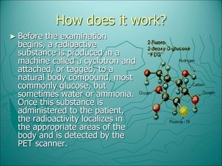
- 4. How does it work? Ō¢║ Before the examination begins, a radioactive substance is produced in a machine called a cyclotron and attached, or tagged, to a natural body compound, most commonly glucose, but sometimes water or ammonia. Once this substance is administered to the patient, the radioactivity localizes in the appropriate areas of the body and is detected by the PET scanner.
- 5. What is a Positron? Ō¢║ A Positron is an anti-matter electron, it is identical in mass but has an apposite charge of +1. Ō¢║ Positron can come from different number of sources, but for PET they are produced by nuclear decay. Ō¢║ Nuclear decay is basically when unstable nuclei are produced in a cyclotron by bombarding the target material with protons, and as a result a neutron is released.
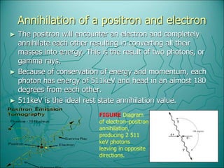
- 6. Annihilation of a positron and electron Ō¢║ The positron will encounter an electron and completely annihilate each other resulting in converting all their masses into energy. This is the result of two photons, or gamma rays. Ō¢║ Because of conservation of energy and momentum, each photon has energy of 511keV and head in an almost 180 degrees from each other. Ō¢║ 511keV is the ideal rest state annihilation value. FIGURE Diagram of electronŌĆōpositron annihilation, producing 2 511 keV photons leaving in opposite directions.
- 7. Coincidence detection Ō¢║Types of coincidence ’é¦ True ’é¦ Scatter ’é¦ random
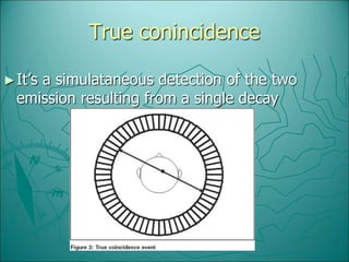
- 8. True conincidence Ō¢║ItŌĆÖs a simulataneous detection of the two emission resulting from a single decay
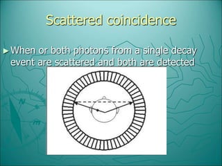
- 9. Scattered coincidence Ō¢║When or both photons from a single decay event are scattered and both are detected
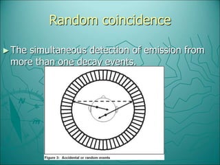
- 10. Random coincidence Ō¢║The simultaneous detection of emission from more than one decay events.
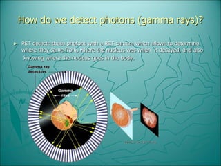
- 11. How do we detect photons (gamma rays)? Ō¢║ PET detects these photons with a PET camera which allows to determine where they came from, where the nucleus was when it decayed, and also knowing where the nucleus goes in the body.
- 12. What are some of the uses for PET Ō¢║Patients with conditions affecting the brain Ō¢║Heart Ō¢║Certain types of Cancer Ō¢║AlzheimerŌĆÖs disease Ō¢║Some neurological disorders
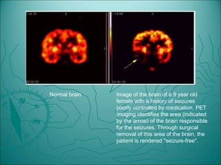
- 13. Normal brain Image of the brain of a 9 year old female with a history of seizures poorly controlled by medication. PET imaging identifies the area (indicated by the arrow) of the brain responsible for the seizures. Through surgical removal of this area of the brain, the patient is rendered "seizure-free".
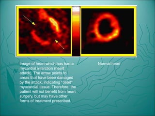
- 14. Image of heart which has had a mycardial infarction (heart attack). The arrow points to areas that have been damaged by the attack, indicating "dead" myocardial tissue. Therefore, the patient will not benefit from heart surgery, but may have other forms of treatment prescribed. Normal heart