Recent advancements in modern x ray tube
Download as PPTX, PDF20 likes7,787 views

All the advancements in X-ray tubes till date are done to increase the Tube heat storage capacity thus increasing the lifetime of x -ray tubes. This slide explains about these recent advancements in x-ray tubes.


More Related Content
What's hot (20)
Similar to Recent advancements in modern x ray tube (20)


Recently uploaded (20)


Recent advancements in modern x ray tube
- 2. History ’üĄ X-ray tubes evolved from experimental Crookes tubes with which X-rays were first discovered on 1895, by the German physicist Wilhelm Conrad R├Čntgen. ’üĄ Also callled Cold cathode type X-ray tubes. ’üĄ Electrons formed by ionization of Gas inside the tube. ’üĄ Platinum Anode Used.
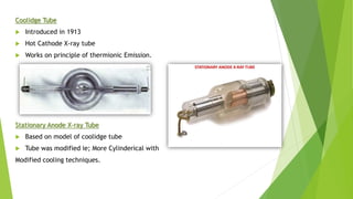
- 3. Coolidge Tube ’üĄ Introduced in 1913 ’üĄ Hot Cathode X-ray tube ’üĄ Works on principle of thermionic Emission. Stationary Anode X-ray Tube ’üĄ Based on model of coolidge tube ’üĄ Tube was modified ie; More Cylinderical with Modified cooling techniques.
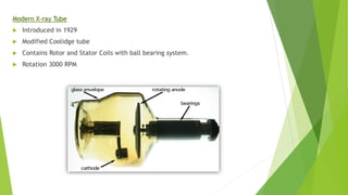
- 4. Modern X-ray Tube ’üĄ Introduced in 1929 ’üĄ Modified Coolidge tube ’üĄ Contains Rotor and Stator Coils with ball bearing system. ’üĄ Rotation 3000 RPM
- 5. Advancement in Rotating Anode X-ray Tube Dual Focus X-ray tube ’üĄ Two filaments Used ’üĄ Most of diagnostic tubes have two focal spots; Large and small ’üĄ Large is used when large body parts are imagined - high heat ’üĄ Small is used when better spatial resolution is desired ŌĆō better detail ’üĄ Small Focal track is superimposed over large focal track
- 6. Advancement in Rotating Anode X-ray Tube ’üĄ New Anode material used Tungsten (90%) ŌĆō Rhenium (10%) Alloy on Molybednum backed on Graphite for good Heat Storage and dissipation. ’üĄ Lubrication material used in bearings of Rotor-stator coil- Silver powder which allows high rotation ’üĄ Increased speed of Rotation 9000 ŌĆō 10,000 RPM allows use of high mA and shorter exposure time. ’üĄ Reduced Anode angle ie; 6┬░
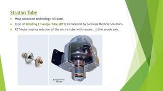
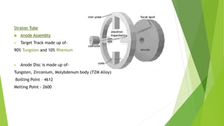
- 7. Straton Tube ’üĄ Most advanced technology till date. ’üĄ Type of Rotating Envelope Tube (RET) introduced by Siemens Medical Solutions ’üĄ RET-tube implies rotation of the entire tube with respect to the anode axis.
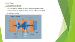
- 8. Straton Tube Characteristic features ’é¦ Electron beam is shaped and controlled by magnetic field. ’é¦ Cooling surface of anode in direct contact with cooling fluid. ’é¦ A rotating envelope
- 9. Straton Tube It consist of four systems I. Tube Envelope system II. Electron Emission system III. Magnetic deflection system IV. Cooling system
- 10. Straton Tube ’üĄ Tube Envelope system ’é¦ Material used ŌĆō Non Magnetic stainless steel ’é¦ Directly attached to anode disc ’é¦ Annular/Circular Window (Thickness ŌĆō 0.2 mm) ’é¦ Maximum rotation speed ŌĆō 9600 RPM
- 11. Straton Tube ’üĄ Anode Assembly ’é¦ Target Track made up of- 90% Tungsten and 10% Rhenium ’é¦ Anode Disc is made up of- Tungsten, Zirconium, Molybdenum body (TZM Alloy) Boilling Point ŌĆō 4612 Melting Point - 2600
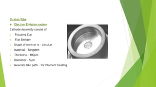
- 12. Straton Tube ’üĄ Electron Emission system Cathode Assembly consist of I. Focusing Cup II. Flat Emitter ’é¦ Shape of emitter is ŌĆō circular ’é¦ Material ŌĆō Tungsten ’é¦ Thickness - 100┬Ąm ’é¦ Diameter - 5┬Ąm ’é¦ Meander like path ŌĆō for filament heating
- 13. Straton Tube ’üĄ Magnetic Deflection System There are three coils present- ’é¦ R ŌĆō Coil ŌĆō Deflect the beam Radial direction onto the focal spot ’é¦ Q ŌĆō Coil ŌĆō Focus beam to determine Size ’é¦ Phi ŌĆō Coils ŌĆō Deflection of flying focal spot intangential direction ’é¦ Microcontroller controls individual coil current. ’é¦ Electronically adjusted focal spot ’é¦ Best Image quality
- 14. Straton Tube ’üĄ Cooling System ’é¦ Unlike other rotating anode x-ray tube which dissipates heat by radiation, Straton tubes dissipates heat by convection process. ’é¦ Anode Disc comes in direct contact with cooling Oil. ’é¦ Oil rotation is turbine flow ’é¦ Flow of oil - during exposure is 25 ltr/Min ’é¦ - during pump running 8 ltr/Min ’é¦ Oil used ŌĆō Mineral Oil ’üĄ When apower of 100 KW is applied , the temprature of focal spot reaches 2500┬░C and temprature on focal track reaches 2000┬░C. But the back of anode has temprature of 200┬░C. ’üĄ This difference in temprature is due to direct cooling.
- 15. ’üĄ Advantages ’é¦ Better Heat Dissipation ’é¦ Various size multiple focal spot ’é¦ Longer tube life ’é¦ Can be used in high KV and high mA technique for prolonged Duration. Ie; High mAs