269034 mediastinum
Download as ppt, pdf0 likes256 views
Examville.com is a website that provides online practice tests, live classes, tutoring, study guides, Q&A, and premium content to help students prepare for exams. The website is free to join.
1 of 26
Downloaded 22 times


Ad
Recommended
Abdominal arota
Abdominal arotaSourabh Ahlawat
╠²
The abdominal aorta begins at the diaphragm and extends down the back of the abdominal cavity, branching off into arteries that supply the abdominal organs and walls. It has two segments - the suprarenal segment above the renal arteries and the infrarenal segment below. The aorta gives off several single and paired visceral arteries like the celiac, superior mesenteric, and renal arteries. It runs parallel to the inferior vena cava and its branches include the lumbar arteries. Abdominal aortic aneurysms are localized enlargements of the aorta over 3cm in diameter, which can rupture and cause life-threatening bleeding.Mediastinum
MediastinumExamville.com LLC
╠²
The document provides a detailed overview of the mediastinum, a central compartment of the thoracic cavity, including its divisions (superior and inferior) and their respective contents. It outlines the anatomical structures and relationships within the mediastinum, such as the thymus, heart, great vessels, esophagus, and lymphatic systems, as well as the nerves and veins involved. Additionally, it discusses specific anatomical boundaries and clinical significance of these contents.Mediastinum 1
Mediastinum 1kooff
╠²
The mediastinum is the central partition of the thoracic cavity that contains structures like the heart, trachea, esophagus and major blood vessels. It is divided into superior, anterior, posterior and middle mediastinum. The superior mediastinum contains structures like the thymus gland, brachiocephalic veins, superior vena cava, arch of the aorta and its branches, trachea, esophagus, phrenic and vagus nerves. It is bounded superiorly by the thoracic inlet and inferiorly by the plane between T4 and T5 vertebrae. The structures pass through the mediastinum and provide blood supply and innervation to the thoracic organs.Mediastinum
MediastinumRawan Shahien
╠²
The mediastinum is the central portion of the thoracic cavity located between the lungs. It is divided into the superior mediastinum above the sternal angle and inferior mediastinum below. The inferior mediastinum is further divided into the anterior, middle, and posterior mediastinum, with the middle mediastinum containing the heart and pericardium.Superior mediastinum
Superior mediastinumJay Patel
╠²
The superior mediastinum is the uppermost division of the mediastinum. It is located posterior to the sternum and anterior to the bodies of the first four thoracic vertebrae. Key structures in the superior mediastinum include the thymus gland, brachiocephalic veins, superior vena cava, arch of aorta and its branches, trachea, esophagus, phrenic nerves, vagus nerves, recurrent laryngeal nerve, thoracic duct, and other smaller structures. The mediastinum is further divided into the superior, inferior anterior, middle and posterior mediastinum.56 mediastinum
56 mediastinumanggreiny
╠²
The mediastinum is the partition between the two pleural cavities and lungs. It is subdivided into the superior, middle, and posterior mediastinum. The superior mediastinum contains structures like the esophagus, trachea, brachiocephalic veins, and vagus nerves. The middle mediastinum contains the heart, pericardium, and pulmonary trunk. The posterior mediastinum contains the esophagus, descending aorta, azygos veins, and vagus nerves.Mediastinum
Mediastinum TAYYAB MUNEER
╠²
The mediastinum is the central compartment of the thoracic cavity that contains the heart, lungs, esophagus, and other structures. It is divided into superior, anterior, middle, and posterior compartments. The superior mediastinum contains the thymus gland, major blood vessels like the aorta and superior vena cava, nerves like the vagus and phrenic nerves, and the esophagus. The middle mediastinum contains the heart surrounded by the pericardium. The anterior and posterior mediastinum contain the esophagus and major blood vessels.ct anantomy lung for rt planning
ct anantomy lung for rt planningvrinda singla
╠²
This document describes the anatomy of various arteries and veins in the thorax as seen on different slices of a CT scan, including:
1) The brachiocephalic artery, left common carotid artery, left subclavian artery, and formation of the brachiocephalic vein by the left subclavian vein and internal jugular vein at the level of T2-T3.
2) The aortic arch and origins of vessels like the brachiocephalic artery and left common carotid artery at a more superior level.
3) Various cardiac and vascular structures seen at different levels moving caudally, including the left atrium, ventricles, pulmonary veins and arteries, venaMediastinum and heart position
Mediastinum and heart positionmed alfarabi
╠²
The mediastinum is divided into superior, inferior, and middle sections. The middle mediastinum contains the heart, which is surrounded by the pericardium. The heart is cone-shaped with its apex pointing downward and to the left. Its surface can be delineated between the upper border of the third right costal cartilage superiorly, the middle of the right sixth costal cartilage laterally, the apex in the left fifth intercostal space midclavicularly, and the inferior border of the left second costal cartilage inferiorly.Mink Dissection ’üŖ
Mink Dissection ’üŖlkat1
╠²
This document provides labels and brief descriptions for various anatomical structures and organs in a dissected mink specimen. Key structures identified and summarized include:
- The panniculus carnosum, platysma, latissimus dorsi, pectoralis major, and pectoralis minor muscles.
- The pharynx, larynx, trachea, esophagus, lungs, bronchi, pulmonary veins and arteries.
- The heart, coronary arteries and veins, right and left atria and ventricles, septum, and pericardium.
- Major blood vessels including the internal and external jugular veins, internal and external carotid arteries, descending and ascending aortacardiovascular system
cardiovascular system Dr Suyashi
╠²
The cardiovascular system consists of the heart, blood vessels, and blood. The heart is a hollow muscular organ made of four chambers separated by septums. It pumps blood through two circuits: systemic circulation pumps blood to the body, while pulmonary circulation pumps blood to and from the lungs. The heart has four valves that ensure one-way blood flow - the tricuspid and mitral valves between the atria and ventricles, and the pulmonary and aortic semilunar valves at the exits of the right and left ventricles. The cardiovascular system transports blood throughout the body, supplying oxygen and nutrients to tissues and removing waste products.Anatomy (middle mediastinum)
Anatomy (middle mediastinum)Osama Al-Zahrani
╠²
The document summarizes the anatomy of the mediastinum. It defines the sternal angle as the joint between the manubrium and sternum. The mediastinum is divided into superior, middle, and posterior sections. The middle mediastinum contains the heart enclosed by the pericardium as well as the great vessels attached to it like the aorta and pulmonary trunk. The document lists the structures found in each section of the mediastinum.Abdominal aorta
Abdominal aortaAzhar Nazir Kiani
╠²
The document discusses the abdominal aorta and its branches. It notes that the abdominal aorta is the largest artery in the abdominal cavity and is a continuation of the thoracic aorta. It terminates at the L4 vertebrae by bifurcating into the right and left common iliac arteries. The major branches discussed include the celiac artery, superior mesenteric artery, inferior mesenteric artery, and renal arteries. Each of these branches and their roles in supplying specific organs are described in 1-2 sentences.mediastinum
mediastinumNepalese army institute of health sciences
╠²
The document discusses the mediastinum, which is the central compartment of the thoracic cavity located between the lungs. It is divided into superior, anterior, middle and posterior mediastinum. The superior mediastinum contains structures such as the thymus gland, great vessels like the superior vena cava and aorta, and nerves like the vagus nerve. The anterior mediastinum contains the thymus gland in children and structures related to the heart. The middle mediastinum contains the heart enclosed in the pericardium. The posterior mediastinum contains the esophagus and descending aorta along with nerves and lymph nodes. Mediastinitis is an infection of the mediastinum which canSuperior and inferior mediastinum
Superior and inferior mediastinumMoamer Gabsa
╠²
The lecture outlines the mediastinum's anatomy, including its boundaries, divisions, and contents, distinguishing between the superior and inferior mediastinum. The superior mediastinum contains structures such as the thymus gland, great vessels, trachea, and esophagus, while the inferior mediastinum is subdivided into anterior, middle, and posterior sections. Key components discussed include the blood supply and innervation of mediastinal organs, as well as the significance of associated structures like the phrenic and vagus nerves.Thoracic aorta
Thoracic aortaAisha Sadaf
╠²
The thoracic aorta begins where the aortic arch ends at the fourth thoracic vertebrae and extends down to the diaphragm. It supplies blood to the thoracic cavity and has several important branches including the bronchial arteries which supply the lungs, esophageal arteries which supply the esophagus, and posterior intercostal arteries which supply the spaces between the ribs. The thoracic aorta also gives off mediastinal and pericardial branches before passing through the diaphragm and becoming the abdominal aorta.ANATOMY OF BLOOD VESSELS
ANATOMY OF BLOOD VESSELSC L GUPTA EYE INSTITUTE MORADABAD UTTER PRADESH
╠²
The document outlines the anatomy and distribution of blood vessels, detailing the structure and functions of arteries, veins, arterioles, venules, and capillaries. It describes the three layers composing blood vessels and highlights differences in structure based on blood pressure, including the role of the circulatory system, systemic and pulmonary circuits, and fetal circulation. Additionally, it explains the hepatic portal circulation and the changes in blood flow dynamics at birth.The Heart And Great Vessels
The Heart And Great VesselsCrisbert Cualteros
╠²
The document provides an overview of the anatomy of the heart and surrounding structures. It describes the location of the heart in the chest and its internal and external structures. These include the four chambers of the heart (right and left atria and ventricles), major blood vessels (veins and arteries), surrounding membranes (pericardium), and other anatomical details.Mediastinum
MediastinumDr Laxman Khanal
╠²
This document outlines the anatomy of the mediastinum, describing its divisions, boundaries, contents, and related pathologies. It explains the significance of structures within the superior, middle, and posterior mediastinum, as well as clinical implications such as mediastinal shift and mediastinal syndrome. Key content includes details about the heart, major vessels, and nerves, and conditions like mediastinitis and mediastinal widening.Great vessels artery
Great vessels arteryRohit Paswan
╠²
The document describes the major blood vessels of the thorax, including the aorta, pulmonary trunk, and superior vena cava. It details the anatomy and branches of each vessel. The aorta originates from the left ventricle and divides into the ascending aorta, aortic arch, and descending thoracic aorta. The pulmonary trunk carries deoxygenated blood from the heart to the lungs, where it divides into right and left pulmonary arteries. The superior vena cava returns oxygenated blood from the upper body to the heart.POWERPOINT
POWERPOINTshamnanoushad
╠²
The heart is a hollow, conical organ located in the chest cavity between the lungs. It has four chambers - two upper atria and two lower ventricles separated by muscular walls. The right atrium receives deoxygenated blood from the body via two large veins and pumps it to the right ventricle. The left atrium receives oxygenated blood from the lungs via pulmonary veins and pumps it to the left ventricle. Blood is then pumped from the ventricles through valves into the arteries - the pulmonary artery from the right ventricle and the aorta from the left ventricle.Ch19bloodvessels
Ch19bloodvesselsRonaldo Paulino
╠²
This document discusses the structure and function of the cardiovascular system. It describes the different types of blood vessels including arteries, veins, and capillaries. It explains the layers of blood vessels and how blood flows from the heart through the arteries, capillaries and veins before returning to the heart. Key concepts covered include blood pressure, resistance, cardiac output, hormones that regulate blood pressure, and normal blood pressure readings.mediastinal imaging and masses
mediastinal imaging and massesArun Singh
╠²
This document discusses the anatomy and radiological imaging of mediastinal masses. It begins with an introduction to the mediastinum and its boundaries. It then describes the different divisions of the mediastinum and contents of each region. Specific anatomical structures discussed include the anterior junction line, right paratracheal stripe, azygoesophageal recess, and paraspinal lines. Common masses that can occur in each mediastinal compartment are mentioned. Radiographic findings that suggest the location of a mediastinal mass are also described.Mediastinum
MediastinumViresh V
╠²
The mediastinum is the space within the thoracic cavity between the lungs, containing structures like the heart, great vessels, esophagus and thoracic duct. It is divided into superior and inferior mediastinum. The superior mediastinum contains the thymus gland, great vessels, nerves and trachea. The inferior mediastinum is further divided into anterior, middle and posterior mediastinum. The middle mediastinum contains the heart and great vessels while the posterior mediastinum contains the descending aorta, esophagus and thoracic duct. Important structures and their relations within the divisions of the mediastinum are described.Thoracic cavity lecture engl.
Thoracic cavity lecture engl.Jay Patel
╠²
The document describes the anatomy of the thorax, including structures within the thoracic wall, mediastinum, and thoracic cavity. It discusses the internal thoracic artery and azygos vein within the thoracic wall. In the mediastinum, it outlines the subdivisions and contents, including the thymus, great vessels, nerves, and lymph nodes. The thoracic cavity contains the lungs, pleurae, and diaphragm.Anatomy of the thorax
Anatomy of the thorax┘åžĄž¦ž▒ ž¦┘Ŗ┘łž©
╠²
The document provides an overview of the anatomy of the thorax, including:
1) The muscles of the thorax including intrinsic and extrinsic muscles.
2) Details on the diaphragm including its shape, origins, insertions, and actions.
3) The major arteries and veins of the thorax including the pulmonary trunk, aorta, superior and inferior vena cava.
4) Nerves associated with the thorax including the phrenic, vagus, and recurrent laryngeal nerves.Thorax and abdomen & pelvis
Thorax and abdomen & pelvisMpdodz
╠²
This document provides an overview of the anatomy of the thorax, abdomen, pelvis, and related structures. It describes the bones and boundaries of the thoracic cage and inlet/outlet. It identifies structures in the mediastinum including the trachea, lungs, pleura, heart and its blood supply. For the abdomen, it describes the organs and their arrangement, abdominal blood supply, and normal abdominal x-ray findings. It also describes the bones, inlet/outlet of the pelvis, differences between the male and female pelvis, pelvic viscera and walls.7-Mediastinum -1.pdrhhbbbnhgbnjkkjkjjjjjkjjjj
7-Mediastinum -1.pdrhhbbbnhgbnjkkjkjjjjjkjjjjjamessesay1999
╠²
The document outlines the anatomy of the mediastinum, defining its boundaries, divisions, and contents, including major structures such as the trachea, heart, and various blood vessels. It details the superior, anterior, middle, and posterior mediastinum, highlighting their specific boundaries and contents, along with the functions of associated nerves like the vagus and phrenic nerves. Additionally, it provides a series of questions and answers to test knowledge on the anatomical features discussed in the lecture.Circulatory System
Circulatory SystemDoc Lorie B
╠²
The circulatory system is responsible for transporting materials like blood, oxygen, carbon dioxide, nutrients, and water throughout the entire body. It consists of the heart, blood vessels, and blood. The heart has four chambers - two upper atria and two lower ventricles. Deoxygenated blood enters the right atrium from the body and is pumped to the lungs where it receives oxygen before reentering the left atrium and being pumped by the left ventricle out to the body through arteries. There are three main types of circulation - pulmonary, coronary, and systemic.Cardiovascular system
Cardiovascular systemrameshdaiya
╠²
The document summarizes the key components and functioning of the cardiovascular system. It describes the heart structures and chambers, circulation paths through arteries, veins and capillaries, and the roles of valves and vessels in pumping blood throughout the body for gas and nutrient exchange as well as immune system functions. Fetal circulation and remnants in adults are also briefly outlined.More Related Content
What's hot (15)
Mediastinum and heart position
Mediastinum and heart positionmed alfarabi
╠²
The mediastinum is divided into superior, inferior, and middle sections. The middle mediastinum contains the heart, which is surrounded by the pericardium. The heart is cone-shaped with its apex pointing downward and to the left. Its surface can be delineated between the upper border of the third right costal cartilage superiorly, the middle of the right sixth costal cartilage laterally, the apex in the left fifth intercostal space midclavicularly, and the inferior border of the left second costal cartilage inferiorly.Mink Dissection ’üŖ
Mink Dissection ’üŖlkat1
╠²
This document provides labels and brief descriptions for various anatomical structures and organs in a dissected mink specimen. Key structures identified and summarized include:
- The panniculus carnosum, platysma, latissimus dorsi, pectoralis major, and pectoralis minor muscles.
- The pharynx, larynx, trachea, esophagus, lungs, bronchi, pulmonary veins and arteries.
- The heart, coronary arteries and veins, right and left atria and ventricles, septum, and pericardium.
- Major blood vessels including the internal and external jugular veins, internal and external carotid arteries, descending and ascending aortacardiovascular system
cardiovascular system Dr Suyashi
╠²
The cardiovascular system consists of the heart, blood vessels, and blood. The heart is a hollow muscular organ made of four chambers separated by septums. It pumps blood through two circuits: systemic circulation pumps blood to the body, while pulmonary circulation pumps blood to and from the lungs. The heart has four valves that ensure one-way blood flow - the tricuspid and mitral valves between the atria and ventricles, and the pulmonary and aortic semilunar valves at the exits of the right and left ventricles. The cardiovascular system transports blood throughout the body, supplying oxygen and nutrients to tissues and removing waste products.Anatomy (middle mediastinum)
Anatomy (middle mediastinum)Osama Al-Zahrani
╠²
The document summarizes the anatomy of the mediastinum. It defines the sternal angle as the joint between the manubrium and sternum. The mediastinum is divided into superior, middle, and posterior sections. The middle mediastinum contains the heart enclosed by the pericardium as well as the great vessels attached to it like the aorta and pulmonary trunk. The document lists the structures found in each section of the mediastinum.Abdominal aorta
Abdominal aortaAzhar Nazir Kiani
╠²
The document discusses the abdominal aorta and its branches. It notes that the abdominal aorta is the largest artery in the abdominal cavity and is a continuation of the thoracic aorta. It terminates at the L4 vertebrae by bifurcating into the right and left common iliac arteries. The major branches discussed include the celiac artery, superior mesenteric artery, inferior mesenteric artery, and renal arteries. Each of these branches and their roles in supplying specific organs are described in 1-2 sentences.mediastinum
mediastinumNepalese army institute of health sciences
╠²
The document discusses the mediastinum, which is the central compartment of the thoracic cavity located between the lungs. It is divided into superior, anterior, middle and posterior mediastinum. The superior mediastinum contains structures such as the thymus gland, great vessels like the superior vena cava and aorta, and nerves like the vagus nerve. The anterior mediastinum contains the thymus gland in children and structures related to the heart. The middle mediastinum contains the heart enclosed in the pericardium. The posterior mediastinum contains the esophagus and descending aorta along with nerves and lymph nodes. Mediastinitis is an infection of the mediastinum which canSuperior and inferior mediastinum
Superior and inferior mediastinumMoamer Gabsa
╠²
The lecture outlines the mediastinum's anatomy, including its boundaries, divisions, and contents, distinguishing between the superior and inferior mediastinum. The superior mediastinum contains structures such as the thymus gland, great vessels, trachea, and esophagus, while the inferior mediastinum is subdivided into anterior, middle, and posterior sections. Key components discussed include the blood supply and innervation of mediastinal organs, as well as the significance of associated structures like the phrenic and vagus nerves.Thoracic aorta
Thoracic aortaAisha Sadaf
╠²
The thoracic aorta begins where the aortic arch ends at the fourth thoracic vertebrae and extends down to the diaphragm. It supplies blood to the thoracic cavity and has several important branches including the bronchial arteries which supply the lungs, esophageal arteries which supply the esophagus, and posterior intercostal arteries which supply the spaces between the ribs. The thoracic aorta also gives off mediastinal and pericardial branches before passing through the diaphragm and becoming the abdominal aorta.ANATOMY OF BLOOD VESSELS
ANATOMY OF BLOOD VESSELSC L GUPTA EYE INSTITUTE MORADABAD UTTER PRADESH
╠²
The document outlines the anatomy and distribution of blood vessels, detailing the structure and functions of arteries, veins, arterioles, venules, and capillaries. It describes the three layers composing blood vessels and highlights differences in structure based on blood pressure, including the role of the circulatory system, systemic and pulmonary circuits, and fetal circulation. Additionally, it explains the hepatic portal circulation and the changes in blood flow dynamics at birth.The Heart And Great Vessels
The Heart And Great VesselsCrisbert Cualteros
╠²
The document provides an overview of the anatomy of the heart and surrounding structures. It describes the location of the heart in the chest and its internal and external structures. These include the four chambers of the heart (right and left atria and ventricles), major blood vessels (veins and arteries), surrounding membranes (pericardium), and other anatomical details.Mediastinum
MediastinumDr Laxman Khanal
╠²
This document outlines the anatomy of the mediastinum, describing its divisions, boundaries, contents, and related pathologies. It explains the significance of structures within the superior, middle, and posterior mediastinum, as well as clinical implications such as mediastinal shift and mediastinal syndrome. Key content includes details about the heart, major vessels, and nerves, and conditions like mediastinitis and mediastinal widening.Great vessels artery
Great vessels arteryRohit Paswan
╠²
The document describes the major blood vessels of the thorax, including the aorta, pulmonary trunk, and superior vena cava. It details the anatomy and branches of each vessel. The aorta originates from the left ventricle and divides into the ascending aorta, aortic arch, and descending thoracic aorta. The pulmonary trunk carries deoxygenated blood from the heart to the lungs, where it divides into right and left pulmonary arteries. The superior vena cava returns oxygenated blood from the upper body to the heart.POWERPOINT
POWERPOINTshamnanoushad
╠²
The heart is a hollow, conical organ located in the chest cavity between the lungs. It has four chambers - two upper atria and two lower ventricles separated by muscular walls. The right atrium receives deoxygenated blood from the body via two large veins and pumps it to the right ventricle. The left atrium receives oxygenated blood from the lungs via pulmonary veins and pumps it to the left ventricle. Blood is then pumped from the ventricles through valves into the arteries - the pulmonary artery from the right ventricle and the aorta from the left ventricle.Ch19bloodvessels
Ch19bloodvesselsRonaldo Paulino
╠²
This document discusses the structure and function of the cardiovascular system. It describes the different types of blood vessels including arteries, veins, and capillaries. It explains the layers of blood vessels and how blood flows from the heart through the arteries, capillaries and veins before returning to the heart. Key concepts covered include blood pressure, resistance, cardiac output, hormones that regulate blood pressure, and normal blood pressure readings.mediastinal imaging and masses
mediastinal imaging and massesArun Singh
╠²
This document discusses the anatomy and radiological imaging of mediastinal masses. It begins with an introduction to the mediastinum and its boundaries. It then describes the different divisions of the mediastinum and contents of each region. Specific anatomical structures discussed include the anterior junction line, right paratracheal stripe, azygoesophageal recess, and paraspinal lines. Common masses that can occur in each mediastinal compartment are mentioned. Radiographic findings that suggest the location of a mediastinal mass are also described.Similar to 269034 mediastinum (20)
Mediastinum
MediastinumViresh V
╠²
The mediastinum is the space within the thoracic cavity between the lungs, containing structures like the heart, great vessels, esophagus and thoracic duct. It is divided into superior and inferior mediastinum. The superior mediastinum contains the thymus gland, great vessels, nerves and trachea. The inferior mediastinum is further divided into anterior, middle and posterior mediastinum. The middle mediastinum contains the heart and great vessels while the posterior mediastinum contains the descending aorta, esophagus and thoracic duct. Important structures and their relations within the divisions of the mediastinum are described.Thoracic cavity lecture engl.
Thoracic cavity lecture engl.Jay Patel
╠²
The document describes the anatomy of the thorax, including structures within the thoracic wall, mediastinum, and thoracic cavity. It discusses the internal thoracic artery and azygos vein within the thoracic wall. In the mediastinum, it outlines the subdivisions and contents, including the thymus, great vessels, nerves, and lymph nodes. The thoracic cavity contains the lungs, pleurae, and diaphragm.Anatomy of the thorax
Anatomy of the thorax┘åžĄž¦ž▒ ž¦┘Ŗ┘łž©
╠²
The document provides an overview of the anatomy of the thorax, including:
1) The muscles of the thorax including intrinsic and extrinsic muscles.
2) Details on the diaphragm including its shape, origins, insertions, and actions.
3) The major arteries and veins of the thorax including the pulmonary trunk, aorta, superior and inferior vena cava.
4) Nerves associated with the thorax including the phrenic, vagus, and recurrent laryngeal nerves.Thorax and abdomen & pelvis
Thorax and abdomen & pelvisMpdodz
╠²
This document provides an overview of the anatomy of the thorax, abdomen, pelvis, and related structures. It describes the bones and boundaries of the thoracic cage and inlet/outlet. It identifies structures in the mediastinum including the trachea, lungs, pleura, heart and its blood supply. For the abdomen, it describes the organs and their arrangement, abdominal blood supply, and normal abdominal x-ray findings. It also describes the bones, inlet/outlet of the pelvis, differences between the male and female pelvis, pelvic viscera and walls.7-Mediastinum -1.pdrhhbbbnhgbnjkkjkjjjjjkjjjj
7-Mediastinum -1.pdrhhbbbnhgbnjkkjkjjjjjkjjjjjamessesay1999
╠²
The document outlines the anatomy of the mediastinum, defining its boundaries, divisions, and contents, including major structures such as the trachea, heart, and various blood vessels. It details the superior, anterior, middle, and posterior mediastinum, highlighting their specific boundaries and contents, along with the functions of associated nerves like the vagus and phrenic nerves. Additionally, it provides a series of questions and answers to test knowledge on the anatomical features discussed in the lecture.Circulatory System
Circulatory SystemDoc Lorie B
╠²
The circulatory system is responsible for transporting materials like blood, oxygen, carbon dioxide, nutrients, and water throughout the entire body. It consists of the heart, blood vessels, and blood. The heart has four chambers - two upper atria and two lower ventricles. Deoxygenated blood enters the right atrium from the body and is pumped to the lungs where it receives oxygen before reentering the left atrium and being pumped by the left ventricle out to the body through arteries. There are three main types of circulation - pulmonary, coronary, and systemic.Cardiovascular system
Cardiovascular systemrameshdaiya
╠²
The document summarizes the key components and functioning of the cardiovascular system. It describes the heart structures and chambers, circulation paths through arteries, veins and capillaries, and the roles of valves and vessels in pumping blood throughout the body for gas and nutrient exchange as well as immune system functions. Fetal circulation and remnants in adults are also briefly outlined.Mediastinum.pptx
Mediastinum.pptxAbhishek840813
╠²
The mediastinum is a central compartment in the thoracic cavity located between the pleural sacs, containing vital structures such as the heart, trachea, and major vessels. It is anatomically divided into the superior, middle, and inferior mediastinum, each with specific borders and contents. Conditions such as mediastinitis and mediastinal syndrome can arise due to various pathologies affecting the mediastinal structures.Thorax anatomy presentation
Thorax anatomy presentationMuhammad Ramzan Ul Rehman
╠²
This document summarizes the muscles, arteries, veins, nerves and other structures of the thorax. It describes the intrinsic and extrinsic muscles of the thorax including the intercostal muscles and diaphragm. It details the major arteries and veins of the thorax such as the pulmonary trunk, aorta, superior vena cava and azygos vein. It also outlines the lymphatic drainage and innervation of the thorax including the phrenic, vagus and recurrent laryngeal nerves. Finally, it provides an overview of the regions and landmarks of the thorax including the mediastinum.1087_Mediastinukrlelmeneklwnenemekwm.pptx
1087_Mediastinukrlelmeneklwnenemekwm.pptxdhruvkathuria8
╠²
The document outlines the anatomy of the mediastinum, detailing its divisions (superior, inferior, anterior, middle, and posterior mediastinum) and the key structures contained within each section, including the esophagus, trachea, and various blood vessels. It also discusses the anatomy and functions of nerves in the mediastinal area, their clinical relevance, and the relationship of these structures to specific thoracic conditions. Additionally, it covers the boundaries, blood supply, and lymphatic drainage of the mediastinum.Anatomy of heart presentation
Anatomy of heart presentationRajeshkumarm16
╠²
The heart is a hollow muscular organ located in the middle mediastinum. It has four chambers - right and left atria and right and left ventricles. The heart is surrounded by membranes including the pericardium, myocardium and endocardium. Blood flows from the right atrium to right ventricle through the tricuspid valve, then to the lungs through the pulmonary semilunar valve. Oxygenated blood returns to the left atrium through pulmonary veins and flows to the left ventricle through the mitral valve and out the aortic semilunar valve to the rest of the body. The heart receives its blood supply from the left and right coronary arteries.thorax.pdf
thorax.pdfsayedkamil06
╠²
The thorax contains the heart, lungs and major blood vessels. It is divided into three compartments - the two pleural cavities containing the lungs and the mediastinum between the lungs containing the heart, trachea and esophagus. The thoracic wall is made up of the vertebrae posteriorly, ribs laterally, and sternum anteriorly. The lungs are further divided into lobes and segments and are supplied by the bronchial tree which is innervated by the pulmonary plexus. Lymph from the lungs drains to tracheobronchial and mediastinal nodes.Anatomy of mediastinum- med student anatomy.pptx
Anatomy of mediastinum- med student anatomy.pptxhirunialoka98
╠²
The mediastinum is a crucial compartment of the thoracic cavity that separates the pleural cavities and contains vital structures including the heart, esophagus, trachea, and major blood vessels. It is divided into superior and inferior mediastinum, with the latter further subdivided into anterior, middle, and posterior spaces. Recent classifications have introduced new compartments based on imaging techniques, delineating prevascular, visceral, and paravertebral sections, each with distinct contents and boundaries.Cardiovascular System
Cardiovascular Systemraj kumar
╠²
The document provides a detailed overview of the cardiovascular system, including the anatomy and function of the heart and circulation. It describes the layers of the heart wall, the cardiac muscle, valves, conducting system, and blood flow through the heart. It also discusses the pulmonary and systemic circuits, blood vessel anatomy, veins, capillaries, and fetal circulation. Finally, it reviews the composition of blood and the functions of red blood cells, white blood cells, and platelets.Esophagus .pdf
Esophagus .pdfssuserbf4af22
╠²
The document summarizes the anatomy and embryology of the esophagus. It discusses the esophagus' location, length, diameter at different ages, layers, blood supply, nerve supply, and more. Some key points:
- The esophagus is a muscular tube that transports food from the pharynx to the stomach. It has an upper and lower sphincter.
- During development, the esophagus derives from the foregut and separates from the trachea by the esophagotracheal septum.
- It has cervical, thoracic, and abdominal portions with different anatomical relationships in each region.
- The wall consists of fibrous, muscular, submucosANA809Lymphatic System lim pics.ppt
ANA809Lymphatic System lim pics.pptAlick12
╠²
The lymphatic system removes foreign material and cell debris from tissues, and returns tissue fluid to the venous system. It is comprised of lymph capillaries, vessels, lymph nodes, and lymphoid tissues including the spleen, thymus, and tonsils. Lymph fluid drains from tissues via capillaries and vessels to lymph nodes, which filter out bacteria and foreign material. The thoracic duct and right lymphatic duct drain lymph from most of the body and empty into large veins in the neck. Regional lymph nodes throughout the body drain specific structures and tissues.anatomy of esophagus by dr ravindra daggupati
anatomy of esophagus by dr ravindra daggupatiRavindra Daggupati
╠²
The document summarizes the anatomy and embryology of the esophagus. It discusses the esophagus' location, length, diameter at different ages, layers, blood supply, nerve supply, and more. Some key points:
- The esophagus is a muscular tube that transports food from the pharynx to the stomach. It has an upper and lower sphincter.
- During development, the esophagus forms from the foregut and separates from the trachea by the esophagotracheal septum.
- It has cervical, thoracic, and abdominal portions with different anatomical relationships in each region.
- The esophagus has four layers - fibrous, muscularanatomy (mediastinum)(0).pptx
anatomy (mediastinum)(0).pptxFunandstudy1
╠²
This document describes the anatomy of the mediastinum, which is divided into superior, anterior, middle, and posterior regions. Each region is defined by its boundaries and contains various structures like blood vessels, nerves, lymph nodes, and organs. Compression of mediastinal structures by tumors can cause a mediastinal syndrome with symptoms like difficulty breathing, swallowing, and chest pain. Infections in the neck can sometimes spread into the superior and posterior mediastinum through fascial planes.anatomy (mediastinum).pptx
anatomy (mediastinum).pptxSalsabeelArif
╠²
This document describes the anatomy of the mediastinum, which is divided into superior, anterior, middle, and posterior regions. Each region is defined by its boundaries and contains various structures like blood vessels, nerves, lymph nodes, and organs. Compression of mediastinal structures by tumors can cause a mediastinal syndrome with symptoms like difficulty breathing, swallowing, and chest pain. Infections in the neck can sometimes spread into the superior and posterior mediastinum through fascial planes.Subclavian vessels.pptx
Subclavian vessels.pptxSundip Charmode
╠²
The subclavian artery and vein originate in the neck and provide blood supply to the upper limbs. The right subclavian artery originates from the brachiocephalic trunk, while the left subclavian artery originates directly from the aortic arch. Key branches of the subclavian artery include the vertebral artery, internal thoracic artery, and thyrocervical trunk. The internal thoracic artery supplies the anterior chest wall, while the vertebral artery supplies the brain. The thyrocervical trunk gives rise to branches including the inferior thyroid artery, which supplies the thyroid gland.Ad
More from abctutor (20)
Review of carbohydrates
Review of carbohydratesabctutor
╠²
Examville.com provides online educational resources like practice tests, live classes, tutoring, study guides, and premium content to help students prepare for exams. The document then reviews carbohydrates, including their classification, structures, functions, examples like monosaccharides, disaccharides, and polysaccharides. It also discusses topics like glycolysis, the citric acid cycle, and the biogenic roles of these metabolic pathways.Human heart
Human heartabctutor
╠²
Examville.com is an online resource that provides practice tests, live classes, tutoring, study guides, Q&A, and premium content to help students prepare for exams. It offers various tools and materials across multiple subjects.Human anatomy
Human anatomyabctutor
╠²
The document provides an overview of the history and approaches to the study of human anatomy. It discusses how anatomy was first formally studied in ancient Egypt and Greece. Key figures like Vesalius, Fabricius, and Harvey made important discoveries and advancements. The document also outlines the different regional, systemic, and clinical approaches to anatomy. It defines important anatomical terminology and positions. Finally, it provides details on the structure and function of skin and fascia.Head and-neck
Head and-neckabctutor
╠²
The document provides detailed information about the anatomy of the head and neck region. It describes the bones that make up the skull, including the neurocranium and facial skeleton. It also discusses landmarks on the anterior, lateral, and posterior aspects of the skull. Additionally, it summarizes the muscles of facial expression and mastication, nerves and vasculature of the head and neck region, as well as structures located in the infratemporal fossa.Cardiac medications
Cardiac medicationsabctutor
╠²
The document provides an overview of various cardiac medications, including their classifications, mechanisms of action, indications, and dosages. It focuses on inotropes like digoxin, chronotropes like atropine, antianginal drugs like nitroglycerin, antidysrhythmics/antiarrhythmics in the four main classes, and discusses specific drugs like quinidine, lidocaine, and flecainide. It includes examples of classroom participation questions and answers about calculating digoxin doses and the preferred route for nitroglycerin during angina attacks.Glycogen metabolism
Glycogen metabolismabctutor
╠²
Examville is an online learning platform that provides free and premium educational resources such as practice tests, live classes, tutoring, study guides, Q&A sessions, and more to help students prepare for exams. Users can access basic functions for free or subscribe to premium content and features. The website aims to help test-takers succeed through comprehensive learning tools and support.12130764 nose
12130764 noseabctutor
╠²
The nasal cavity is divided by the nasal septum into left and right cavities. Each cavity contains 4 passages formed by the nasal conchae: the sphenoethmoidal recess, superior meatus, middle meatus, and inferior meatus. The nasal cavities are lined with mucous membrane and contain paranasal sinuses. Epistaxis or nosebleeds can occur due to various causes like trauma, infections, or anatomical abnormalities. Posterior nosebleeds from Woodruff's plexus are difficult to treat due to its inaccessible location.7938186 envelopes-of-the-brain
7938186 envelopes-of-the-brainabctutor
╠²
The document discusses the three meninges - the outer dura mater, middle arachnoid mater, and inner pia mater. It describes the layers of the dura mater, venous sinuses within the dura mater including the superior sagittal sinus, transverse sinus and sigmoid sinus, arteries that supply the dura mater, and the subarachnoid space between the arachnoid mater and pia mater. It also mentions the choroid plexus which is involved in cerebrospinal fluid production.7877402 male reproductive system
7877402 male reproductive systemabctutor
╠²
The document provides information about the male reproductive system. It describes the penis, scrotum, testes, epididymis, vas deferens, seminal vesicles, prostate gland, bulbourethral glands and other structures. It discusses the layers, blood supply, functions and some medical issues related to these organs.997610 anatomy-head
997610 anatomy-headabctutor
╠²
The document summarizes the anatomy of the head and mandible bone. It describes the different parts of the skull that make up the head, including the cranium, facial bones, and three ossicles in the middle ear. It then provides a detailed overview of the mandible bone, describing its body and ramus, as well as processes like the coronoid process, condylar process, and angle. It also discusses landmarks like the mental foramen and mandibular canal.997430 body-fluids
997430 body-fluidsabctutor
╠²
The document summarizes the body's fluid compartments and regulation of fluid balance. It describes the extracellular and intracellular fluid compartments, how fluid moves between compartments via osmosis, and factors that can disrupt fluid balance and cause edema. Specifically, it outlines three major factors that can increase capillary fluid filtration into tissues and cause edema: increased capillary pressure, decreased plasma protein levels, and increased capillary permeability.997520 edema
997520 edemaabctutor
╠²
Examville.com is an online learning platform that provides practice tests, live classes, tutoring, study guides, Q&A, and premium content to help students prepare for exams. The document then discusses the presence of excess fluid in body tissues (edema), its intracellular and extracellular causes, and factors that can increase capillary filtration leading to extracellular edema such as increased capillary pressure, decreased plasma protein, and increased capillary permeability. Safety factors that prevent edema are also summarized.997399 adrenal-glands
997399 adrenal-glandsabctutor
╠²
The document discusses the adrenal glands and their hormones. It describes that the adrenal glands sit above the kidneys and contain an adrenal cortex and medulla. The cortex secretes corticosteroids like mineralocorticoids and glucocorticoids. Aldosterone is the main mineralocorticoid produced in the zona glomerulosa, while cortisol is the primary glucocorticoid from the zona fasciculata. Cortisol regulates blood glucose and its secretion is controlled by ACTH. The document also outlines the functions of aldosterone in regulating sodium, potassium, and blood pressure.997388 the-pancreas
997388 the-pancreasabctutor
╠²
The document discusses the pancreas and its role in regulating blood glucose levels through the hormones insulin, glucagon, and somatostatin. It describes how insulin promotes glucose and fat storage while glucagon has the opposite effect of raising blood glucose. Diabetes results from insufficient insulin or insensitivity to its effects and can cause various health complications if not managed properly through diet, exercise, medication and insulin administration. The key roles of the pancreas and these hormones in maintaining blood glucose homeostasis are summarized.914909 female-reproductive
914909 female-reproductiveabctutor
╠²
The document provides information about the female reproductive system, including the ovaries, fallopian tubes, uterus, vagina, and mammary glands. It describes the structure and functions of these organs, such as follicular development in the ovaries, changes in the endometrium through the menstrual cycle, roles of the placenta in pregnancy, and hormonal control of lactation.907472 renal-autoregulation
907472 renal-autoregulationabctutor
╠²
The document discusses perspectives on the protective and regulatory roles of renal autoregulation mechanisms. It suggests the primary purpose of the myogenic response is to protect the kidney from hypertension, not regulate renal function. Evidence indicates autoregulation protects against hypertensive injury by maintaining glomerular capillary pressure. While regulatory functions aim to stabilize renal blood flow and filtration, protection has different requirements and the myogenic response directly senses pressure changes.907471 properties-of-triacylglycerols
907471 properties-of-triacylglycerolsabctutor
╠²
The document discusses various topics related to lipids and their metabolism, including the properties and reactions of triacylglycerols, cholesterol metabolism and its role in atherosclerosis, ketone body formation and their role as an energy source, particularly for the brain during periods of starvation. Atherosclerosis results from elevated cholesterol levels, especially LDL cholesterol, and diet and exercise can help lower cholesterol. Ketone bodies are produced from fatty acids and certain amino acids in the liver during periods of fasting or low carbohydrate availability and serve as an energy source.823998 structure-of-ampiphatic-lipids-and-the-different-complexes
823998 structure-of-ampiphatic-lipids-and-the-different-complexesabctutor
╠²
Examville.com provides online practice tests, live classes, tutoring, study guides, Q&A, and premium content to help students prepare for exams. Lipids are molecules that contain both hydrophobic and hydrophilic groups, making them amphipathic. In an aqueous solution, amphipathic lipids will orient themselves with their polar heads towards the water and nonpolar tails pointing away from the water, allowing them to form micelles and lipid bilayers that are important for processes like lipid digestion and formation of cell membranes.823987 urinary-system
823987 urinary-systemabctutor
╠²
The urinary system removes waste from the body via the kidneys, ureters, bladder, and urethra. The kidneys filter blood to form urine via nephrons, which consist of a renal corpuscle and renal tubule. Urine passes from nephrons to the renal pelvis and ureters into the bladder, then exits via the urethra. The kidneys also regulate electrolytes and blood pressure by producing hormones like erythropoietin and renin.823984 gluconeo-glycogen-metabolism
823984 gluconeo-glycogen-metabolismabctutor
╠²
Examville.com is a website that provides online practice tests, live classes, tutoring, study guides, Q&A, and premium content to help students prepare for exams. The document then discusses various topics related to carbohydrate metabolism including glycogen metabolism, gluconeogenesis, glycolysis, and glycogen storage diseases. It provides details on the pathways and regulation of glucose synthesis and breakdown in the body.Ad
269034 mediastinum
- 1. www.Examville.com Online practice tests, live classes, tutoring, study guides Q&A, premium content and more .
- 2. MEDIASTINUM
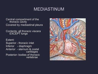
- 3. MEDIASTINUM Central compartment of the thoracic cavity Covered by mediastinal pleura Contents: all thoracic viscera EXCEPT lungs Extent: Superior - thoracic inlet Inferior - diaphragm Anterior - sternum & costal cartilages Posterior- bodies of thoracic vertebrae
- 4. MEDIASTINUM Surrounded by blood and lymphatic vessels Lymph nodes, nerves and adipose tissues Looseness of structures enable mediastinum to accommodate changes in movement, volume & pressure in the thoracic cavity
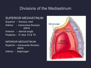
- 5. Divisions of the Mediastinum SUPERIOR MEDIASTINUM Superior - thoracic inlet Inferior - transverse thoracic plane Anterior - sternal angle Posterior - IV disc T4 & T5 INFERIOR MEDIASTINUM Superior ŌĆō transverse thoracic plane Inferior - diaphragm
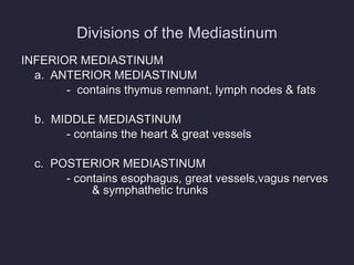
- 6. Divisions of the Mediastinum INFERIOR MEDIASTINUM a. ANTERIOR MEDIASTINUM - contains thymus remnant, lymph nodes & fats b. MIDDLE MEDIASTINUM - contains the heart & great vessels c. POSTERIOR MEDIASTINUM - contains esophagus, great vessels,vagus nerves & symphathetic trunks
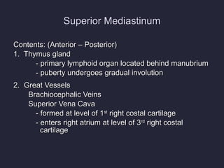
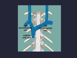
- 7. Superior Mediastinum Contents: (Anterior ŌĆō Posterior) 1. Thymus gland - primary lymphoid organ located behind manubrium - puberty undergoes gradual involution 2. Great Vessels Brachiocephalic Veins Superior Vena Cava - formed at level of 1 st right costal cartilage - enters right atrium at level of 3 rd right costal cartilage
- 8. ╠²
- 9. ╠²
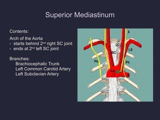
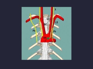
- 10. Superior Mediastinum Contents: Arch of the Aorta - starts behind 2 nd right SC joint - ends at 2 nd left SC joint Branches: Brachiocephalic Trunk Left Common Carotid Artery Left Subclavian Artery
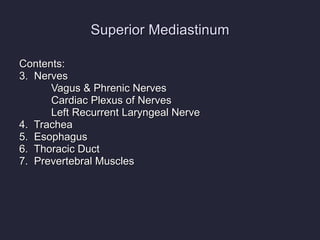
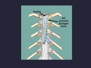
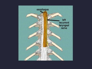
- 11. Superior Mediastinum Contents: 3. Nerves Vagus & Phrenic Nerves Cardiac Plexus of Nerves Left Recurrent Laryngeal Nerve 4. Trachea 5. Esophagus 6. Thoracic Duct 7. Prevertebral Muscles
- 12. ╠²
- 13. ╠²
- 14. ╠²
- 15. ╠²
- 16. ╠²
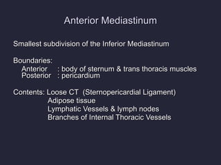
- 17. Anterior Mediastinum Smallest subdivision of the Inferior Mediastinum Boundaries: Anterior : body of sternum & trans thoracis muscles Posterior : pericardium Contents: Loose CT (Sternopericardial Ligament) Adipose tissue Lymphatic Vessels & lymph nodes Branches of Internal Thoracic Vessels
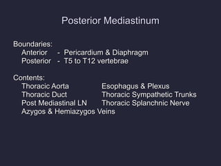
- 18. Posterior Mediastinum Boundaries: Anterior - Pericardium & Diaphragm Posterior - T5 to T12 vertebrae Contents: Thoracic Aorta Esophagus & Plexus Thoracic Duct Thoracic Sympathetic Trunks Post Mediastinal LN Thoracic Splanchnic Nerve Azygos & Hemiazygos Veins
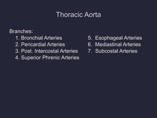
- 19. Thoracic Aorta Branches: 1. Bronchial Arteries 5. Esophageal Arteries 2. Pericardial Arteries 6. Mediastinal Arteries 3. Post. Intercostal Arteries 7. Subcostal Arteries 4. Superior Phrenic Arteries
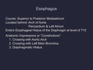
- 20. Esophagus Course: Superior to Posterior Mediastinum Located behind: Arch of Aorta Pericardium & Left Atrium Enters Esophageal Hiatus of the Diaphragm at level of T10 Anatomic Impressions or ŌĆ£ConstrictionsŌĆØ: 1. Crossing with Aortic Arch 2. Crossing with Left Main Bronchus 3. Diaphragmatic Hiatus
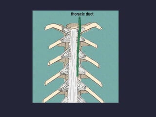
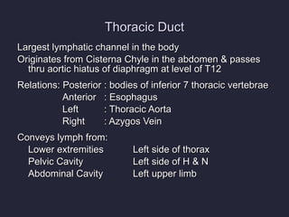
- 21. Thoracic Duct Largest lymphatic channel in the body Originates from Cisterna Chyle in the abdomen & passes thru aortic hiatus of diaphragm at level of T12 Relations: Posterior : bodies of inferior 7 thoracic vertebrae Anterior : Esophagus Left : Thoracic Aorta Right : Azygos Vein Conveys lymph from: Lower extremities Left side of thorax Pelvic Cavity Left side of H & N Abdominal Cavity Left upper limb
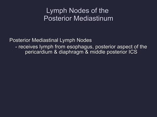
- 22. Lymph Nodes of the Posterior Mediastinum Posterior Mediastinal Lymph Nodes - receives lymph from esophagus, posterior aspect of the pericardium & diaphragm & middle posterior ICS
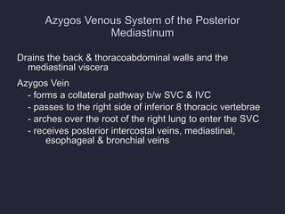
- 23. Azygos Venous System of the Posterior Mediastinum Drains the back & thoracoabdominal walls and the mediastinal viscera Azygos Vein - forms a collateral pathway b/w SVC & IVC - passes to the right side of inferior 8 thoracic vertebrae - arches over the root of the right lung to enter the SVC - receives posterior intercostal veins, mediastinal, esophageal & bronchial veins
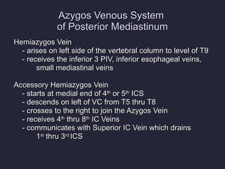
- 24. Azygos Venous System of Posterior Mediastinum Hemiazygos Vein - arises on left side of the vertebral column to level of T9 - receives the inferior 3 PIV, inferior esophageal veins, small mediastinal veins Accessory Hemiazygos Vein - starts at medial end of 4 th or 5 th ICS - descends on left of VC from T5 thru T8 - crosses to the right to join the Azygos Vein - receives 4 th thru 8 th IC Veins - communicates with Superior IC Vein which drains 1 st thru 3 rd ICS
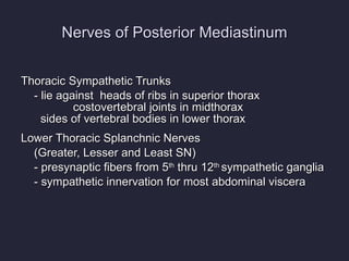
- 25. Nerves of Posterior Mediastinum Thoracic Sympathetic Trunks - lie against heads of ribs in superior thorax costovertebral joints in midthorax sides of vertebral bodies in lower thorax Lower Thoracic Splanchnic Nerves (Greater, Lesser and Least SN) - presynaptic fibers from 5 th thru 12 th sympathetic ganglia - sympathetic innervation for most abdominal viscera
- 26. ItŌĆÖs FREE to join. http://www.examville.com