The nose and paranasal sinuses have complex anatomy. The nose is divided into an external cartilaginous portion and internal bony nasal cavities separated by a nasal septum. The nasal cavities contain turbinates and drain into various meatuses. Several pairs of paranasal sinuses surround the nasal cavities. The sinuses are lined with ciliated respiratory epithelium and drain into the nasal cavities. The nose has roles in breathing, warming, and filtering air as well as detecting smells. Congenital anomalies like choanal atresia can cause nasal obstruction.
The document provides an overview of the anatomy of the nose and paranasal sinuses. It describes the external nose, nasal cavity, and paranasal sinuses. The nasal cavity has a floor, roof, medial wall and lateral walls. The lateral walls have three projections called conchae that divide the nasal cavity into inferior, middle, and superior meatuses. The paranasal sinuses include the maxillary, frontal, sphenoid, and ethmoid sinuses. They communicate with the nasal cavity and have functions such as resonating voice and reducing skull weight.
The document summarizes the anatomy of the nose. It describes the external nose, nasal cavity, and structures within including bones, cartilages, walls, openings, blood supply, nerves and lymph drainage. Key points are the pyramidal external nose composed of bones and cartilage, the nasal cavity divided into three regions and outlined by four walls, and various sinuses and structures that open into the nasal cavity.
Dentist in pune.(BDS. MDS) - Dr. Amit T. Suryawanshi. Nose & Paranasal sinuses.All Good Things
油
Dentist in pune. (BDS. MDS) - Dr. Amit T. Suryawanshi. Seminar- Nose & Paranasal sinuses.
Email ID- amitsuryawanshi999@gmail.com
Contact -Ph no.-9405622455
Subscribe our channel on youtube - Copy and paste this URL. https://www.youtube.com/channel/UC_gylEXTrjmEbbOTSXjuZ4Q/videos?view_as=public
Follow us on slideshare
The nasal cavity is divided by the nasal septum into left and right cavities. Each cavity contains 4 passages formed by the nasal conchae: the sphenoethmoidal recess, superior meatus, middle meatus, and inferior meatus. The nasal cavities are lined with mucous membrane and contain paranasal sinuses. Epistaxis or nosebleeds can occur due to various causes like trauma, infections, or anatomical abnormalities. Posterior nosebleeds from Woodruff's plexus are difficult to treat due to its inaccessible location.
The document provides an overview of the anatomy of the nose. It discusses the external and internal structures of the nose, including the nasal cavity and paranasal sinuses. The external nose has bony and cartilaginous parts that provide structure. Internally, the nasal cavity is divided by the nasal septum and lined by various types of epithelium. The document outlines the blood supply, nerve innervation, lymphatic drainage and musculature of the nose. It provides details on the four paired paranasal sinuses within the facial bones that are connected to the nasal cavity.
The document describes the anatomy of the nose and paranasal sinuses. It details the external nose, nasal cavity, and paranasal sinuses. The nasal cavity is divided by the nasal septum into right and left halves and has a floor, roof, lateral walls, and medial wall. It is connected to the paranasal sinuses which include the maxillary, frontal, ethmoid, and sphenoid sinuses. The document outlines the openings, blood supply, nerve supply, and lymph drainage of the nose and paranasal sinuses.
The document discusses the anatomy of the nose and paranasal sinuses. It describes the external nose, nasal cavity, and paranasal sinuses. The nasal cavity contains the floor, roof, lateral and medial walls. It is divided by the nasal septum into right and left passages. The paranasal sinuses include the maxillary, frontal, sphenoid, and ethmoid sinuses. They open into the nasal cavity and drain mucus through their openings. The document outlines the blood supply, nerve supply, and lymph drainage of the nose and sinuses.
The document discusses the anatomy of the nose and paranasal sinuses. It describes the external nose, nasal cavity, and paranasal sinuses. The nasal cavity is divided by the nasal septum into right and left halves and contains three meatuses - the inferior, middle, and superior. The four paranasal sinuses are the maxillary, frontal, sphenoid, and ethmoid sinuses. Each sinus opens into a meatus of the nasal cavity and drains mucus through small openings. The document outlines the bones, blood supply, nerve supply, and functions of the nose and paranasal sinuses.
The document summarizes the anatomy of the nose and paranasal sinuses. It describes the external nose, nasal cavity, paranasal sinuses, blood supply, nerve supply, and functions of the nose. The external nose has a bony and cartilaginous framework that provides structure. Internally, the nasal cavity is divided by the nasal septum and contains the paranasal sinuses which include the maxillary, frontal, ethmoid, and sphenoid sinuses. The nose functions include filtration, warming and humidifying air, olfaction, and vocal resonance.
The framework of the nose consists of bone and cartilage. Two small nasal bones and extensions of the maxillae form the bridge of the nose, which is the bony portion. The remainder of the framework is cartilage and is the flexible portion. Connective tissue and skin cover the framework.
Air enters the nasal cavity from the outside through two openings: the nostrils or external nares. The openings from the nasal cavity into the pharynx are the internal nares. Nose hairs at the entrance to the nose trap large inhaled particles.
Paranasal sinuses are air-filled cavities in the frontal, maxilae, ethmoid, and sphenoid bones. These sinuses, which have the same names as the bones in which they are located, surround the nasal cavity and open into it. They function to reduce the weight of the skull, to produce mucus, and to influence voice quality by acting as resonating chambers.
This document provides an overview of the nasal septum and paranasal sinuses. It describes the anatomy of the external nose, nasal cavity, and paranasal sinuses. Key points include that the nasal septum divides the nose into right and left halves, the nasal cavity has floors, walls and openings that connect to the paranasal sinuses, and the paranasal sinuses include the maxillary, frontal, sphenoid and ethmoid sinuses within facial bones. Clinical correlations regarding examining and treating conditions like sinusitis are also discussed.
The nose has several functions including smelling, breathing, filtering air, and draining secretions. It is divided into the external nose and nasal cavity. The nasal cavity contains convoluted bones and cartilages that warm and humidify air before it reaches the lungs. It is lined with mucosa and divided by the nasal septum into left and right sides. The nasal cavity is further divided by projections called conchae into passages and recesses where various sinuses open. Infections can spread from the nasal cavity to nearby areas like the sinuses, ears, or eyes.
The Respiratory System in the Head and NeckHadi Munib
油
The document describes the anatomy and structures of the respiratory system in the head and neck region, including the nose, nasal cavity, paranasal sinuses, pharynx, and larynx. It discusses the external features, internal structures, blood supply, nerve innervation, and functions of these areas. The nasal cavity is divided by the septum and contains three meatuses for drainage of the paranasal sinuses. The larynx contains cartilages like the thyroid and cricoid that support the vocal cords.
The nose has several important functions including respiration, olfaction, conditioning of inspired air, and protection of the lower airways. It has external and internal structures. The external nose consists of bone and cartilage while the internal nose includes nasal cavities divided by nasal conchae into passages that lead to paranasal sinuses. The nasal mucosa conditions air and supports smell. Olfactory neurons detect smells and relay signals to brain areas involved in processing odors.
Nose, nasal cavity and paranasal sinuses.pptxSundip Charmode
油
This document discusses the nose, nasal cavity, and paranasal sinuses. It begins by outlining the boundaries and features of the nasal cavity, including its roof, floor, and lateral walls. It then describes the nasal septum in detail, including its bony, cartilaginous, and vascular components. Finally, it briefly introduces the paranasal sinuses, noting their locations, characteristics, and functions in humidifying and warming inhaled air.
Nose is the part of respiratory system. External nose and nasal cavity. The visible portion that project from the face. It's skeleton is mainly cartilaginous ( small bony contributions are present). The superior bony part of the nose, including it's root, is covered by thin skin. Nasal septum has bony part vomer bone . Sometimes the deviation is so severe that the nasal septum is in contact with the lateral wall of the nasal cavity and often obstruct breathing or except snoring........................
Boundaries of the nasal cavity and often do I get to know about you guys are present for the nasal cavity and often do I need some of you thank god I was in my heart and soul mate is the nasal cavity and I can is wider than the roof. Inferior concha is the longest and border and is formed by an independent bone covered by
The Nose and nasal cavity, anatomy, and clinical diseases of nasal cavity and...HamzehKYacoub
油
Nasal cavity is the most superior part of the respiratory system.
Blood supply of nose and Cavernous Sinus.
Epistaxis causes and locations.
Allergic Rhinitis and Non-allergic rhinitis with eosinophilia (NARES).
Ostiomeatal complex (OMC)
Sinusitis.
Nasal polyps.
Headaches types.
Neuralgia.
The nasal cavity and paranasal air sinuses are described. The nasal cavity has 3 regions and is separated from other structures by bones. Each side has a roof, floor, medial and lateral wall. The lateral wall contains conchae that increase surface area. Paranasal sinuses include the frontal, ethmoidal, sphenoidal and maxillary sinuses. They are described in terms of location, openings, blood supply and innervation.
The nose has several functions including smelling, breathing, filtering air, and draining secretions. It is made up of an external cartilage portion and internal nasal cavities divided by a nasal septum. The nasal cavities contain turbinate bones that increase their surface area and are lined with mucosa. They open anteriorly through the nostrils and posteriorly into the nasopharynx. The nose receives blood supply from several arteries and drains venous blood. Lymph drains from the nose to cervical lymph nodes. The nose has both general sensory innervation and a specialized olfactory area in the upper nasal cavity.
Non-Invasive ICP Monitoring for NeurosurgeonsDhaval Shukla
油
This presentation delves into the latest advancements in non-invasive intracranial pressure (ICP) monitoring techniques, specifically tailored for neurosurgeons. It covers the importance of ICP monitoring in clinical practice, explores various non-invasive methods, and discusses their accuracy, reliability, and clinical applications. Attendees will gain insights into the benefits of non-invasive approaches over traditional invasive methods, including reduced risk of complications and improved patient outcomes. This comprehensive overview is designed to enhance the knowledge and skills of neurosurgeons in managing patients with neurological conditions.
Invasive systems are commonly used for monitoring intracranial pressure (ICP) in traumatic brain injury (TBI) and are considered the gold standard. The availability of invasive ICP monitoring is heterogeneous, and in low- and middle-income settings, these systems are not routinely employed due to high cost or limited accessibility. The aim of this presentation is to develop recommendations to guide monitoring and ICP-driven therapies in TBI using non-invasive ICP (nICP) systems.
More Related Content
Similar to anatomy of nose¶nasal sinuses.pdf (20)
The document discusses the anatomy of the nose and paranasal sinuses. It describes the external nose, nasal cavity, and paranasal sinuses. The nasal cavity contains the floor, roof, lateral and medial walls. It is divided by the nasal septum into right and left passages. The paranasal sinuses include the maxillary, frontal, sphenoid, and ethmoid sinuses. They open into the nasal cavity and drain mucus through their openings. The document outlines the blood supply, nerve supply, and lymph drainage of the nose and sinuses.
The document discusses the anatomy of the nose and paranasal sinuses. It describes the external nose, nasal cavity, and paranasal sinuses. The nasal cavity is divided by the nasal septum into right and left halves and contains three meatuses - the inferior, middle, and superior. The four paranasal sinuses are the maxillary, frontal, sphenoid, and ethmoid sinuses. Each sinus opens into a meatus of the nasal cavity and drains mucus through small openings. The document outlines the bones, blood supply, nerve supply, and functions of the nose and paranasal sinuses.
The document summarizes the anatomy of the nose and paranasal sinuses. It describes the external nose, nasal cavity, paranasal sinuses, blood supply, nerve supply, and functions of the nose. The external nose has a bony and cartilaginous framework that provides structure. Internally, the nasal cavity is divided by the nasal septum and contains the paranasal sinuses which include the maxillary, frontal, ethmoid, and sphenoid sinuses. The nose functions include filtration, warming and humidifying air, olfaction, and vocal resonance.
The framework of the nose consists of bone and cartilage. Two small nasal bones and extensions of the maxillae form the bridge of the nose, which is the bony portion. The remainder of the framework is cartilage and is the flexible portion. Connective tissue and skin cover the framework.
Air enters the nasal cavity from the outside through two openings: the nostrils or external nares. The openings from the nasal cavity into the pharynx are the internal nares. Nose hairs at the entrance to the nose trap large inhaled particles.
Paranasal sinuses are air-filled cavities in the frontal, maxilae, ethmoid, and sphenoid bones. These sinuses, which have the same names as the bones in which they are located, surround the nasal cavity and open into it. They function to reduce the weight of the skull, to produce mucus, and to influence voice quality by acting as resonating chambers.
This document provides an overview of the nasal septum and paranasal sinuses. It describes the anatomy of the external nose, nasal cavity, and paranasal sinuses. Key points include that the nasal septum divides the nose into right and left halves, the nasal cavity has floors, walls and openings that connect to the paranasal sinuses, and the paranasal sinuses include the maxillary, frontal, sphenoid and ethmoid sinuses within facial bones. Clinical correlations regarding examining and treating conditions like sinusitis are also discussed.
The nose has several functions including smelling, breathing, filtering air, and draining secretions. It is divided into the external nose and nasal cavity. The nasal cavity contains convoluted bones and cartilages that warm and humidify air before it reaches the lungs. It is lined with mucosa and divided by the nasal septum into left and right sides. The nasal cavity is further divided by projections called conchae into passages and recesses where various sinuses open. Infections can spread from the nasal cavity to nearby areas like the sinuses, ears, or eyes.
The Respiratory System in the Head and NeckHadi Munib
油
The document describes the anatomy and structures of the respiratory system in the head and neck region, including the nose, nasal cavity, paranasal sinuses, pharynx, and larynx. It discusses the external features, internal structures, blood supply, nerve innervation, and functions of these areas. The nasal cavity is divided by the septum and contains three meatuses for drainage of the paranasal sinuses. The larynx contains cartilages like the thyroid and cricoid that support the vocal cords.
The nose has several important functions including respiration, olfaction, conditioning of inspired air, and protection of the lower airways. It has external and internal structures. The external nose consists of bone and cartilage while the internal nose includes nasal cavities divided by nasal conchae into passages that lead to paranasal sinuses. The nasal mucosa conditions air and supports smell. Olfactory neurons detect smells and relay signals to brain areas involved in processing odors.
Nose, nasal cavity and paranasal sinuses.pptxSundip Charmode
油
This document discusses the nose, nasal cavity, and paranasal sinuses. It begins by outlining the boundaries and features of the nasal cavity, including its roof, floor, and lateral walls. It then describes the nasal septum in detail, including its bony, cartilaginous, and vascular components. Finally, it briefly introduces the paranasal sinuses, noting their locations, characteristics, and functions in humidifying and warming inhaled air.
Nose is the part of respiratory system. External nose and nasal cavity. The visible portion that project from the face. It's skeleton is mainly cartilaginous ( small bony contributions are present). The superior bony part of the nose, including it's root, is covered by thin skin. Nasal septum has bony part vomer bone . Sometimes the deviation is so severe that the nasal septum is in contact with the lateral wall of the nasal cavity and often obstruct breathing or except snoring........................
Boundaries of the nasal cavity and often do I get to know about you guys are present for the nasal cavity and often do I need some of you thank god I was in my heart and soul mate is the nasal cavity and I can is wider than the roof. Inferior concha is the longest and border and is formed by an independent bone covered by
The Nose and nasal cavity, anatomy, and clinical diseases of nasal cavity and...HamzehKYacoub
油
Nasal cavity is the most superior part of the respiratory system.
Blood supply of nose and Cavernous Sinus.
Epistaxis causes and locations.
Allergic Rhinitis and Non-allergic rhinitis with eosinophilia (NARES).
Ostiomeatal complex (OMC)
Sinusitis.
Nasal polyps.
Headaches types.
Neuralgia.
The nasal cavity and paranasal air sinuses are described. The nasal cavity has 3 regions and is separated from other structures by bones. Each side has a roof, floor, medial and lateral wall. The lateral wall contains conchae that increase surface area. Paranasal sinuses include the frontal, ethmoidal, sphenoidal and maxillary sinuses. They are described in terms of location, openings, blood supply and innervation.
The nose has several functions including smelling, breathing, filtering air, and draining secretions. It is made up of an external cartilage portion and internal nasal cavities divided by a nasal septum. The nasal cavities contain turbinate bones that increase their surface area and are lined with mucosa. They open anteriorly through the nostrils and posteriorly into the nasopharynx. The nose receives blood supply from several arteries and drains venous blood. Lymph drains from the nose to cervical lymph nodes. The nose has both general sensory innervation and a specialized olfactory area in the upper nasal cavity.
Non-Invasive ICP Monitoring for NeurosurgeonsDhaval Shukla
油
This presentation delves into the latest advancements in non-invasive intracranial pressure (ICP) monitoring techniques, specifically tailored for neurosurgeons. It covers the importance of ICP monitoring in clinical practice, explores various non-invasive methods, and discusses their accuracy, reliability, and clinical applications. Attendees will gain insights into the benefits of non-invasive approaches over traditional invasive methods, including reduced risk of complications and improved patient outcomes. This comprehensive overview is designed to enhance the knowledge and skills of neurosurgeons in managing patients with neurological conditions.
Invasive systems are commonly used for monitoring intracranial pressure (ICP) in traumatic brain injury (TBI) and are considered the gold standard. The availability of invasive ICP monitoring is heterogeneous, and in low- and middle-income settings, these systems are not routinely employed due to high cost or limited accessibility. The aim of this presentation is to develop recommendations to guide monitoring and ICP-driven therapies in TBI using non-invasive ICP (nICP) systems.
Local Anesthetic Use in the Vulnerable PatientsReza Aminnejad
油
Local anesthetics are a cornerstone of pain management, but their use requires special consideration in vulnerable groups such as pediatric, elderly, diabetic, or obese patients. In this presentation, well explore how factors like age and physiology influence local anesthetics' selection, dosing, and safety. By understanding these differences, we can optimize patient care and minimize risks.
PERSONALITY DEVELOPMENT & DEFENSE MECHANISMS.pptxPersonality and environment:...ABHAY INSTITUTION
油
Personality theory is a collection of ideas that explain how a person's personality develops and how it affects their behavior. It also seeks to understand how people react to situations, and how their personality impacts their relationships.
Key aspects of personality theory
Personality traits: The characteristics that make up a person's personality.
Personality development: How a person's personality develops over time.
Personality disorders: How personality theories can be used to study personality disorders.
Personality and environment: How a person's personality is influenced by their environment.
Solubilization in Pharmaceutical Sciences: Concepts, Mechanisms & Enhancement...KHUSHAL CHAVAN
油
This presentation provides an in-depth understanding of solubilization and its critical role in pharmaceutical formulations. It covers:
Definition & Mechanisms of Solubilization
Role of surfactants, micelles, and bile salts in drug solubility
Factors affecting solubilization (pH, polarity, particle size, temperature, etc.)
Methods to enhance drug solubility (Buffers, Co-solvents, Surfactants, Complexation, Solid Dispersions)
Advanced approaches (Polymorphism, Salt Formation, Co-crystallization, Prodrugs)
This resource is valuable for pharmaceutical scientists, formulation experts, regulatory professionals, and students interested in improving drug solubility and bioavailability.
Rabies Bali 2008-2020_WRD Webinar_WSAVA 2020_Final.pptxWahid Husein
油
A decade of rabies control programmes in Bali with support from FAO ECTAD Indonesia with Mass Dog Vaccination, Integrated Bite Case Management, Dog Population Management, and Risk Communication as the backbone of the programmes
Cardiac Arrhythmia definition, classification, normal sinus rhythm, characteristics , types and management with medical ,surgical & nursing, health education and nursing diagnosis for paramedical students.
Creatines Untold Story and How 30-Year-Old Lessons Can Shape the FutureSteve Jennings
油
Creatine burst into the public consciousness in 1992 when an investigative reporter inside the Olympic Village in Barcelona caught wind of British athletes using a product called Ergomax C150. This led to an explosion of interest in and questions about the ingredient after high-profile British athletes won multiple gold medals.
I developed Ergomax C150, working closely with the late and great Dr. Roger Harris (1944 2024), and Prof. Erik Hultman (1925 2011), the pioneering scientists behind the landmark studies of creatine and athletic performance in the early 1990s.
Thirty years on, these are the slides I used at the Sports & Active Nutrition Summit 2025 to share the story, the lessons from that time, and how and why creatine will play a pivotal role in tomorrows high-growth active nutrition and healthspan categories.
Chair and Presenters Sara A. Hurvitz, MD, FACP, Carey K. Anders, MD, FASCO, and Vyshak Venur, MD, discuss metastatic HER2-positive breast cancer in this CME/NCPD/CPE/AAPA/IPCE activity titled Fine-Tuning the Selection and Sequencing of HER2-Targeting Therapies in HER2-Positive MBC With and Without CNS Metastases: Expert Guidance on How to Individualize Therapy Based on Latest Evidence, Disease Features, Treatment Characteristics, and Patient Needs and Preferences. For the full presentation, downloadable Practice Aids, and complete CME/NCPD/CPE/AAPA/IPCE information, and to apply for credit, please visit us at https://bit.ly/4f8sUs7. CME/NCPD/CPE/AAPA/IPCE credit will be available until March 2, 2026.
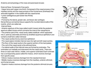
1. Anatomy and physiology of the nose and paranasal sinuses
External Nose: Composed of two parts:
Upper bony part (upper one third): Composed of the nasal process of the
maxillary bones and the nasal process of the frontal bone (forehead) that
articulate together in the middle with two nasal bones
Lower cartilaginous part (lower two thirds)
Flexible
Framed by the lateral, greater alar, and lesser alar cartilages
Dense fibrous connective tissue supports the rounded lateral walls of the
nostrils, which are called ala
Internal Nose
Anterior aperture of the nose called nostril (nares) while the posterior
aperture called choana which connects the nose to the nasopharynx
The anterior part of the nasal cavity called vestibule which separated
into 2 parts by collumella and lines by stratified squamous epithelium and
contains small hairs called vibrissae
The nasal cavities line by columnar epithelium
The nasal septum divides internal nose into right and left nasal cavities.
The floor of the nasal cavity is the superior portion of the hard palate.
The roof of the nasal cavity is the ethmoid bone.
The lateral walls of the internal nose are formed by turbinates. The
turbinates curl medially and downward into the nasal cavity. There are
three on each side inferior, middle, and superior turbinates.Each turbinate
has a corresponding space called the meatus, which receives drainage
from specific areas:
Inferior meatus receives drainage from the nasolacrimal duct.
Middle meatus receives drainage from the maxillary, anterior ethmoid,
and frontal sinuses.
Superior meatus receives drainage from the posterior ethmoid sinuses.
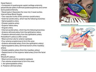
2. Nasal Septum:
Composed of quadrangular septal cartilage anteriorly,
perpendicular plate of ethmoid posterosuperiorly and vomer
bone posteroinferiorly.
Nasal septum Separates the nose into 2 nasal cavities.
Internal Nose Blood Supply
Two vascular trunks of the common carotid artery:
External carotid artery, which has the following branches:
Sphenopalatine artery
Greater palatine artery
Superior labial artery
Angular artery
Internal carotid artery, which has the following branches:
Anterior ethmoid artery from the ophthalmic artery
Posterior ethmoid artery from the ophthalmic artery
Kiesselbachs plexus (Littles area):
Most common site for anterior epistaxis
Four arteries anastomose to form this area:
Anterior ethmoidal artery (from the ophthalmic artery)
Sphenopalatine artery (terminal branch of the maxillary
artery)
Greater palatine artery (from the maxillary artery)
Septal branch of the superior labial artery (from the facial
artery)
Woodruffs plexus:
Most common site for posterior epistaxis
Two arteries anastomose to form this area:
1. the sphenopalatine artery
2. Posterior ethmoid artery
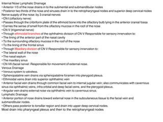
3. Internal Nose Lymphatic Drainage
Anterior 1/3 of the nose drains in to the submental and submandibular nodes
Posterior two thirds of the nose and sinuses drain in to the retropharyngeal nodes and superior deep cervical nodes
Nerve supply of the nose: by 3 cranial nerves
CN I (olfactory nerve)
Passes through the cribriform plate of the ethmoid bone into the olfactory bulb lying in the anterior cranial fossa
Carries the sense of smell from the olfactory mucosa in the roof of the nose
CN V (trigeminal nerve)
Through ethmoidal branches of the ophthalmic division of CN V:Responsible for sensory innervation to:
The lining of the anterior part of the nasal cavity
To the surrounding olfactory mucosa in the roof of the nose
To the lining of the frontal sinus
Through Maxillary division of CN V:Responsible for sensory innervation to:
The lateral wall of the nose
The nasal septum
The maxillary sinus
CN VII (facial nerve|: Responsible for movement of external nose.
Venous Drainage
Venous system is valveless.
Sphenopalatine vein drains via sphenopalatine foramen into pterygoid plexus.
Ethmoidal veins drain into superior ophthalmic vein
Anterior facial vein drains through common facial vein to internal jugular vein; also communicates with cavernous
sinus via ophthalmic veins, infra orbital and deep facial veins, and the pterygoid plexus.
Angular vein drains external nose via ophthalmic vein to cavernous sinus.
Lymphatic Drainage
Anterior portion of nose drains toward external nose in the subcutaneous tissue to the facial vein and
submandibular nodes.
Others pass posterior to tonsillar region and drain into upper deep cervical nodes.
Most drain into pharyngeal plexus and then to the retropharyngeal nodes
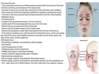
4. Paranasal Sinuses
The paranasal sinuses are air-filled spaces located within the bones of the skull
and face and they are centered on the nasal cavity
Includes 4 pairs one on each side: sphenoid, frontal, ethmoid, and maxillary
All sinuses are lined with respiratory epithelium capable of producing mucus.
The cilia in the sinus cavities help to empty secretions into the nasal cavities.
Maxillary Sinus
Present at birth
Largest of the paranasal sinuses (15 ml its volume)
Lateral wall of the nose separates the sinus from the nose
Roof of the sinus is also the floor of the orbit
Drains into the middle meatus of the nasal cavity
Roots of the posterior molar teeth may extend into the floor of the sinus
The anterior maxillary sinus wall houses the infraorbital nerve, which runs along
the roof of the sinus and supplies sensory innervation to the lower eyelid, the
side of the nose, and the upper lip.
Ethmoidal sinus
Paired bony scaffolds, connected by cribriformplate
Boundaries:
Lamina papyracea of orbit
Orbital process of the frontal bone
Middle and superior turbinates medially
Lateral cribriform plate lamella (also medially)
Sphenoid sinus posterioriy
Nerve supply: anterior and posterior ethmoidal nerves
Blood supply: anterior and posterior ethmoidal arteries from the ophthalmic A.
Ant. cells: drain into middle meatus, the Post. cells drain into superior meatus.
5. Frontal sinus:-
. abscent at birth
Occupy the frontal bone, 2 in number
Relations: The floor of the frontal sinus corresponds to the roof of the orbit. The frontal
sinus is defined anteriorly by the thicker anterior table of the frontal bone and the thinner
posterior table which separates the frontal sinus from the anterior horns of the frontal lobes
of the brain.
The pneumatization patterns of the frontal sinus widely vary. An intersinus septation
generally divides the sinus, albeit asymmetrically.
5-15% of adults, the frontal sinus is completely aplastic.
The frontal sinus ostium opens inferiorly into middle meatus
Sphenoid Sinus
Occupies sphenoid bone, 2 in number separated by Intersinus septum (not usually midline)
Post-ethmoid A and N give vascular and sensory supplies, respectively
Ostium ~1 cm above choana, and drains into sphenoethmoidal recess
Relations: 1. Pituitary fossa and middle cranial fossa superiorly 2. Cavernous sinus and ICA
laterally 3. Pons and posterior cranial fossa posteriorly 4. Optic nerve lies next to or even
within the sinus 5. Pterygoid canal with nerve inferiorly. Sphenoid sinus forms the roof of
nasopharynx.
Ostiomeatal Complex (OMC)
Area that drains the frontal, anterior ethmoid, and maxillary sinuses.
The OMC drains into the middle meatus and then into the nasopharynx
Functions of the Nose
Airway: conduit for air
Filtration: trap and remove airborne particulate matter
Humidification: increases relative humidity
Heating: provides radiant heat of inspired air
Nasal reflex: causes periodic nasal congestion, rhinorrhea, or sneezing
A. Postural reflex: increased congestion with supine position; congestion on the side
of dependence upon lying on the side
B. Hot or cold temperature reflex: sneezing upon sudden exposure of skin to
dramatic temperature extremes
Chemosensation: detects irritants and temperature changes
Olfaction
6. Functions of paranasal sinuses
The following are the possible functions of paranasal sinuses:
Imparting resonance to the voice.
Humidification and warming of inspired air.
Increasing the olfactory area of nose.
Providing thermal insulation to vital parts of head.
Shock absorber function.
Aiding facial growth: frontal and maxillary sinuses were designed to
assist forward and downward growth of the face.
Lightening the skull bones to maintain head position.
Secreting mucous to moisten the nasal cavity.
Aiding nasal cavity immune defense and production of nitric oxide.
CONGENITAL ANOMALIES
CHOANAL ATRESIA
Choanal atresia (CA) is a rare congenital disorder where the back of the nasal
passage (choana) is blocked, usually by abnormal bony or soft tissue. Although it
is rare, it is the most common cause of nasal obstruction in neonates and young
infants. It can be
unilateral or bilateral. When it is bilateral, it can cause immediate respiratory
compromise in the newborn and is considered a medical emergency. The
diagnosis can be made on physical examination by trying to pass a small catheter
through the patients nose. Surgery is the treatment of choice.
Congenital Midline masses include 1. Dermoid cyst 2. Glioma 3. Encephalocele:
Congenital herniation of central nervous system (CNS) tissue through skull base