


![Low Tidal Volume Ventilation
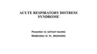
• Calculate ideal body weight (IBW) in pounds
1. Males- 106 + [ 6 x (height in inches – 60 inches)]
2. Females- 105 + [ 5 x (height in inches – 60 inches)]
• Convert into kg by multiplying with 0.453
• Set initial tidal volume to 6 ml/kg IBW
• Set respiratory rate to < 35 per minute to match baseline minute ventilation](https://image.slidesharecdn.com/ards-satya-220821142805-1bb19916/85/ARDS-Satya-pptx-37-320.jpg)


More Related Content
Similar to ARDS- Satya.pptx (20)
Recently uploaded (20)


ARDS- Satya.pptx
- 1. ACUTE RESPIRATORY DISTRESS SYNDROME Presenter: Dr. SATYAJIT HAJONG Moderator: Dr .Th. BROJENDRO
- 2. The Berlin Definition Of ARDS • A clinical syndrome of acute onset dyspnea , severe hypoxemia, diffuse pulmonary infiltrates and decreased respiratory system compliance leading to respiratory failure in absence of evidence of congestive heart failure
- 3. ÔÅΩFirst described in 1967 by Ashbaugh and Petty in The Lancet ÔÅΩIncidence (per 1,00,000 person years)- ÔÅΩTotal- 86.2 ÔÅΩModerate and severe- 64 ÔÅΩDramatically increases between age 75 to 84 yrs - 306
- 4. OLDER DEFINITION According to American European Consensus Conference – acute onset of illness , bilateral chest radiographic infiltrates consistent with pulmonary Oedema , poor systemic oxygenation and absence of evidence for left atrial hypertension • PaO2/FiO2 if is < 300- Acute Lung Injury, if < 200 then ARDS • Cardiogenic pulmonary oedema must be excluded by clinical criteria/ PCWP lower than 18 mm Hg • Limitations- subjective variability of CXR interpretation, variable PaO2/FiO2 with PEEP
- 5. THE BERLIN DEFINITION OF ARDS 2012 Clinical variables Parameters Onset Within 1 week of inciting event Chest radiograph (CXR/ CT chest) B/L opacities, not explained by effusion, atelectasis or nodules Non-cardiac aetiology Respiratory failure not fully explained by cardiac failure or fluid overload Hypoxemia (PaO2/FiO2) </= 300 mmHg at PEEP >/= 5 cm H2O
- 6. ARDS Severity ARDS Severity PaO2/FiO2 in mmHg (at PEEP>/= 5cm H2O) Mortality Mild 200-300 26% Moderate 100-200 32% Severe <100 45-58%
- 7. Etiology Direct causes Indirect causes • Pneumonia(40 to 50)% • Aspiration of gastric contents • Pulmonary contusion • Fat , amniotic fluid or air embolism • Near drowning • Inhalational injury • Reperfusion injury • High altitude pulmonary oedema • Neurogenic pulmonary oedema • Re-expansion pulmonary oedema • Sepsis- most common cause • Multiple Trauma • Burns • Acute pancreatitis • Post cardiopulmonary bypass • Toxic ingestions- aspirin, TCAs • Transfusion of blood products
- 8. Factors Influencing The Risk and Mortality • Advanced age • Chronic liver disease • Hypoproteinaemia • Increased severity and extent of illness as measured by APACHE score • Hyper transfusion of blood products • Chronic alcohol abuse • Cigarette smoking • Long hospital stay prior to the development of ARDS
- 9. The natural history of ARDS is marked by three phases  1.Exudative (First 7 days)  2.Proliferative (After 7-21 days)  3.Fibrotic (After 3-4 weeks) Pathophysiology
- 10. Initial “Exudative" phase-diffuse alveolar damage within the first week
- 11. Alveolar capillary membrane(ACM) integrity is lost, interstitial and alveolus fills with proteinaceous fluid, surfactant can no longer support alveolus Ware et al. NEJM 2000; 342:1334
- 12. Direct or indirect injury to the alveolus causes alveolar macrophages to release pro- inflammatory cytokines Ware et al. NEJM 2000; 342:1334
- 13. Cytokines attract neutrophils into the alveolus and interstitum, where they damage the alveolar-capillary membrane (ACM). Ware et al. NEJM 2000; 342:1334
- 14. Most patient recover in this stage  Signs of resolution and lung repair * Neutrophil to lymphocytes predominant infiltrates * Type II pneumocytes proliferation and differentiation into type I pneumatocytes * New pulmonary surfactant Proliferative phase
- 15. Require long term airway support(mechanical/supplemental O2) Histologically-alveolar duct and interstitial fibrosis Pulmonary hypertension from intimal fibroproliferation Pulmonary fibrosis= increased mortality Fibrotic phase
- 16. Stages of ARDS EXUDATIVE PROLIFERATIVE FIBROTIC • 0-6 days • Accumulation of excess fluid in the lung • Hypoxemia is maximum at this stage • Some individuals recover from this stage • 3 stages of oedema- • Stage 1- drained by lymphatic • Stage2- interstitial • Stage 3- alveolar • 7-21 days • Connective tissue, fibroblasts proliferation • Termed as stiff lung/ shock lung • Abnormally enlarged air spaces and fibrotic tissue are increasingly apparent • > 21 days • Oxygenation improves and extubation becomes possible • Lung function continue to improve over as long as 6-12 months • Varying levels of residual fibrotic changes remain
- 17. Resolution of ARDS Removal of alveolar oedema fluid (10-20% per hour) via 5 routes- lymphatics, blood vessels, airways, pleural space and mediastinum • Mechanism- activated NaCl transporter • Protein removal- (1-2% per hour)- mostly as intact- f/b phagocytosis • Removal of interstitial fluid- in blood vessels or mediastinum • 25-30% of oedema fluid- leaves into pleural space- pleural effusion
- 20. Clinical Presentation • Acute dyspnoea within 7 days of an inciting event • Hypoxemia resistant to oxygen therapy (d/t large R->L shunt)- Need for high fraction of inspired O2(*FiO2) to maintain O2 saturation • Tachypnoea (earliest sign), tachycardia, cyanosis • Restlessness, confusion, disorientation • Hypertension or low BP when in shock • Bilateral fine crepitation on chest auscultation • Other manifestations of the underlying cause
- 22. Differential Diagnosis Most common Less frequent • Cardiogenic pulmonary oedema • Diffuse pneumonia • Alveolar haemorrhage • Acute interstitial lung disease • Hypersensitivity pneumonitis • Radiation pneumonitis • B/L lower lobe atelectasis • Severe U/L lower lobe atelectasis • Massive pulmonary embolism
- 23. Chest Radiograph in ARDS • Diffuse bilateral lung infiltrates extending to the lung margins • Early in the course infiltrates may have patchy peripheral distribution • CT scan of chest- more informative • Drawbacks- oedema may not be visible until lung water increases upto 30% and upto 12 hours after onset of dyspnoea
- 28. Chest x ray in ARDS
- 29. Arterial Blood Gas • Hypoxemia- PaO2/FiO2 <300 mm Hg • Initially respiratory alkalosis • However if ARDS is due to sepsis- metabolic acidosis with or without respiratory compensation • As the condition progresses the work of breathing increases and pCO2 begins to rise- respiratory acidosis
- 30. Broncho alveolar lavage • Most reliable to confirm or exclude ARDS • Neutrophils- in normal individuals <5% whereas in ARDS upto 80% • Total protein- lavage fluid rich in protein is evidence of inflammation • When protein in lavage fluid expressed as a fraction of serum protein concentration- • If <0.5 indicates hydrostatic oedema • If >0.7 indicates lung inflammation
- 31. Other Investigations • CBC- leukopenia/ leucocytosis, thrombocytopenia (in DIC) • RFT and LFT- deranged in acute tubular necrosis and hepatocellular injury respectively • Cytokines- IL-1, IL-6, IL-8 levels are elevated • Pro calcitonin- marker of sepsis • Von Willebrand Factor & plasma angiopoietin 2 : markers of impending ARDS with non pulmonary sepsis- poor outcome • Plasma BNP- to exclude cardiogenic pulmonary oedema • <100 pg/ml- unlikely heart failure • >500 pg/ml- heart failure is likely • Echocardiography - to exclude cardiogenic pulmonary edema
- 32. Complications COMMON OTHERS • Nosocomial pneumonia • Barotrauma • SIRS and MODS 1. Renal- 40-55% 2. Liver- reversible , enzyme changes in 95%, fulminant hepatitis in 10% 3. Myocardial depression by TNF- 10-23% 4. DIC- 26% 5. GIT- 7-30% as haemorrhage, ileus, malabsorbtion • Oxygen toxicity • Stress ulcers • Tracheal ulcerations • Deep vein thrombosis • Pulmonary embolism • Pressure sores
- 33. Management of ARDS Goals : • To diagnose and treat the precipitating cause • To maintain oxygenation • To prevent ventilator induced lung injury (VILI) • Hemodynamic management • To keep pH in normal range without compromising goal to prevent VILI • Prevention of other complications
- 35. Mechanical Ventilation • No role of NIPPV (Only one comparative study by Ferrer et.al) • To enhance patient ventilator synchrony and patient comfort by sedation, amnesia, opioid analgesia, antipyretics and pharmacological paralysis- also decreases oxygen consumption • To apply PEEP to maximize alveolar recruitment & minimize cyclic atelectasis • Wean from mechanical ventilation when patient can breathe without assisted ventilation to the earliest
- 36. 1. Maintaining adequate oxygenation- • Positive end expiratory pressure (PEEP) is employed • When utilized in sufficient amount PEEP allows lowering of FiO2 from high, potentially toxic concentrations • Lung protective mechanical ventilation- • Mechanical ventilation using limited tidal volume • The goal is to avoid injury to the alveoli 1. By overexpansion during inspiration (volu-trauma) 2. Due to repetitive opening and closing during inspiration and expiration (atelecta-trauma)
- 37. Low Tidal Volume Ventilation • Calculate ideal body weight (IBW) in pounds 1. Males- 106 + [ 6 x (height in inches – 60 inches)] 2. Females- 105 + [ 5 x (height in inches – 60 inches)] • Convert into kg by multiplying with 0.453 • Set initial tidal volume to 6 ml/kg IBW • Set respiratory rate to < 35 per minute to match baseline minute ventilation
- 38. Ventillator Mode - Volume Assist / control Tidal Volume (VT)– 6ml/kg of IBW Measure pleatue pressure (Pplat 0.5 sec inspiratory pause ) every 4hrs and after each change in PEEP and VT • Goal is to maintain plateau pressure(Pplat) </= 30 cm of H2O If Pplat rises above 30cm of H2O decrease tidal volume to 5 or to 4ml/kg IBW • If Pplat is below 25cm of H2O increase tidal volume by 1ml /kg IBW to keep Pplat >25cm of H2O
- 39. • Positive end expiratory pressure (PEEP) is employed • I:E ratio -1: to 1:3 • Maintain PaO2 : ( 50 to 60) mm of Hg • SpO2 : ( 88 to 95) % FiO2 30 to 40 % 40% 50% 60% 70% 80% 90% 100 % PEEP 5to8 8 to 14 8 to 16 10 to 20 10 to 20 14 to 22 16 to 22 18 to 25
- 40. Acidosis management If pH < 7.3 increase RR until pH >7.3 or RR 35 If pH remains < 7.3 with RR 35 consider bicarbonate infusion If pH < 7.15, VT may be increased( (Pplat may exceed 30cm of water)
- 41. Alkalosis management If pH >7.4 and paient not triggering ventillator decrease set RR but not below 6/min
- 42. Weaning from Mechanical Ventillation • Daily interruption of sedation • Daily screen for spontenous breathing trial • Give spontenous breathing trial when all of the following criteria are present • 1. FiO2<40% PEEP <8cm of H2O • 2. Not receiving neuro mascular blocking agent • 3.Patient is awake and following commands • 4.Systolic arterial pressure >90mm of Hg without vasopressors • 5.Tracheal secretions are minimal and patient have a good cough and gag reflex
- 43. Spontaneous Breathing Trial • 1. Place patient on 5mm Hg pressure support with 5mm of Hg PEEP • 2. Monitor HR , RR, SPO2 for 20 to 30 min • 3. Extubate if there is no signs of distress ( tachycardia , tachypnea , agitation , diaphoresis )
- 46. Open Lung Ventilation • Combines LTVV with enough applied PEEP • LTTV- mitigates alveolar over distention and PEEP- minimizes cyclic atelectasis • PEEP is set at least 2 cm above the lower inflection point of the pressure volume curve • If the point is uncertain- PEEP of 16 cm of H2O is to be applied • Drawback -it has the potential to cause barotrauma and decrease cardiac output
- 47. Adjuncts to lung protective ventilation 1. Permissive hypercapnia • LTVV causes reduction of CO2 elimination- allowing this to persist in favour of maintaining lung protective LTVV is known as permissive hypercapnia • Can be minimized by highest respiratory rate that does not induce auto PEEP • Also by changing from a heat & moisture exchanger to a heated humidifier • Can cause hyperventilation (brainstem stimulation)- neuromuscular blockade • Data shows arterial pCO2 levels of 60-70 mmHg is safe
- 48. 2. Recruitment manoeuvres- intermittent increase of PEEP, by maintaining CPAP of 35 to 40cm of H2O for 30 seconds 3. Prone positioning- • About 66% of the patients improve oxygenation with this • Mechanisms - 1. Increase in functional residual capacity 2. Change in regional diaphragmatic motion 3. Perfusion redistribution 4. Improve clearance of secretions
- 49. Extra Corporeal Membrane Oxygenation • Modified heart lung machine- gas exchange and circulatory support • Veno-venous(VV) ECMO- for gas exchange • Veno-arterial(VA) ECMO- for both gas exchange and circulatory support • ARDS with pneumonia is the commonest situation needing ECMO
- 50. Newer therapeutic strategies 1. Intra venous mesenchymal stromal (stem) cells(START Trial) – being tested in phase 2 clinical trial – have pleotropic protective and reparative effect in the lungs 2. Use of pulmonary vasodialators - inhaled nitric oxide and prostacyclin 3. Glucocorticoids ( methylprednisolone ) – hastens resolution of late fibroproliferative ARDS – not recommended for routine use
- 51. Fluid management ‚Ä¢ Golden rule- hydrostatic pressure to be kept as low as possible provided that oxygen delivery to the tissues is not compromised ‚Ä¢ Tailored systemic fluid restriction to achieve lowest intravascular volume that maintain adequate tissue perfusion ‚Ä¢ The lung infiltration in ARDS is an inflammatory process- diuretics don‚Äôt reduce this , but helps in lowering intra vascular volume ‚Ä¢ To maintain CVP < 4ùëêùëö ùëúùëì ùêª2ùëÇ ,MAP> 65 mmHg and Urine output > 0.5 ml/kg/hour
- 52. Nutrition • High fat and low carbohydrate diet reduces the duration of mechanical ventilation by reduction in carbon dioxide production and reduction in respiratory quotient
- 53. Other agents Agent Mechanism of action Recommendation Glucocorticoids Anti-inflammatory NO Neuromuscular blockers Promotes ventricular synchrony, decrease oxygen consumption YES Statins Antagonising inflammatory cytokines Under experimentation Beta-agonists Fluid removal by activated sodium pump via cAMP NO Macrolide antibiotics Unknown Under experimentation
- 54. Mortality and Functional Recovery Mortality- ARDS Network published in 2012 , 60 day mortality was 23% 1 year mortality is 41% 32% for mild ARDS , 58 to 60 % for moderate to severe ARDS Early death - due to the underlying cause of ARDS most commonly • Death in later course- nosocomial pneumonia and sepsis causing MODS
- 55. Mortality and Functional Recovery • Functional recovery- • Maximum lung function- by 6 months • One year after extubation over 33% survivors have normal spirometry and diffusion capacity • Most of the remaining survivors have only mild abnormalities- others low exercise capacity • Significant rate of post traumatic stress disorder in the form of depression and anexity
- 56. Conclusion • ARDS is a multisystem syndrome • Characterised by accumulation of fluid in the lungs with resulting hypoxemia and some degree of fibrotic changes • The most frequent causes - sepsis, pneumonia, aspiration and severe trauma • Treatment- supportive, ventilation and oxygenation strategies • Despite many theoretical therapies the best proven strategy to improve survival is LOW TIDAL VOLUME VENTILATION • Despite all advances mortality is still very high ranging from 26-58%
- 57. References • Fishman’s Pulmonary Diseases and Disorders 5th Edition • Harrison’s Principles of Internal Medicine 20th edition • The ICU book,3rd edition- Paul L. Marino • JAMA, June 20th, 2012- vol 307, no 23: Berlin definition • Finks Textbook of Critical care 7th edition
- 58. THANK YOU
- 59. Pathophysiology • Normally small amount of fluid leaking into the interstitium is drained by lymphatic system • In ARDS- diffuse alveolar injury (T1 pneumocytes)-> release of cytokines-> recruitment and activation of neutrophils • Neutrophils release toxic mediators (ROS, proteases)-> capillary endothelium and alveolar epithelium damage-> protein loss into interstitium • Loss of oncotic gradient-> fluid pours into interstitium overwhelming the lymphatic system • Epithelial breakage- alveoli get filled with oedema fluid and debris
- 60. • In addition surfactants are lost resulting in alveolar collapse • These result in impaired gas exchange, decreased compliance and increased pulmonary arterial pressure • Physiological shunt- continued perfusion of non ventilated alveoli (V/Q=0) • Microscopically- 1. Widespread alveolar and interstitial oedema, inflammation & haemorrhage 2. Hyaline membrane composed of plasma proteins, fibrin & necrotic debris- the footprint of a pathologic finding- diffuse alveolar damage (DAD) • MODS- due to increased concentration of biologically active soluble Fas ligand (sFasL 45 kD)- apoptosis
- 62. Time course of evolution of ARDS
- 65. • Effective anti sepsis interventions • Antibodies against macrophage migration inhibitory factor • Antibodies against high mobility group B1 protein • Stem cell therapy- Human Mesenchymal Stem Cells for Acute Respiratory Distress Syndrome (START trial)
Editor's Notes
- #3: Synonyms- sponge lung/ shock lung/ non cardiogenic pulmonary oedema/ capillary leak syndrome/ adult hyaline membrane disease/ wet lung/ da nang lung
- #4: Non-cardiogenic pulmonary edema Profound hypoxemia – difference between ali and ards.
- #21: due to hyper adrenergic state),
- #23: Alveolar haemorrhage (especially in post bone marrow transplantation) pneumonia (often with autoimmune diseases/post bone marrow transplantation ) Severe U/L lower lobe atelectasis (especially when on vasodilators- blunting hypoxic vasoconstriction)