white matter of cerebrum
Association tracts (fibers)- different regions in cerebral cortex
Projection tracts (fibers)- cerebral cortex other masses of gray matter
Commissures fibers- R L
Short association fibers:
Connect nearer gyri on the same hemisphere.
B- Long association fibers:
Connect distant gyri on the same hemisphere.
Example:
1-Superior longitudinal fasciculus:
Connects the frontal & occipital lobes.
It lies above the insula.
It has a subsidiary bundle known as arcuate fasciculus, which connects the frontal & temporal lobes.
It is important for language function
The basal ganglia are a group of subcortical structures located deep within the brain that play a crucial role in coordinating and fine-tuning voluntary motor activity and are involved in higher cortical functions. The basal ganglia include the corpus striatum, substantia nigra, and subthalamic nucleus. They have extensive connections with the cerebral cortex and thalamus. The basal ganglia help regulate muscle tone and smooth voluntary motor activities through direct and indirect neural pathways. Disorders of the basal ganglia can cause conditions like Parkinson's disease, Wilson's disease, chorea, athetosis, Huntington's disease, and hemiballismus.
The brain is one of the most complex organs in the human body. It controls muscle movements, gland secretions, breathing, temperature regulation and processes sensory data. The brain and spinal cord begin as an embryonic structure called the neural tube which forms the three primary brain vesicles - forebrain, midbrain and hindbrain. These vesicles further differentiate into the adult brain structures. The major components of the adult brain are the cerebrum, cerebellum and brainstem. The cerebrum contains lobes, gyri, sulci and ventricles. It also contains white matter, basal ganglia and diencephalon structures like the thalamus.
The document discusses the different types of white fibres in the brain, including association fibres, commissural fibres, and projection fibres. It describes key association fibre bundles like the cingulum and fornix. It also explains commissural fibres like the corpus callosum and anterior commissure. Projection fibres including the corticospinal tract are discussed, with details about fibre arrangement in the internal capsule and how lesions can cause motor or sensory deficits. Ascending and descending fibres passing through different parts of the internal capsule are identified.
This document outlines a sheep brain dissection lab. It begins by comparing sheep and human brain structures like the olfactory bulb and cerebellum. Students will dissect a preserved sheep brain to identify structures like the cerebrum, cerebellum, ventricles, hippocampus, and cranial nerves. They will make mid-sagittal and coronal cuts, diagramming their findings. The goal is to study sheep brain anatomy and compare it to human functions.
The cerebellum is located behind the brain stem and is divided into three lobes - anterior, posterior, and flocculonodular. It receives input from the spinal cord, vestibular system, and cerebral cortex. The cerebellar cortex consists of molecular, purkinje, and granular layers. Purkinje cells are the sole output, projecting to deep cerebellar nuclei which connect to motor and premotor areas. The cerebellum is involved in coordination, precision of movement, and maintaining balance and posture.
The cerebellum is located in the posterior cranial fossa. It consists of two hemispheres united by a midline vermis. The cerebellum receives inputs from the cerebral cortex, spinal cord, and vestibular system. It is divided into three lobes - the archicerebellum, paleocerebellum, and neocerebellum - which are involved in balance, muscle tone, and voluntary movement coordination respectively. The cerebellar cortex has three layers - molecular, purkinje cell, and granular. Purkinje cells are the main output neurons. The cerebellum regulates posture, balance, and muscle coordination through its connections with the brainstem and thalamus.
The Human Brain lectures Brain structures parts functionsChZaheer3
油
The brain is one of the most complex organs in the human body. It controls muscle movements, gland secretions, breathing, temperature regulation and processes sensory data. The brain develops from the neural tube which forms the three primary brain vesicles - forebrain, midbrain and hindbrain. These vesicles further differentiate into the structures of the adult brain. The major parts of the adult brain are the cerebrum, cerebellum and brainstem. The cerebrum is divided into two hemispheres and four lobes. It controls higher functions such as emotion, memory and speech. The brain also contains ventricles that produce cerebrospinal fluid and structures such as the basal ganglia and diencephalon.
The cerebellum is located in the posterior cranial fossa. It consists of two hemispheres united by a midline vermis. The primary fissure divides the cerebellum into anterior and posterior lobes. The cerebellum receives afferent fibers from the spinal cord, vestibular system, and cerebral cortex. It contains three lobes - anterior, posterior, and flocculonodular - as well as deep cerebellar nuclei including the dentate, emboliform, fastigial, and globose nuclei. The cerebellum is connected to the brainstem via three peduncles and plays a role in motor coordination and balance.
The cerebellum is located behind the brain stem and contains half of the brain's neurons. It consists of a central vermis and two lateral lobes. The cerebellum is divided into three lobes - anterior, posterior, and flocculonodular. It receives input and sends output via three peduncles. The cerebellum coordinates movement, balance, and posture through feedforward processing in its modular circuitry and plays a role in motor learning. Damage impairs coordination, balance, and movement control.
The document provides information about the cerebellum including its anatomy, development, functions, and disorders. It describes the cerebellum's role in motor control and coordination. Anatomically, it discusses the cerebellum's lobes, zones, layers and internal structures. It also outlines the cerebellum's input pathways from various sensory systems and its output pathways to motor control centers. Regarding development, it notes the cerebellum arises from the rhombic lip and progresses through four histogenesis stages. Finally, it lists some common cerebellar disorders like ataxias and degenerations.
This document provides information about the parts of the brain and their development. It discusses the main divisions of the brain including the cerebrum, diencephalon, brainstem, and cerebellum. It describes the layers of the cerebral cortex as well as structures within the cerebrum like the basal ganglia. The document also covers the development of the central nervous system from the neural tube and discusses structures derived from the embryonic brain such as the midbrain, pons, medulla, and cerebellum.
OBJECTIVES
1.To understand anatomy cerebellum and brainstem
2.To explore the functional role of the cerebellum &brainstem and clinical relevances
3.Highlights the neural pathways involving the cerebellum and brainstem
4.Understand clinical implication of damaged to the cerebellum and brain stem
Cerebellum by dr.gourav thakre 20 03-2012Gourav Thakre
油
This document provides an overview of the cerebellum, including its external features, subdivisions, cytoarchitecture, connections, circuitry, development, blood supply, and functions. Key points include:
- The cerebellum is located in the posterior cranial fossa and is divided into hemispheres, vermis, and lobes.
- It contains three layers - molecular, Purkinje cell, and granular layers. Purkinje cells are the sole output.
- Afferents include mossy and climbing fibers from the spinal cord, brainstem, and cortex. Efferents project to brainstem and thalamus.
- Functions include maintenance of posture and coordination of voluntary movement. It is involved
The basal ganglia are a group of subcortical nuclei located at the base of the forebrain that help control posture and voluntary movement. They include the striatum (caudate nucleus and putamen), globus pallidus, substantia nigra, and subthalamic nucleus. The basal ganglia have direct and indirect pathways that use GABA and glutamate to influence motor, cognitive, and emotional functions through closed loops with the cortex and thalamus.
Applied Surgical Anatomy of the Brain and Spinal CordLiew Boon Seng
油
The document summarizes key anatomy related to the spinal cord and scalp. It describes the layers of the scalp from superficial to deep. It then discusses the skull vault and base, identifying several surface landmarks. It details the anatomy of the spinal cord, including its length, weight, segments, internal configuration of grey and white matter, and surrounding meninges. Finally, it lists the objectives which are to identify brain and spinal cord anatomy and relate it to surgical procedures and surface markings.
The brain stem consists of the medulla, pons, and midbrain. It is situated in the posterior cranial fossa. The medulla is the lowest part and connects with the spinal cord. It contains nuclei for cranial nerves and tracts for sensory and motor functions. The pons is in the middle and connects the midbrain with the medulla. It contains pontine nuclei and transverse fibers. The midbrain connects the hindbrain and forebrain. It contains the cerebral peduncles and tectum including the superior and inferior colliculi.
The basal ganglia are a group of subcortical nuclei that control movement and behavior. They include the striatum, globus pallidus, subthalamic nucleus, and substantia nigra. The basal ganglia use direct, indirect, and hyperdirect circuits to facilitate and inhibit motor signals from the cortex. Deep brain stimulation of the basal ganglia can help treat movement disorders like Parkinson's disease by modulating these circuits.
This document provides an overview of the anatomy of the brain. It discusses the central nervous system and its major components including the brain, spinal cord, and meninges. It describes the protective coverings of the brain including the cranium, meninges, and cerebrospinal fluid. It then details the specific structures of the brain including the cerebrum, diencephalon, brainstem, cerebellum, and their lobes, sulci, gyri and surfaces. Key structures like the corpus callosum, ventricles, and basal ganglia are also summarized.
This document provides an overview of the anatomy of the brain. It describes the central nervous system including the brain and spinal cord. It then discusses the protective coverings of the brain including the cranium, meninges, and cerebrospinal fluid. The document proceeds to describe the various parts of the brain in detail, including the cerebrum, diencephalon, brainstem, cerebellum, and ventricles. It discusses the protective coverings, blood supply via the circle of Willis, and functions of different regions.
10 Critical Skills Kids Need in the AI EraRachelDines1
油
What skills do the next generation need to thrive in the age of AI? Exploring the benefits of AI and the potential risks when it comes to the next generation.
Learn the key differences between the Internet and WAN. Understand how high Internet plans and private networks can serve different purposes for businesses.
Building a Multiplatform SDKMAN in JavaFX.pdfJago de Vreede
油
SDKMAN is one of the most popular ways to install/upgrade Java or other build tooling on your system. It works great from the command line, but what if you could bring its power to a graphical interface? And what if it worked seamlessly on Windows too? In this talk, we will use SDKMAN as an example of how to build a multiplatform native application using JavaFX for the UI and GraalVM to compile native images. We will dive into the process of creating native apps with GraalVM, distributing them with GitHub, and identifying some limitations of native Java applications. Plus, well explore alternative methods for shipping native apps across platforms. By the end of this session, you will have practical insights on how to build and distribute native apps with or without JavaFX.
cyber hacking and cyber fraud by internet online moneyVEENAKSHI PATHAK
油
Cyber fraud is a blanket term to describe crimes committed by cyberattacks via the internet. These crimes are committed with the intent to illegally acquire and leverage an individual's or businesss sensitive information for monetary gain
JACKPOT TANGKI4D BERMAIN MENGGUNAKAN ID PRO 2025 TEPERCAYA LISENSI STAR GAMIN...TANGKI4D
油
MODAL 50RIBU JACKPOT 10JUTA
BERMAIN DI STARLIGHT PRINCESS
TUNGGU APA LAGI MAIN KAN SEKARANG
GUNAKAN POLA BERMAIN REKOMENDASI KAMI
3x MANUAL SPIN DC ON-OFF
10x TURBO Spin DC OFF
2x MANUAL Spin DC ON-OFF
20x CEPAT Spin DC OFF
COMBO DENGAN BUY FITURE SPIN
#Tangki4dexclusive #tangki4dlink #tangki4dvip #bandarsbobet #idpro2025 #stargamingasia #situsjitu #jppragmaticplay
Shopify API Integration for Custom Analytics_ Advanced Metrics & Reporting Gu...CartCoders
油
CartCoders offers specialized Shopify integration services to enhance your eCommerce store's functionality and user experience. Connect your Shopify store seamlessly with essential software and applications. Perfect for businesses aiming to streamline operations and boost efficiency.
RIRs and the Next Chapter of Internet Growth - from IPv4 to IPv6APNIC
油
Subha Shamarukh, Internet Resource Analyst at APNIC, presented on 'RIRs and the Next Chapter of Internet Growth - from IPv4 to IPv6' at the Bangladesh Internet Governance Forum held in Dhaka on 29 January 2025.
RIRs and the Next Chapter of Internet Growth - from IPv4 to IPv6APNIC
油
Basaaaaaaaaaaaaaaaaaaaaaaaaaaal gangli.ppt
1. Basal Ganglia
Prof. Saleh M. Al-Dhaheri
Head of Human Anatomy & Histology Department
Sanaa University
2. Introduction
Definition: It is a collection of nuclear
(grey matter) masses which lie within
white matter of the cerebral
hemisphere.
Components:
Extrapyramidal centres:
Caudate nucleus.
lentiform or lenticular nucleus (putamen &
globus pallidus).
A small centre:
Amygdaloid nucleus.
Unkown function:
Claustrum.
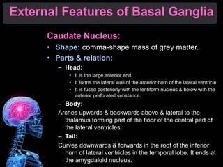
3. External Features of Basal Ganglia
Caudate Nucleus:
Shape: comma-shape mass of grey matter.
Parts & relation:
Head:
It is the large anterior end.
It forms the lateral wall of the anterior horn of the lateral ventricle.
It is fused posteriorly with the lentiform nucleus & below with the
anterior perforated substance.
Body:
Arches upwards & backwards above & lateral to the
thalamus forming part of the floor of the central part of
the lateral ventricles.
Tail:
Curves downwards & forwards in the roof of the inferior
horn of lateral ventricles in the temporal lobe. It ends at
the amygdaloid nucleus.
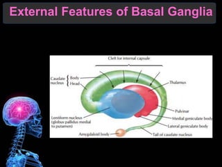
5. Amygdaloid Nucleus:
It is a small rounded mass lying in the uncus of the
temporal lobe, joined to the tail of caudate
nucleus.
It is a small centre.
Lentiform Nucleus:
Shape: it resembles a biconvex lens.
Parts it consists of:
Putamen: the larger, darker lateral part.
Globus pollidus: the smaller, paler medial part.
Surface & relations:
Medial surface: is highly convex & is related to the
internal capsule which separated the lentiform nucleus
from head of cuadate (anteriorly)
External Features of Basal Ganglia
6. & the thalamus posteriorly.
Lateral surface: is slightly convex & is related to the
external capsule which separates the lentiform nucleus
from the claustrum & insula.
Claustrum: it is a thin layer of gray matter lying
between the external capsule (medially) & the
white matter of the insula (laterally). Its antero-
inferior part fuses with the amygdaloid nucleus &
anterior perforated substance. Its function is
unknown.
The corpus Striatum it is a name given to:
Caudate nucleus.
The lentiform nucleus
Intervening anterior limb of internal capsule.
They are called so, because they show a striated appearance.
External Features of Basal Ganglia
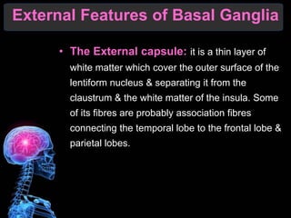
7. The External capsule: it is a thin layer of
white matter which cover the outer surface of the
lentiform nucleus & separating it from the
claustrum & the white matter of the insula. Some
of its fibres are probably association fibres
connecting the temporal lobe to the frontal lobe &
parietal lobes.
External Features of Basal Ganglia
9. The corpus Striatum
It is an important extrapyramidal centre.
The caudate nucleus putamen represent the
receptor nuclei which receive most afferent fibres.
The globus pallidus represent the affector part
giving rise to most efferent fibres.
Connection of corpus Striatum:
Afferent fibres derived from:
Cerebral cortex via corticostriate fibres.
Thalamus via thalamostriate fibres.
Substantia nigra via nigrostriate fibres.
Efferent fibres emerge from the globus pallidus which
pass to other extrapyramidal centres as follow:
Ansa lenticularis. Fasciculus lenticularis.
Subthalamic fasciculus.
Internal Features of Basal Ganglia
10. Amygdaloid Nucleus: part of the limbic system.
Afferents: it receives fibres from the olfactory tract.
Efferent: its fibres constitute the stria termenalis which
passes first backwards in the roof of the inferior horn of the
lateral ventricles then curves forwards in the floor of the
central part of the lateral ventricles. The fibre ends in:
Septal Nuclei.
Habenular nuclei.
Internal Features of Basal Ganglia
11. Level of Section A
Trough cerebral hemisphere
Internal Features of Basal Ganglia
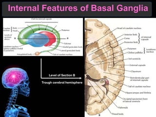
12. Level of Section B
Trough cerebral hemisphere
Internal Features of Basal Ganglia
13. Limbic System
It is formerly name rhinecephalon.
Definition: it is the name applied to number of
cortical & subcortical structure lying on the medial
surface of the cerebral hemisphere in the form of
an arch (limbus).
Structure it consists of:
Olfactory components:
1. Olfactory bulb tract which reach the following parts of
the limbic system:
Anterior perforated substance which fibres proceed to the
amygdaloid & habenular nucleus
The piriform area including the uncus & anterior part of the
hippocampal gyrus.
2. Hippocampal formation: which include: hippocampus,
dentate gyrus, indusium griseum, medial & lateral
longutidunal striae.
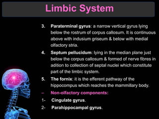
14. 3. Paraterminal gyrus: a narrow vertical gyrus lying
below the rostrum of corpus callosum. It is continuous
above with indusium griseum & below with medial
olfactory stria.
4. Septum pellucidum: lying in the median plane just
below the corpus callosum & formed of nerve fibres in
adition to collection of septal nuclei which constitute
part of the limbic system.
5. The fornix: it is the efferent pathway of the
hippocompus which reaches the mammillary body.
Non-olfactory components:
1- Cingulate gyrus.
2- Parahippocampal gyrus.
Limbic System
15. Connections of the limbic system: it
interconnected with the following centers (cerebral
cortex, thalamus, hypothalamus, epithalamus).
Functions of the limbic system:
1. Reception of olfactory stimuli.
2. Integration of olfactory, visceral sensation.
Limbic System