Crossectional anatomy of Head
Download as PPTX, PDF8 likes1,719 views
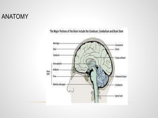
This document provides an overview of the cross sectional anatomy of the head at different levels, from below the 4th ventricle to above the ventricular level. It discusses the anatomy, blood supply, and various pathologies that can be seen in different imaging modalities like T1 sagittal, flair, and CT angiogram scans, such as epidural hematoma, subdural hematoma, subarachnoid hemorrhage, MCA infarct, lacunar infarct, neurocysticercosis, tuberculoma, and meningioma.
1 of 23
Downloaded 59 times


Recommended
magnetic resonance in angiography 
magnetic resonance in angiography BISHAL KHANAL
Ěý
This document discusses magnetic resonance angiography (MRA) and its advantages and disadvantages compared to catheter angiography. It describes different MRA techniques including contrast enhanced MRA, time of flight angiography, phase contrast angiography, and non-contrast techniques. It also discusses artifacts that can appear on MRA such as metal artifacts and blooming artifacts. Key features and images of each technique are provided.magnetic resonance angiography
magnetic resonance angiographyqavi786
Ěý
1. Magnetic resonance angiography (MRA) is a non-invasive imaging technique that uses magnetic resonance imaging to visualize blood vessels and evaluate vascular anatomy and blood flow without using ionizing radiation or iodinated contrast material.
2. There are different MRA techniques including time-of-flight MRA, phase contrast MRA, and contrast-enhanced MRA. Time-of-flight MRA relies on differences in flowing and stationary blood signal while phase contrast MRA assesses velocity and direction of flow. Contrast-enhanced MRA uses gadolinium contrast to improve vessel depiction.
3. MRA has various clinical applications for evaluating carotid and intracranial arterial stenosis, aneurysms,Portable n mobile unit
Portable n mobile unitSudil Paudyal
Ěý
Portable and mobile radiographic units can be either portable or mobile. Portable units are small enough to be carried by one person for use outside of a radiology department. Mobile units are larger and mounted on wheels, able to be moved throughout a hospital. Both use an X-ray tube, generator, and control unit to produce radiographic images. Newer units are using high frequency generators, computed radiography, or direct radiography for more efficient and higher quality imaging. Mobile C-arm units are important for fluoroscopy in operating rooms.MR ANGIOGRAPHY PHYSICS
MR ANGIOGRAPHY PHYSICSAvinashDahatre
Ěý
Everything regarding the physics of MRA is given along with flow charts and images. Also have covered new advances and refrences taken from MR made easy and some articles related to MRIX ray imaging intensifier
X ray imaging intensifierSurgicaltechie.com
Ěý
Types Fluoroscopy used in operation theatre, interventional radiology, Medical gastroenterology etc. Physics of Multidetector CT Scan
Physics of Multidetector CT ScanDr Varun Bansal
Ěý
Basic physics of multidetector computed tomography ( CT Scan) - how ct scan works, different generations of ct, how image is generated and displayed and image artifacts related to CT Scan.MRI Parameters for Imaging
MRI Parameters for ImagingAnjan Dangal
Ěý
This document discusses various magnetic resonance imaging parameters and how they affect image contrast and quality. It describes intrinsic parameters like repetition time and echo time that modify tissue signal, and extrinsic parameters like field of view and slice thickness that influence data collection. For each parameter, it explains how changes affect T1 and T2 weighting, signal-to-noise ratio, spatial resolution, scan time, and other characteristics. Examples of images with different parameter values are also shown to demonstrate the effects.CT Angiography presentation 
CT Angiography presentation Shatha M
Ěý
This document outlines the protocol for performing CT angiography (CTA) from the cerebral arteries to the lower limbs. It discusses indications for CTA including aneurysms, stenosis, dissections, and more. The preparation, positioning, and scanning protocols are provided for CTA of the head to lower limbs as well as the subclavian arteries. Pediatric protocols are also summarized. The document concludes with examples of CTA findings and references.SELDINGER TECHNIQUE & INTERVENTIONAL RADIOLOGY
SELDINGER TECHNIQUE & INTERVENTIONAL RADIOLOGYRiyas M K
Ěý
its a basic introduction about Seldinger technique and Intervetional radiology.In interventional radiology, procedures generally start with the Seldinger technique to access the vasculature, using a needle through which a guidewire is inserted, followed by navigation of catheters within the vessels.CT Image reconstruction
CT Image reconstructionSantosh Ojha
Ěý
This slide best explains the introduction of CT, basis and types of CT image reconstructions with detailed explanation about Interpolation, convolution, Fourier slice theorem, Fourier transformation and brief explanation about the image domain i.e digital image processing. Chest x ray positioning
Chest x ray positioningairwave12
Ěý
This document provides guidance on chest X-ray positioning and interpretation. It outlines different chest X-ray views including PA, lateral, AP, decubitus, and inspiratory-expiratory views. For a PA view, the patient faces the cassette with the tube 6 feet away. Proper inspiration is important, with the diaphragm at the 8th-10th posterior or 5th-6th anterior rib. Key areas to examine include the trachea, heart, diaphragm, lungs, pleural spaces, and bones. Paired inspiratory-expiratory views can demonstrate air trapping and diagnose foreign bodies.Angiography basics and seldinger technique
Angiography basics and seldinger techniqueSamuel Hernandez
Ěý
1. Angiography is performed by inserting a catheter into an artery or vein using the Seldinger technique, which involves inserting a needle and guidewire before threading the catheter.
2. The angiography team includes a radiologist, nurse, and technologists who prepare equipment like catheters, guidewires, and injectors to visualize blood vessels and perform interventional procedures.
3. Digital subtraction angiography uses computer algorithms to subtract bone structures from images, clearly showing blood vessel anatomy for diagnostic and therapeutic purposes like angioplasty.CT Generations and Artefacts
CT Generations and ArtefactsDr. Yash Kumar Achantani
Ěý
The document summarizes the history and development of computed tomography (CT) scanning technology. It describes the key events and innovations such as the development of the first CT scanner by Godfrey Hounsfield in 1972 (1), the introduction of whole body scanning in 1975 (2), and Hounsfield and Cormack being awarded the Nobel Prize in 1979 (3). Subsequent generations of CT scanners incorporated improvements like faster scanning speeds, multiple detectors, and eliminating moving parts to enable ultra-fast scanning.Interventional radiology part 2 final-Dr Chandni Wadhwani
Interventional radiology part 2 final-Dr Chandni WadhwaniChandni Wadhwani
Ěý
Role of IR in treatment of Varicose veins and Bone lesions.
Newer modality: HIFU
Videos on Embolization techniques, role of IR in hepatobiliary system and in portal hypertension.
Gradient echo pulse sequence swastik
Gradient echo pulse sequence swastikSwastik Poudel
Ěý
The document describes gradient echo pulse sequences. It discusses how gradients are used for spatial encoding by dephasing and rephasing magnetic moments. It explains slice selection, frequency encoding, and phase encoding. It describes how gradient echo sequences differ from spin echo by using variable flip angles and gradients instead of RF pulses to generate echoes. It discusses various gradient echo techniques including coherent, spoiled, and balanced sequences. It provides details on sequence parameters and how they control T1, T2, and PD weightings.Loopogram
LoopogramSam Shaikh
Ěý
Brief review of urinary diversion, loopogram, Loopogram Procedure, Benefits and associated risks, Bladder cancer,
Abnormal loopogram findings
Image Contrast, Noise, Resolution
Image Contrast, Noise, ResolutionMiami Cancer Institute
Ěý
This document discusses the effects of kVp and mAs on various properties of x-ray images. It explains that kVp determines the highest x-ray energy and quality, while mAs determines the quantity of photons and exposure time. Higher kVp and mAs increase spatial resolution, contrast, and signal-to-noise ratio, but also increase radiation dose. The document covers these parameters for screen-film radiography as well as computed tomography, and how they impact visible properties like image noise, contrast, and resolution.MRI spin echo pulse sequences
MRI spin echo pulse sequencesShiva Prakash
Ěý
This document discusses different pulse sequences used in MRI, including spin echo sequences that rephase spins using 180 degree pulses. It focuses on conventional spin echo, fast spin echo, and inversion recovery sequences. Conventional spin echo uses 90 and 180 degree pulses to generate a spin echo. Fast spin echo reduces scan time by acquiring multiple phase-encoding steps per repetition time. Inversion recovery begins with a 180 degree inversion pulse to suppress signals from fat (STIR) or cerebrospinal fluid (FLAIR).CT procedure of neck
CT procedure of neckSabitaMandal1
Ěý
CT imaging of the neck provides detailed anatomical information and is useful for evaluating neck masses, lymphadenopathy, thyroid diseases and trauma. The neck is divided into triangles and spaces which radiologists use to characterize abnormalities. CT protocols involve intravenous contrast administration and thin slices through the neck. MRI is also used and has advantages over CT such as better soft tissue contrast without radiation, though CT remains superior for assessing bone.Mri gradient coils
Mri gradient coilsShahnawaz Khan
Ěý
Gradient coils are used in MRI to spatially encode the MRI signal by creating linear magnetic field gradients in the x, y, and z directions. They are made of conducting loops or strips arranged in patterns like fingerprints that produce calibrated distortions of the main magnetic field. Modern scanners typically use distributed windings in copper sheets etched into complex patterns. Gradient coils are driven by powerful gradient amplifiers and require cooling to handle the heat from switching high currents rapidly. Proper gradient design and performance is crucial for achieving good spatial resolution and image quality in MRI.Catheters $ guidewires
Catheters $ guidewiresEmeka Ubah
Ěý
Guide wires and catheters are medical devices used in angiographic procedures. Guide wires are stainless steel wires that guide catheters through blood vessels. They have flexible tips and come in various lengths and diameters. Catheters are hollow tubes inserted into the body and come in different materials, sizes, and tip configurations. Common uses are angiography, drainage of fluids, and placement of stents and balloons. Both must be carefully sterilized between uses to prevent infection.CT Procedure of Thorax (CT Chest)
CT Procedure of Thorax (CT Chest)Upakar Paudel
Ěý
This document summarizes the key points about chest CT protocols and techniques:
1. Chest CT is used to further evaluate abnormalities found on chest x-rays and can diagnose many lung disorders due to its high resolution images. Proper patient positioning, administration of intravenous contrast, and adjusting scanning parameters are important for high quality images.
2. Chest CT protocols involve scanning from the thoracic inlet to the dome of the diaphragm with thin slices and reconstructions to visualize the lungs, mediastinum, chest wall, and upper abdomen. Contrast is used for certain indications to enhance visibility of vessels and lesions.
3. Indications for chest CT include evaluating lung tumors, pulmonary nodules, infectionsmr angiography.pptx
mr angiography.pptxVanshikaGarg76
Ěý
MR angiography is a type of MRI scan that uses magnetic fields and radio waves to provide pictures of blood vessels without using a catheter. It has advantages over conventional angiography in that it is less invasive, less expensive, and faster. Disadvantages include not depicting small vessels or slow blood flow as well. There are various techniques used in MR angiography including contrast enhanced MRA, which uses gadolinium contrast to visualize vascular structures, and non-contrast MRA such as time-of-flight angiography and phase contrast angiography. Artifacts that can occur include metal artifacts from implanted devices and blooming artifacts around paramagnetic substances.
Diffusion Weighted Imaging- Avinesh Shrestha
Diffusion Weighted Imaging- Avinesh ShresthaAvinesh Shrestha
Ěý
Diffusion-weighted imaging is a method of MRI imaging capable of showing the diffusion in the tissue. Venography
VenographySUJAN KARKI
Ěý
This document provides an overview of venography, which is an imaging technique used to examine veins. It discusses the basic principles of venography, including ascending and descending techniques. It describes the anatomy of veins and provides diagrams. It also covers indications, contraindications, techniques, and potential complications of lower limb, upper limb, and peripheral varicography venography procedures. The goal of venography is to accurately diagnose conditions like deep vein thrombosis.Radiological anatomy of the Carotid arteries
Radiological anatomy of the Carotid arteriesMohamed M.A. Zaitoun
Ěý
This document provides information on imaging of the carotid arteries and carotid angiography. It discusses various imaging modalities used to image the carotid arteries including ultrasound, CT, MRI, CT angiography, MR angiography, duplex ultrasound, and plain films. It then provides detailed information on carotid angiography including definitions, indications, complications, techniques, and how to avoid complications. Transcranial ultrasound in premature infants is also briefly discussed.Gradient Recalled Echo(GRE)
Gradient Recalled Echo(GRE)SUJAN KARKI
Ěý
This document provides an overview of MRI gradient echo pulse sequences, types, and applications. It discusses the basics of spatial encoding using slice selection, phase encoding, and frequency encoding gradients. It describes coherent gradient echo sequences which maintain transverse magnetization between excitations, and incoherent sequences which eliminate residual transverse magnetization. Spoiling techniques are discussed which remove signal from residual transverse magnetization to enhance T1 contrast. Applications include angiography, myelography and fast imaging where T1 or proton density contrast is desired.Cross sectional anatomy of the neck
Cross sectional anatomy of the neckSahil Chaudhry
Ěý
The document discusses the anatomy of the neck region. It begins by outlining the gross anatomy including the extent and boundaries of the neck. It then describes the divisions of the neck created by the sternocleidomastoid muscle and details the contents of the anterior and posterior triangles. Next, it discusses the layers of cervical fascia and the spaces they enclose, including the visceral, retropharyngeal, parapharyngeal, danger, and prevertebral spaces. It notes the clinical importance of understanding the neck spaces for localizing lesions, differential diagnosis, and guided procedures. Finally, it briefly summarizes some of the key structures contained within the neck, such as the thyroid gland, larynx, and parathyContrast Media
Contrast MediaJayanti Gyawali
Ěý
Contrast media are agents used to enhance the visibility of structures in medical imaging. There are several types including positive contrast media which make structures appear brighter on scans, and negative contrast media which make structures seem darker. Common contrast agents contain iodine and can be ionic monomers, ionic dimers, non-ionic monomers, or non-ionic dimers. While contrast imaging provides important medical information, the agents sometimes cause side effects from mild reactions like nausea to more severe issues like pulmonary edema. Care must be taken with patients having risk factors for complications.More Related Content
What's hot (20)
SELDINGER TECHNIQUE & INTERVENTIONAL RADIOLOGY
SELDINGER TECHNIQUE & INTERVENTIONAL RADIOLOGYRiyas M K
Ěý
its a basic introduction about Seldinger technique and Intervetional radiology.In interventional radiology, procedures generally start with the Seldinger technique to access the vasculature, using a needle through which a guidewire is inserted, followed by navigation of catheters within the vessels.CT Image reconstruction
CT Image reconstructionSantosh Ojha
Ěý
This slide best explains the introduction of CT, basis and types of CT image reconstructions with detailed explanation about Interpolation, convolution, Fourier slice theorem, Fourier transformation and brief explanation about the image domain i.e digital image processing. Chest x ray positioning
Chest x ray positioningairwave12
Ěý
This document provides guidance on chest X-ray positioning and interpretation. It outlines different chest X-ray views including PA, lateral, AP, decubitus, and inspiratory-expiratory views. For a PA view, the patient faces the cassette with the tube 6 feet away. Proper inspiration is important, with the diaphragm at the 8th-10th posterior or 5th-6th anterior rib. Key areas to examine include the trachea, heart, diaphragm, lungs, pleural spaces, and bones. Paired inspiratory-expiratory views can demonstrate air trapping and diagnose foreign bodies.Angiography basics and seldinger technique
Angiography basics and seldinger techniqueSamuel Hernandez
Ěý
1. Angiography is performed by inserting a catheter into an artery or vein using the Seldinger technique, which involves inserting a needle and guidewire before threading the catheter.
2. The angiography team includes a radiologist, nurse, and technologists who prepare equipment like catheters, guidewires, and injectors to visualize blood vessels and perform interventional procedures.
3. Digital subtraction angiography uses computer algorithms to subtract bone structures from images, clearly showing blood vessel anatomy for diagnostic and therapeutic purposes like angioplasty.CT Generations and Artefacts
CT Generations and ArtefactsDr. Yash Kumar Achantani
Ěý
The document summarizes the history and development of computed tomography (CT) scanning technology. It describes the key events and innovations such as the development of the first CT scanner by Godfrey Hounsfield in 1972 (1), the introduction of whole body scanning in 1975 (2), and Hounsfield and Cormack being awarded the Nobel Prize in 1979 (3). Subsequent generations of CT scanners incorporated improvements like faster scanning speeds, multiple detectors, and eliminating moving parts to enable ultra-fast scanning.Interventional radiology part 2 final-Dr Chandni Wadhwani
Interventional radiology part 2 final-Dr Chandni WadhwaniChandni Wadhwani
Ěý
Role of IR in treatment of Varicose veins and Bone lesions.
Newer modality: HIFU
Videos on Embolization techniques, role of IR in hepatobiliary system and in portal hypertension.
Gradient echo pulse sequence swastik
Gradient echo pulse sequence swastikSwastik Poudel
Ěý
The document describes gradient echo pulse sequences. It discusses how gradients are used for spatial encoding by dephasing and rephasing magnetic moments. It explains slice selection, frequency encoding, and phase encoding. It describes how gradient echo sequences differ from spin echo by using variable flip angles and gradients instead of RF pulses to generate echoes. It discusses various gradient echo techniques including coherent, spoiled, and balanced sequences. It provides details on sequence parameters and how they control T1, T2, and PD weightings.Loopogram
LoopogramSam Shaikh
Ěý
Brief review of urinary diversion, loopogram, Loopogram Procedure, Benefits and associated risks, Bladder cancer,
Abnormal loopogram findings
Image Contrast, Noise, Resolution
Image Contrast, Noise, ResolutionMiami Cancer Institute
Ěý
This document discusses the effects of kVp and mAs on various properties of x-ray images. It explains that kVp determines the highest x-ray energy and quality, while mAs determines the quantity of photons and exposure time. Higher kVp and mAs increase spatial resolution, contrast, and signal-to-noise ratio, but also increase radiation dose. The document covers these parameters for screen-film radiography as well as computed tomography, and how they impact visible properties like image noise, contrast, and resolution.MRI spin echo pulse sequences
MRI spin echo pulse sequencesShiva Prakash
Ěý
This document discusses different pulse sequences used in MRI, including spin echo sequences that rephase spins using 180 degree pulses. It focuses on conventional spin echo, fast spin echo, and inversion recovery sequences. Conventional spin echo uses 90 and 180 degree pulses to generate a spin echo. Fast spin echo reduces scan time by acquiring multiple phase-encoding steps per repetition time. Inversion recovery begins with a 180 degree inversion pulse to suppress signals from fat (STIR) or cerebrospinal fluid (FLAIR).CT procedure of neck
CT procedure of neckSabitaMandal1
Ěý
CT imaging of the neck provides detailed anatomical information and is useful for evaluating neck masses, lymphadenopathy, thyroid diseases and trauma. The neck is divided into triangles and spaces which radiologists use to characterize abnormalities. CT protocols involve intravenous contrast administration and thin slices through the neck. MRI is also used and has advantages over CT such as better soft tissue contrast without radiation, though CT remains superior for assessing bone.Mri gradient coils
Mri gradient coilsShahnawaz Khan
Ěý
Gradient coils are used in MRI to spatially encode the MRI signal by creating linear magnetic field gradients in the x, y, and z directions. They are made of conducting loops or strips arranged in patterns like fingerprints that produce calibrated distortions of the main magnetic field. Modern scanners typically use distributed windings in copper sheets etched into complex patterns. Gradient coils are driven by powerful gradient amplifiers and require cooling to handle the heat from switching high currents rapidly. Proper gradient design and performance is crucial for achieving good spatial resolution and image quality in MRI.Catheters $ guidewires
Catheters $ guidewiresEmeka Ubah
Ěý
Guide wires and catheters are medical devices used in angiographic procedures. Guide wires are stainless steel wires that guide catheters through blood vessels. They have flexible tips and come in various lengths and diameters. Catheters are hollow tubes inserted into the body and come in different materials, sizes, and tip configurations. Common uses are angiography, drainage of fluids, and placement of stents and balloons. Both must be carefully sterilized between uses to prevent infection.CT Procedure of Thorax (CT Chest)
CT Procedure of Thorax (CT Chest)Upakar Paudel
Ěý
This document summarizes the key points about chest CT protocols and techniques:
1. Chest CT is used to further evaluate abnormalities found on chest x-rays and can diagnose many lung disorders due to its high resolution images. Proper patient positioning, administration of intravenous contrast, and adjusting scanning parameters are important for high quality images.
2. Chest CT protocols involve scanning from the thoracic inlet to the dome of the diaphragm with thin slices and reconstructions to visualize the lungs, mediastinum, chest wall, and upper abdomen. Contrast is used for certain indications to enhance visibility of vessels and lesions.
3. Indications for chest CT include evaluating lung tumors, pulmonary nodules, infectionsmr angiography.pptx
mr angiography.pptxVanshikaGarg76
Ěý
MR angiography is a type of MRI scan that uses magnetic fields and radio waves to provide pictures of blood vessels without using a catheter. It has advantages over conventional angiography in that it is less invasive, less expensive, and faster. Disadvantages include not depicting small vessels or slow blood flow as well. There are various techniques used in MR angiography including contrast enhanced MRA, which uses gadolinium contrast to visualize vascular structures, and non-contrast MRA such as time-of-flight angiography and phase contrast angiography. Artifacts that can occur include metal artifacts from implanted devices and blooming artifacts around paramagnetic substances.Diffusion Weighted Imaging- Avinesh Shrestha
Diffusion Weighted Imaging- Avinesh ShresthaAvinesh Shrestha
Ěý
Diffusion-weighted imaging is a method of MRI imaging capable of showing the diffusion in the tissue. Venography
VenographySUJAN KARKI
Ěý
This document provides an overview of venography, which is an imaging technique used to examine veins. It discusses the basic principles of venography, including ascending and descending techniques. It describes the anatomy of veins and provides diagrams. It also covers indications, contraindications, techniques, and potential complications of lower limb, upper limb, and peripheral varicography venography procedures. The goal of venography is to accurately diagnose conditions like deep vein thrombosis.Radiological anatomy of the Carotid arteries
Radiological anatomy of the Carotid arteriesMohamed M.A. Zaitoun
Ěý
This document provides information on imaging of the carotid arteries and carotid angiography. It discusses various imaging modalities used to image the carotid arteries including ultrasound, CT, MRI, CT angiography, MR angiography, duplex ultrasound, and plain films. It then provides detailed information on carotid angiography including definitions, indications, complications, techniques, and how to avoid complications. Transcranial ultrasound in premature infants is also briefly discussed.Gradient Recalled Echo(GRE)
Gradient Recalled Echo(GRE)SUJAN KARKI
Ěý
This document provides an overview of MRI gradient echo pulse sequences, types, and applications. It discusses the basics of spatial encoding using slice selection, phase encoding, and frequency encoding gradients. It describes coherent gradient echo sequences which maintain transverse magnetization between excitations, and incoherent sequences which eliminate residual transverse magnetization. Spoiling techniques are discussed which remove signal from residual transverse magnetization to enhance T1 contrast. Applications include angiography, myelography and fast imaging where T1 or proton density contrast is desired.Viewers also liked (20)
Cross sectional anatomy of the neck
Cross sectional anatomy of the neckSahil Chaudhry
Ěý
The document discusses the anatomy of the neck region. It begins by outlining the gross anatomy including the extent and boundaries of the neck. It then describes the divisions of the neck created by the sternocleidomastoid muscle and details the contents of the anterior and posterior triangles. Next, it discusses the layers of cervical fascia and the spaces they enclose, including the visceral, retropharyngeal, parapharyngeal, danger, and prevertebral spaces. It notes the clinical importance of understanding the neck spaces for localizing lesions, differential diagnosis, and guided procedures. Finally, it briefly summarizes some of the key structures contained within the neck, such as the thyroid gland, larynx, and parathyContrast Media
Contrast MediaJayanti Gyawali
Ěý
Contrast media are agents used to enhance the visibility of structures in medical imaging. There are several types including positive contrast media which make structures appear brighter on scans, and negative contrast media which make structures seem darker. Common contrast agents contain iodine and can be ionic monomers, ionic dimers, non-ionic monomers, or non-ionic dimers. While contrast imaging provides important medical information, the agents sometimes cause side effects from mild reactions like nausea to more severe issues like pulmonary edema. Care must be taken with patients having risk factors for complications.Miniatlas of Human Cross-Sectional Anatomy
Miniatlas of Human Cross-Sectional AnatomyPhilip Tate
Ěý
A cadaver was sectioned and photographed. The cadaver photographs were correlated with MRI and CT images. Photoshop and Quark Express were used to produce a 62 page atlas that presents sectional anatomy using a systemic approach.
Coauthored with James Kennedy and John LampiganoCt brain by prof. Wael samir
Ct brain by prof. Wael samirFaculty of Medicine Ain Shams University
Ěý
The document discusses the following:
1) Intended learning objectives which include discussing radiation hazards of CT, use of contrast agents, and interpretation of CT perfusion and angiography in cerebral ischemia.
2) Radiation dose considerations from various radiographic studies including effective dose ranges.
3) Contrast agent facts including types, dosing, and safety issues.
4) Preventive measures for contrast media emergencies.MRI Brain
MRI BrainJayanti Gyawali
Ěý
This document outlines an MRI brain protocol. It begins with an introduction to MRI brain imaging and its advantages over CT, such as lack of radiation exposure and greater soft tissue contrast. Common indications for MRI brain are then listed. The document describes patient preparation, contrast usage, coil positioning, imaging sequences including T1, T2, FLAIR, DWI, and advanced techniques like MRS and fMRI. Specific protocols are provided for conditions like MS, trauma, pituitary imaging, and CSF flow studies.Sectional anatomy
Sectional anatomyWafa Alfaleh
Ěý
The document summarizes the major facial bones seen on CT imaging, including the maxillary, zygomatic, lacrimal, nasal, palatine, vomer, and mandible bones. It describes the location and anatomical features of each bone, such as how the maxillary bones form the oral, nasal, and orbital cavities and how the nasal bones fuse to form the bridge of the nose. Examples of CT images are provided to demonstrate the anatomical relationship between the different facial bones.Nmt 405 abdomen and pelvis ppt
Nmt 405 abdomen and pelvis pptClassyCJ
Ěý
This document discusses cross sectional anatomy of the abdomen through coronal and transverse 3D scans. It provides a link to view a 3D coronal scan of the abdomen and pelvis as well as mentions a transverse scan. The scans allow viewing internal structures of the abdomen from different angles.Cone beam computerized tomography mamita
Cone beam computerized tomography mamitaMamita Sakhakarmi
Ěý
This document discusses cone beam computed tomography (CBCT), including its history developing in 1982 for angiography and maxillofacial imaging, principles using a cone-shaped radiation source and 2D detector to acquire multiple projections, uses in assessing pathologies, deformities, implants and treatment planning, advantages of shorter scan times and improved efficiency over other techniques, and artifacts that can occur.Mri evaluation of pediatric white matter lesions 
Mri evaluation of pediatric white matter lesions DrBhishm Sevendra
Ěý
1. Canavan disease, Alexander disease, vanishing white matter disease, and megalencephalic leukoencephalopathy involve sub cortical and deep white matter and are characterized by macrocephaly. Zellweger syndrome and Kearn sayer disease involve the normocephalic deep white matter and cerebral atrophy.
2. Krabbe's disease involves the thalamus and basal ganglia with increased choline in the centrum semiovale, while adrenoleucodystrophy affects the corticospinal tract with a posterior fossa tripe layer appearance and enhancement. Maple syrup urine disease shows restricted diffusion in the internal capsule and posterior fossa.
3. MetachromaticMRI OF GIT SECTIONAL ANATOMY
MRI OF GIT SECTIONAL ANATOMYVipin Kumar
Ěý
The document provides a sectional anatomy of the gastrointestinal tract and renal system. It includes detailed labeled diagrams of the structures of the oral cavity, esophagus, stomach, small intestine, large intestine, liver, pancreas, and kidneys. The diagrams show the location and relationships between these organs and surrounding blood vessels.Basics of neuroimaging
Basics of neuroimagingMehdi Nasr Isfahani
Ěý
1. The document discusses the basics of neuroimaging using CT and MRI. It explains how different tissues appear on CT and MRI scans and provides examples of normal anatomy.
2. It then covers the systematic approach to interpreting head CT scans and analyzing different areas of the brain. Examples of cross-sectional anatomy at different brain levels are shown on CT scans.
3. The document also discusses the physics behind MRI and how tissues appear differently on T1-weighted, T2-weighted, and FLAIR sequences. Multiple images demonstrate normal brain anatomy on post-contrast MRI scans.Doppler Physics
Doppler PhysicsSahil Chaudhry
Ěý
Beginners guide to understand the concepts and techniques of Doppler USG imaging -types , technical info,subtypes ,artefacts and interpretationProton magnetic resonance spectroscopy
Proton magnetic resonance spectroscopyhazem youssef
Ěý
This document discusses proton magnetic resonance spectroscopy (1H MRS) of the central nervous system. 1H MRS is a noninvasive technique that provides information about brain biochemistry and metabolism. It works by detecting different resonant frequencies of hydrogen nuclei in molecules, which are influenced by chemical bonds. The document outlines the basic principles and techniques of 1H MRS, including single voxel spectroscopy and magnetic resonance spectroscopic imaging. It discusses the clinical applications of 1H MRS for characterizing different brain pathologies such as tumors, infections, and treatment monitoring. Specific metabolic patterns are described for analyzing different brain tumors, meningiomas, and primary CNS lymphoma.Solid state detector mamita
Solid state detector mamitaMamita Sakhakarmi
Ěý
This document discusses different types of solid state radiation detectors, including scintillation detectors, thermoluminescent dosimeters (TLD), and semiconductor detectors. Scintillation detectors detect radiation via light emission in inorganic crystal materials like NaI or organic crystals like anthracene. TLDs "capture" radiation dose information and release light when heated, allowing dose measurement. Common TLD materials are LiF:Mg,Ti and Li2B4O7:Mn. Semiconductor detectors like silicon and germanium act as solid state ionization chambers and are used for high resolution energy measurement of alpha and beta particles.5lab components of ct scanner
5lab components of ct scannerKhaleeque Memon
Ěý
This document describes the key components of a CT scanner, including the gantry, x-ray tube, detector array, high voltage generator, and patient support couch. The gantry houses the x-ray tube, detector array, and other components and rotates around the patient. The x-ray tube produces x-rays, while the detector array detects the x-rays that pass through the patient and produces images. A high voltage generator supplies power to the x-ray tube. The patient lies on a support couch that positions them for imaging and must be made of material that does not interfere with the x-rays.MR spectroscopy
MR spectroscopyairwave12
Ěý
MR spectroscopy is a non-invasive technique that uses MRI to measure brain chemistry. It provides information about metabolites like NAA, creatine, and choline to help characterize lesions and diseases. Single-voxel MRS is less advanced but faster, while multi-voxel MRS examines more areas but takes longer. MRS is an additive test that is interpreted along with conventional MRI images to aid diagnosis.Principles of Doppler ultrasound
Principles of Doppler ultrasoundSamir Haffar
Ěý
This document discusses the principles of Doppler ultrasound. It begins with a brief history of Doppler and how the Doppler effect was discovered. It then covers the basic physics of Doppler ultrasound including the Doppler equation. The remainder of the document discusses specific Doppler parameters and how to optimize the Doppler examination including:
- Adjusting spectral and color Doppler parameters
- Normal arterial and venous flow patterns
- Changes in flow related to stenosisCross Sectional Anatomy Of The Abdomen Annotated
Cross Sectional Anatomy Of The Abdomen AnnotatedDebby Edney
Ěý
This document discusses cross sectional anatomy of the abdomen through coronal and transverse 3D scans. It provides a link to view a 3D coronal scan of the abdomen and pelvis on RadiologyInfo's image gallery for an expanded view. It also mentions a transverse scan.Head And Neck
Head And Neckjo Han
Ěý
The document describes the anatomy of several muscles in the head and neck region. It provides details on the origin, insertion, innervation and actions of muscles like the sternocleidomastoid, scalene muscles, masseter, temporalis, occipitofrontalis, splenius muscles, suboccipital muscles including the rectus capitis posterior major and minor, and oblique capitis superior and inferior. It also provides instructions on palpating and testing some of these muscles.The gross anatomy of the head and neck lecture 3
The gross anatomy of the head and neck lecture 3Lucidante1
Ěý
The document provides an overview of the gross anatomy of the head and neck, focusing on the osteology of the skull. It describes the two main parts of the skull - the neurocranium and viscerocranium. The neurocranium comprises the bones that form the brain case and calvarium. The viscerocranium comprises the facial bones. It provides detailed descriptions of the individual skull bones and their features, including landmarks, foramina, and sutures. It also discusses variations in skull anatomy between infants and adults, common fractures, and other clinical considerations.Recently uploaded (20)
PERSONALITY DEVELOPMENT & DEFENSE MECHANISMS.pptxPersonality and environment:...
PERSONALITY DEVELOPMENT & DEFENSE MECHANISMS.pptxPersonality and environment:...ABHAY INSTITUTION
Ěý
Personality theory is a collection of ideas that explain how a person's personality develops and how it affects their behavior. It also seeks to understand how people react to situations, and how their personality impacts their relationships.
Key aspects of personality theory
Personality traits: The characteristics that make up a person's personality.
Personality development: How a person's personality develops over time.
Personality disorders: How personality theories can be used to study personality disorders.
Personality and environment: How a person's personality is influenced by their environment. Optimization in Pharmaceutical Formulations: Concepts, Methods & Applications
Optimization in Pharmaceutical Formulations: Concepts, Methods & ApplicationsKHUSHAL CHAVAN
Ěý
This presentation provides a comprehensive overview of optimization in pharmaceutical formulations. It explains the concept of optimization, different types of optimization problems (constrained and unconstrained), and the mathematical principles behind formulation development. Key topics include:
Methods for optimization (Sequential Simplex Method, Classical Mathematical Methods)
Statistical analysis in optimization (Mean, Standard Deviation, Regression, Hypothesis Testing)
Factorial Design & Quality by Design (QbD) for process improvement
Applications of optimization in drug formulation
This resource is beneficial for pharmaceutical scientists, R&D professionals, regulatory experts, and students looking to understand pharmaceutical process optimization and quality by design approaches.MLS 208 - UNIT 4 A - Tissue Processing - ETANDO AYUK - SANU 1 - Secured.pdf
MLS 208 - UNIT 4 A - Tissue Processing - ETANDO AYUK - SANU 1 - Secured.pdfEswatini Medical Christian University - EMCU / Southern Nazarene University - SANU
Ěý
The course covers the steps undertaken from tissue collection, reception, fixation,
sectioning, tissue processing and staining. It covers all the general and special
techniques in histo/cytology laboratory. This course will provide the student with the
basic knowledge of the theory and practical aspect in the diagnosis of tumour cells
and non-malignant conditions in body tissues and for cytology focusing on
gynaecological and non-gynaecological samples.
Diabetic Ketoacidosis (DKA) & Its Management Protocol
Diabetic Ketoacidosis (DKA) & Its Management ProtocolDr Anik Roy Chowdhury
Ěý
Dr. Anik Roy Chowdhury
MBBS, BCS(Health), DA, MD (Resident)
Department of Anesthesiology, ICU & Pain Medicine
Shaheed Suhrawardy Medical College Hospital (ShSMCH)Acute & Chronic Inflammation, Chemical mediators in Inflammation and Wound he...
Acute & Chronic Inflammation, Chemical mediators in Inflammation and Wound he...Ganapathi Vankudoth
Ěý
A complete information of Inflammation, it includes types of Inflammation, purpose of Inflammation, pathogenesis of acute inflammation, chemical mediators in inflammation, types of chronic inflammation, wound healing and Inflammation in skin repair, phases of wound healing, factors influencing wound healing and types of wound healing.Macafem Reviews 2024 - Macafem for Menopause Symptoms
Macafem Reviews 2024 - Macafem for Menopause SymptomsMacafem Supplement
Ěý
At Macafem, we provide 100% natural support for women navigating menopause. For over 20 years, we've helped women manage symptoms, and in 2024, we're proud to share their heartfelt experiences.
PRODUCTION OF HB VACCINE AND INTERFERONS BY rDNA - Copy.pptx
PRODUCTION OF HB VACCINE AND INTERFERONS BY rDNA - Copy.pptxkarishmaduhijod1
Ěý
APPLICATION of RECOMBINANAT DNA TECHNOLOGY : IN THE PRODUCTION OF HEPATITIS B VACCINE ,INSULIN and INTERFERONNon-Invasive ICP Monitoring for Neurosurgeons
Non-Invasive ICP Monitoring for NeurosurgeonsDhaval Shukla
Ěý
This presentation delves into the latest advancements in non-invasive intracranial pressure (ICP) monitoring techniques, specifically tailored for neurosurgeons. It covers the importance of ICP monitoring in clinical practice, explores various non-invasive methods, and discusses their accuracy, reliability, and clinical applications. Attendees will gain insights into the benefits of non-invasive approaches over traditional invasive methods, including reduced risk of complications and improved patient outcomes. This comprehensive overview is designed to enhance the knowledge and skills of neurosurgeons in managing patients with neurological conditions.
Invasive systems are commonly used for monitoring intracranial pressure (ICP) in traumatic brain injury (TBI) and are considered the gold standard. The availability of invasive ICP monitoring is heterogeneous, and in low- and middle-income settings, these systems are not routinely employed due to high cost or limited accessibility. The aim of this presentation is to develop recommendations to guide monitoring and ICP-driven therapies in TBI using non-invasive ICP (nICP) systems.
HUMAN SEXUALITY AND SEXUAL RESPONCE CYCLE
HUMAN SEXUALITY AND SEXUAL RESPONCE CYCLEdaminipatel37
Ěý
It is all about topic of obg for new semester students Biography of Dr. Vincenzo Giordano
Biography of Dr. Vincenzo GiordanoDr. Vincenzo Giordano
Ěý
Dr. Vincenzo Giordano began his medical career 2011 at Aberdeen Royal Infirmary in the Department of Cardiothoracic Surgery. Here, he performed complex adult cardiothoracic surgical procedures, significantly enhancing his proficiency in patient critical care, as evidenced by his FCCS certification.BIOMECHANICS OF THE MOVEMENT OF THE SHOULDER COMPLEX.pptx
BIOMECHANICS OF THE MOVEMENT OF THE SHOULDER COMPLEX.pptxdrnidhimnd
Ěý
The shoulder complex acts as in coordinated fashion to provide the smoothest and greatest range of motion possible of the upper limb.
Combined motion of GH and ST joint of shoulder complex helps in:
Distribution of motion between other two joints.
Maintenance of glenoid fossa in optimal position.
Maintenance of good length tension
Although some amount of glenohumeral motion may occur while the other shoulder articulations remain stabilized, movement of the humerus more commonly involves some movement at all three shoulder joints.
Renal Physiology - Regulation of GFR and RBF
Renal Physiology - Regulation of GFR and RBFMedicoseAcademics
Ěý
1. Explain the physiological control of glomerular filtration and renal blood flow
2. Describe the humoral and autoregulatory feedback mechanisms that mediate the autoregulation of renal plasma flow and glomerular filtration rate
Solubilization in Pharmaceutical Sciences: Concepts, Mechanisms & Enhancement...
Solubilization in Pharmaceutical Sciences: Concepts, Mechanisms & Enhancement...KHUSHAL CHAVAN
Ěý
This presentation provides an in-depth understanding of solubilization and its critical role in pharmaceutical formulations. It covers:
Definition & Mechanisms of Solubilization
Role of surfactants, micelles, and bile salts in drug solubility
Factors affecting solubilization (pH, polarity, particle size, temperature, etc.)
Methods to enhance drug solubility (Buffers, Co-solvents, Surfactants, Complexation, Solid Dispersions)
Advanced approaches (Polymorphism, Salt Formation, Co-crystallization, Prodrugs)
This resource is valuable for pharmaceutical scientists, formulation experts, regulatory professionals, and students interested in improving drug solubility and bioavailability.
Stability of Dosage Forms as per ICH Guidelines
Stability of Dosage Forms as per ICH GuidelinesKHUSHAL CHAVAN
Ěý
This presentation covers the stability testing of pharmaceutical dosage forms according to ICH guidelines (Q1A-Q1F). It explains the definition of stability, various testing protocols, storage conditions, and evaluation criteria required for regulatory submissions. Key topics include stress testing, container closure systems, stability commitment, and photostability testing. The guidelines ensure that pharmaceutical products maintain their identity, purity, strength, and efficacy throughout their shelf life. This resource is valuable for pharmaceutical professionals, researchers, and regulatory experts.The influence of birth companion in mother care and neonatal outcome
The influence of birth companion in mother care and neonatal outcomeLokesh Kumar Sharma
Ěý
this content related to birth companionship, role of birth companion in care of mother and neonatal Sudurpaschim logsewa aayog Medical Officer 8th Level Curriculum
Sudurpaschim logsewa aayog Medical Officer 8th Level CurriculumDr Ovels
Ěý
Sudurpaschim province psc ( lok sewa aayog) medical officer 8th level syllabusCorrelation of vitamin D level with prediabetes status_Dr Ahmed Al Montasir_f...
Correlation of vitamin D level with prediabetes status_Dr Ahmed Al Montasir_f...zilkerapurbo
Ěý
Correlation of vitamin D level with prediabetes statusMLS 208 - UNIT 4 A - Tissue Processing - ETANDO AYUK - SANU 1 - Secured.pdf
MLS 208 - UNIT 4 A - Tissue Processing - ETANDO AYUK - SANU 1 - Secured.pdfEswatini Medical Christian University - EMCU / Southern Nazarene University - SANU
Ěý
Acute & Chronic Inflammation, Chemical mediators in Inflammation and Wound he...
Acute & Chronic Inflammation, Chemical mediators in Inflammation and Wound he...Ganapathi Vankudoth
Ěý
Crossectional anatomy of Head
- 1. CROSS SECTIONAL ANATOMY OF HEAD Jayanti Gyawali B.Sc.MIT 2nd year Third Batch
- 2. ANATOMY
- 3. BLOOD SUPPLY
- 12. T1 sag
- 13. flair
- 14. CT ANGIOGRAM
- 16. sdh
- 17. SAH
- 18. MCA infarct
- 19. Lacunar infarct
- 20. NCC
- 21. tuberculoma
- 22. NCC
- 23. Meningioma