E mri mr_signal_slides
Download as ppt, pdf0 likes70 views

The document contains multiple copyright notices from 2006 attributed to Denis Hoa et al and Campus Medica with a web address. It provides basic information about the copyright and ownership but no other substantive content in 3 sentences or less.
1 of 19
Download to read offline

Ad
Recommended
E mri spatialencoding_slides
E mri spatialencoding_slidesMagde Gad
╠²
The document is copyrighted material from 2006 by Denis Hoa et al. for Campus Medica. It provides basic information about magnetic resonance imaging (MRI) technology and its medical applications. The copyright and source are attributed 9 separate times throughout the short document.Introduction to Magnetic resonance imaging (mri)
Introduction to Magnetic resonance imaging (mri)CHINENYE OZOTA
╠²
Magnetic resonance imaging (MRI) is a non-invasive medical imaging technique using magnetic fields and radio waves to create detailed images of organs, distinguishing it from other modalities like CT scans. Since its inception in the 1970s, MRI has evolved to become a versatile tool in various medical applications, including neuroimaging, cardiac imaging, and oncology. Despite its advantages, safety considerations exist, particularly concerning contraindications with certain medical devices and the potential discomfort associated with the scanning process.Introduction to MRI, a Short Summary
Introduction to MRI, a Short SummaryHabibUllah182
╠²
This document provides an introduction to magnetic resonance imaging (MRI). It discusses the history, instrumentation, working principles, and applications of MRI. MRI uses strong magnetic fields and radio waves to generate detailed images of organs and tissues in the body. Felix Bloch, Edward Purcell, and Raymond Damadian contributed to early discoveries around MRI. Key MRI machine components include the main magnet, gradient coils, and radiofrequency coils. MRI works by aligning hydrogen protons in tissues when a magnetic field is applied, and using radio waves to elicit signals to form images. MRI has many medical applications including neuroimaging, cardiology, musculoskeletal imaging, and angiography.Neuroimaging in psychiatry
Neuroimaging in psychiatrySantanu Ghosh
╠²
The document provides an overview of neuroimaging techniques used in psychiatry such as MRI, CT, PET, SPECT, fMRI, DTI, and MRS. It discusses the basic principles, milestones in development, and applications of these techniques. Specifically, it summarizes research using these neuroimaging methods that have found abnormalities in brain structure and function in patients with obsessive-compulsive disorder (OCD), such as reduced serotonin transporter binding in fronto-striatal circuits and differences in brain activity in regions like the thalamus and orbitofrontal cortex.Medical Image Registration 1st Edition Joseph V. Hajnal
Medical Image Registration 1st Edition Joseph V. Hajnalkupersowlely
╠²
Medical Image Registration 1st Edition Joseph V. Hajnal
Medical Image Registration 1st Edition Joseph V. Hajnal
Medical Image Registration 1st Edition Joseph V. HajnalDiscuss Imaging techniques in Orthopaedics
Discuss Imaging techniques in OrthopaedicsNafiuAbdullateef
╠²
The document discusses the role of imaging in orthopaedics, detailing historical advancements and various imaging techniques such as X-rays, MRI, CT scans, and ultrasound. It emphasizes the importance of understanding each modality's applications, advantages, and limitations for effective patient diagnosis and treatment. Additionally, it highlights the use of imaging in surgical planning, intraoperative monitoring, and treatment evaluation.MR MAMMOGRAPHY shdjsjsjdjsjdjdhdhdhdhdhs
MR MAMMOGRAPHY shdjsjsjdjsjdjdhdhdhdhdhsvarunraj362196
╠²
The document provides a comprehensive overview of MR mammography, detailing indications, contraindications, patient preparation, and techniques employed in the examination. It emphasizes the importance of appropriate timing in relation to the menstrual cycle, as well as considerations for previous breast interventions and patient positioning to minimize artifacts. Additionally, it explains the various imaging techniques and sequences used in MR mammography, focusing on achieving high sensitivity for detecting breast cancer.Magnetic resonance
Magnetic resonanceEmina Dzaferovic
╠²
This document provides an overview of magnetic resonance imaging (MRI) and discusses its history, technical aspects, medical uses, safety considerations, comparisons to other imaging techniques, regulations, underlying physics principles, ongoing developments, and potential job opportunities. MRI is a medical imaging technique that produces detailed images of the internal structures and functions of the body without using ionizing radiation. It was developed in the 1970s and continues to be an area of active research and clinical application.BREAST IMAGING IN DIFFERENT CONDITIONS OF BREAST
BREAST IMAGING IN DIFFERENT CONDITIONS OF BREASTDharmendraThakur26
╠²
This document summarizes various breast imaging modalities. It discusses the role of mammography, ultrasound, MRI, PET, and other techniques in evaluating the breast. Mammography remains the primary screening tool but has limitations related to breast density. Ultrasound helps diagnose palpable lesions and differentiate cysts from solid masses. MRI detects additional cancers but has limitations of cost and availability. Combined modalities provide improved evaluation of the breast compared to single techniques alone.Mri breast,indication,contra,prtocol,techniq,curves
Mri breast,indication,contra,prtocol,techniq,curvesDr;Mohamed alkenawy
╠²
MRI of the breast has certain contraindications including the presence of metallic implants, those unable to lie prone, or with large body habitus. Normal breast tissue may enhance asymmetrically, so scheduling during days 7-20 of the menstrual cycle can provide less enhancement. Dedicated breast coils are used with patients lying prone, and protocols involve unilateral or bilateral imaging with pre-and post-contrast sequences to analyze enhancement kinetics. Morphological features like irregular shapes and enhancement kinetics help identify lesions, with Type I curves associated with benign lesions and Type III with malignancy. MRI is useful for screening, determining tumor extent, assessing recurrence or residual disease, and providing information not available from other imaging methods.Image guided surgery
Image guided surgeryShivram Gautaam
╠²
This document discusses image guided surgery, which uses computer technology and 3D imaging like CT and MRI scans to guide surgical interventions. Key aspects covered include:
- Image guidance allows surgeons to view a patient's anatomy during surgery to locate structures hidden from direct vision.
- Registration aligns the 3D imaging with the patient's actual anatomy using tracking devices and fiducial markers.
- Volume rendering and surface rendering techniques are used to visualize 3D models of the patient's anatomy overlaid during surgery.
- Accuracy depends on factors like registration error and tracking device precision. Image guidance is useful for locating small structures in complex areas like the skull base.Mri by talha
Mri by talhaTalha Javed
╠²
MRI produces detailed images of the body's internal structures using magnetic fields and radio waves rather than ionizing radiation. It was developed in the 1970s and the first human images were published in 1977. MRI is commonly used to diagnose conditions affecting organs and tissues like the brain, heart, muscles, joints, and blood vessels. It works by aligning hydrogen atoms with magnetic fields and using radio waves to produce signals that are formed into detailed images.MRI (Magnetic resonance imaging)
MRI (Magnetic resonance imaging)Summu Thakur
╠²
MRI has become an integral imaging tool over the last 20 years. It uses magnetic fields and radio waves to create detailed images of organs and tissues without exposing patients to ionizing radiation. Different pulse sequences (T1, T2, proton density etc.) along with contrast agents allow MRI to characterize soft tissues and pathology. It is commonly used to image the brain, spine, joints, soft tissues, and for angiography. Recent advances include diffusion MRI, spectroscopy, and functional MRI. MRI has good soft tissue contrast but is more expensive than other modalities.MEDICAL IMAGING NOTES
MEDICAL IMAGING NOTESAsifullahSamim
╠²
The document provides an overview of medical imaging, detailing various techniques such as x-rays, CT scans, MRI, ultrasound, and nuclear medicine, which are essential for diagnosing and treating medical conditions. It discusses specific modalities like projection radiography, highlighting its use, limitations, and the effects of magnification on image quality. Additionally, the document explains the advantages and applications of imaging technologies like nuclear medicine and digital radiography, emphasizing their roles in modern healthcare.3d Printing For The Radiologist 1st Edition 1st Edition Nicole Wake
3d Printing For The Radiologist 1st Edition 1st Edition Nicole Wakemekdesemrani
╠²
3d Printing For The Radiologist 1st Edition 1st Edition Nicole Wake
3d Printing For The Radiologist 1st Edition 1st Edition Nicole Wake
3d Printing For The Radiologist 1st Edition 1st Edition Nicole WakeBiomedical Imaging Applications and Advances 1st Edition Peter Morris
Biomedical Imaging Applications and Advances 1st Edition Peter Morrisguielhorref0
╠²
Biomedical Imaging Applications and Advances 1st Edition Peter Morris
Biomedical Imaging Applications and Advances 1st Edition Peter Morris
Biomedical Imaging Applications and Advances 1st Edition Peter MorrisBody MRI
Body MRIPatty G.
╠²
This document provides an overview of body MRI techniques and MRI of the liver. It discusses MRI field strength and surface coils, common pulse sequences including T1-weighted, T2-weighted, STIR, and contrast-enhanced imaging. It also summarizes key findings of many primary and secondary liver lesions and diseases that can be evaluated by MRI. The goal is to familiarize radiologists with liver MRI findings to establish tissue diagnoses and accurately diagnose and stage neoplasms.Surgical radiology and its uses in the hospital
Surgical radiology and its uses in the hospitalSalarKhanJadoon1
╠²
Surgical radiology and its uses in the hospitalBasicunderstandingonmagneticresonanceimagingmri 141231045409-conversion-gate01
Basicunderstandingonmagneticresonanceimagingmri 141231045409-conversion-gate01Hannah Rajsekhar
╠²
The document discusses the importance of physiotherapists understanding basic MRI interpretation. It notes that some patients come for treatment without their MRI interpretation sheet, leaving blank images for the physiotherapist to interpret. Having a basic understanding of MRI principles, machine components, image types and interpretation helps physiotherapists confirm assessments when the interpretation sheet is missing. The document aims to provide this foundational MRI knowledge for physiotherapists.Pattern recognition in medical images
Pattern recognition in medical imagesJayabalanRajalakshmi
╠²
This document provides an overview of pattern recognition in medical images. It discusses several medical imaging modalities like computed tomography (CT), magnetic resonance imaging (MRI), ultrasound, and single photon emission computed tomography (SPECT). CT and MRI can provide detailed cross-sectional images of the body with good soft tissue contrast. Ultrasound imaging provides real-time images and is widely used during procedures due to its lack of radiation. SPECT involves injecting radioactive tracers to produce tomographic images based on physiological processes. Overall, medical image analysis applies general image processing and computer vision techniques to medical images to supplement expert diagnosis and provide quantitative information.Functional MRI Techniques in modern MRI.pdf
Functional MRI Techniques in modern MRI.pdfrajaarjunan74
╠²
This document discusses various MRI techniques including functional MRI (fMRI), magnetic resonance angiography (MRA), magnetic resonance cholangiopancreatography (MRCP), MR urography, and basic MRI concepts like k-space. It provides details on the principles, sequences, and clinical applications of each technique. fMRI maps brain activity during tasks using the BOLD response. MRA noninvasively images blood vessels using techniques like time-of-flight. MRCP visualizes the biliary and pancreatic ducts without contrast. These techniques allow evaluation of various anatomical and functional systems in a noninvasive manner.How 3D Technology Is Transforming Medical Imaging
How 3D Technology Is Transforming Medical ImagingApparao Mukkamala
╠²
The document discusses the advancements in 3D medical imaging technologies, highlighting their impact on clarity, detail, and efficiency in medical diagnoses. It outlines significant improvements in imaging techniques such as cinematic rendering, tomosynthesis, and AI integration that enhance the accuracy of diagnoses and treatment planning. The future of medical imaging is expected to be driven by increased computational power, AI, and cloud hosting, which will further streamline processes and improve patient care.MRI for Surgeons introduction and basics
MRI for Surgeons introduction and basicsrohitsharma19711
╠²
The document provides an extensive overview of Magnetic Resonance Imaging (MRI), detailing its history, mechanisms, imaging techniques, and various applications including brain, liver, and breast imaging. It highlights the principles of MRI, such as resonance and signal creation, and discusses the use of contrast agents and imaging sequences to enhance diagnostic accuracy. The conclusion emphasizes the significance of MRI knowledge for surgical training, particularly in identifying neoplastic tissues and distinguishing scar from recurrence.Mammography
MammographySumanjali Anjali
╠²
This document provides an overview of mammography presented by Sumanjali N. of Manipal Hospital in Whitefield, Bengaluru. It begins with an introduction to mammography and breast anatomy. It then discusses breast cancer and various imaging modalities used including mammogram, ultrasound, tomosynthesis, PET mammogram, MR mammogram, and thermography. The role of a mammography technologist is outlined. Standard mammographic views and the breast imaging reporting and data system (BI-RADS) for assessing findings are described. Common mammographic artifacts are also reviewed. The presentation concludes by emphasizing the importance of screening mammography in early breast cancer detection and reassurance of patients.Basic Science for the MRCS: A revision guide for surgical trainees 4th Editio...
Basic Science for the MRCS: A revision guide for surgical trainees 4th Editio...aikeebakle
╠²
Basic Science for the MRCS: A revision guide for surgical trainees 4th Edition Michael S. Delbridge & Wissam Al-Jundi
Basic Science for the MRCS: A revision guide for surgical trainees 4th Edition Michael S. Delbridge & Wissam Al-Jundi
Basic Science for the MRCS: A revision guide for surgical trainees 4th Edition Michael S. Delbridge & Wissam Al-JundiRadiology ppt
Radiology pptQURATULAIN MUGHAL
╠²
This document provides an overview of magnetic resonance imaging (MRI). It begins by defining MRI as a medical imaging technique that uses magnetic fields and radio waves to produce detailed images of the body. The document then covers the history, components, principles, and types of MRI images. It discusses how MRI works to detect tissue properties using relaxation times and how varying pulse sequences produces different contrasts. The document concludes by outlining the clinical applications and benefits of MRI, such as its ability to clearly image soft tissues without radiation, as well as some limitations like expense and the enclosed scanner.Magnetic resonance imaging
Magnetic resonance imagingvishal gupta
╠²
MRI uses strong magnetic fields and radio waves to generate images of the inside of the body. It is a medical imaging technique widely used in radiology to visualize anatomy and physiological processes. MRI has many medical uses and applications across different body systems. It is generally a safe technique but there are some risks needing consideration for things like implants, projectile effects, and claustrophobia. Guidelines and certifications aim to standardize roles and ensure safe MRI practices._3 very important.ppt
_3 very important.pptMagde Gad
╠²
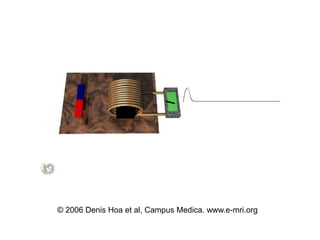
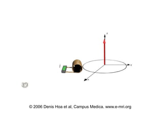
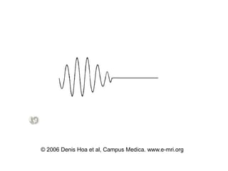
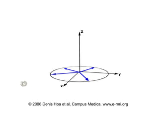
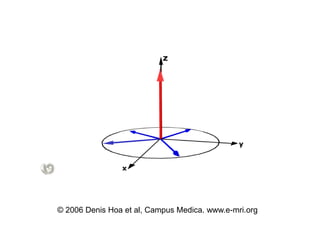
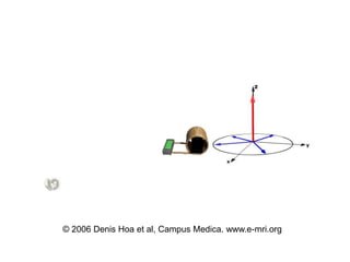
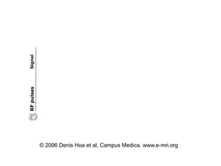
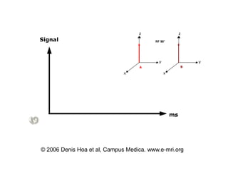
The document discusses the physical principles behind signal generation in magnetic resonance imaging (MRI). It describes how hydrogen protons generate a signal when placed in a strong magnetic field. The protons precess around the field at their Larmor frequency. A radiofrequency pulse excites the protons, which then relax back to equilibrium and emit a magnetic resonance signal that is detected by a receiver coil. The signal decays over time according to the T1 longitudinal relaxation time and T2* transverse relaxation time, which are tissue properties that can provide contrast for MRI.Basics of CT- Lecture 4.ppt
Basics of CT- Lecture 4.pptMagde Gad
╠²
The document provides details on the key components of a computed tomography (CT) scanner. It discusses the gantry, which houses the x-ray tube, detector array, and high voltage generator. It describes different types of x-ray tubes, detectors, including scintillation and gas ionization detectors, and collimators. The computer is also highlighted as a crucial component, processing the data collected and representing the cross-sectional images.More Related Content
Similar to E mri mr_signal_slides (20)
BREAST IMAGING IN DIFFERENT CONDITIONS OF BREAST
BREAST IMAGING IN DIFFERENT CONDITIONS OF BREASTDharmendraThakur26
╠²
This document summarizes various breast imaging modalities. It discusses the role of mammography, ultrasound, MRI, PET, and other techniques in evaluating the breast. Mammography remains the primary screening tool but has limitations related to breast density. Ultrasound helps diagnose palpable lesions and differentiate cysts from solid masses. MRI detects additional cancers but has limitations of cost and availability. Combined modalities provide improved evaluation of the breast compared to single techniques alone.Mri breast,indication,contra,prtocol,techniq,curves
Mri breast,indication,contra,prtocol,techniq,curvesDr;Mohamed alkenawy
╠²
MRI of the breast has certain contraindications including the presence of metallic implants, those unable to lie prone, or with large body habitus. Normal breast tissue may enhance asymmetrically, so scheduling during days 7-20 of the menstrual cycle can provide less enhancement. Dedicated breast coils are used with patients lying prone, and protocols involve unilateral or bilateral imaging with pre-and post-contrast sequences to analyze enhancement kinetics. Morphological features like irregular shapes and enhancement kinetics help identify lesions, with Type I curves associated with benign lesions and Type III with malignancy. MRI is useful for screening, determining tumor extent, assessing recurrence or residual disease, and providing information not available from other imaging methods.Image guided surgery
Image guided surgeryShivram Gautaam
╠²
This document discusses image guided surgery, which uses computer technology and 3D imaging like CT and MRI scans to guide surgical interventions. Key aspects covered include:
- Image guidance allows surgeons to view a patient's anatomy during surgery to locate structures hidden from direct vision.
- Registration aligns the 3D imaging with the patient's actual anatomy using tracking devices and fiducial markers.
- Volume rendering and surface rendering techniques are used to visualize 3D models of the patient's anatomy overlaid during surgery.
- Accuracy depends on factors like registration error and tracking device precision. Image guidance is useful for locating small structures in complex areas like the skull base.Mri by talha
Mri by talhaTalha Javed
╠²
MRI produces detailed images of the body's internal structures using magnetic fields and radio waves rather than ionizing radiation. It was developed in the 1970s and the first human images were published in 1977. MRI is commonly used to diagnose conditions affecting organs and tissues like the brain, heart, muscles, joints, and blood vessels. It works by aligning hydrogen atoms with magnetic fields and using radio waves to produce signals that are formed into detailed images.MRI (Magnetic resonance imaging)
MRI (Magnetic resonance imaging)Summu Thakur
╠²
MRI has become an integral imaging tool over the last 20 years. It uses magnetic fields and radio waves to create detailed images of organs and tissues without exposing patients to ionizing radiation. Different pulse sequences (T1, T2, proton density etc.) along with contrast agents allow MRI to characterize soft tissues and pathology. It is commonly used to image the brain, spine, joints, soft tissues, and for angiography. Recent advances include diffusion MRI, spectroscopy, and functional MRI. MRI has good soft tissue contrast but is more expensive than other modalities.MEDICAL IMAGING NOTES
MEDICAL IMAGING NOTESAsifullahSamim
╠²
The document provides an overview of medical imaging, detailing various techniques such as x-rays, CT scans, MRI, ultrasound, and nuclear medicine, which are essential for diagnosing and treating medical conditions. It discusses specific modalities like projection radiography, highlighting its use, limitations, and the effects of magnification on image quality. Additionally, the document explains the advantages and applications of imaging technologies like nuclear medicine and digital radiography, emphasizing their roles in modern healthcare.3d Printing For The Radiologist 1st Edition 1st Edition Nicole Wake
3d Printing For The Radiologist 1st Edition 1st Edition Nicole Wakemekdesemrani
╠²
3d Printing For The Radiologist 1st Edition 1st Edition Nicole Wake
3d Printing For The Radiologist 1st Edition 1st Edition Nicole Wake
3d Printing For The Radiologist 1st Edition 1st Edition Nicole WakeBiomedical Imaging Applications and Advances 1st Edition Peter Morris
Biomedical Imaging Applications and Advances 1st Edition Peter Morrisguielhorref0
╠²
Biomedical Imaging Applications and Advances 1st Edition Peter Morris
Biomedical Imaging Applications and Advances 1st Edition Peter Morris
Biomedical Imaging Applications and Advances 1st Edition Peter MorrisBody MRI
Body MRIPatty G.
╠²
This document provides an overview of body MRI techniques and MRI of the liver. It discusses MRI field strength and surface coils, common pulse sequences including T1-weighted, T2-weighted, STIR, and contrast-enhanced imaging. It also summarizes key findings of many primary and secondary liver lesions and diseases that can be evaluated by MRI. The goal is to familiarize radiologists with liver MRI findings to establish tissue diagnoses and accurately diagnose and stage neoplasms.Surgical radiology and its uses in the hospital
Surgical radiology and its uses in the hospitalSalarKhanJadoon1
╠²
Surgical radiology and its uses in the hospitalBasicunderstandingonmagneticresonanceimagingmri 141231045409-conversion-gate01
Basicunderstandingonmagneticresonanceimagingmri 141231045409-conversion-gate01Hannah Rajsekhar
╠²
The document discusses the importance of physiotherapists understanding basic MRI interpretation. It notes that some patients come for treatment without their MRI interpretation sheet, leaving blank images for the physiotherapist to interpret. Having a basic understanding of MRI principles, machine components, image types and interpretation helps physiotherapists confirm assessments when the interpretation sheet is missing. The document aims to provide this foundational MRI knowledge for physiotherapists.Pattern recognition in medical images
Pattern recognition in medical imagesJayabalanRajalakshmi
╠²
This document provides an overview of pattern recognition in medical images. It discusses several medical imaging modalities like computed tomography (CT), magnetic resonance imaging (MRI), ultrasound, and single photon emission computed tomography (SPECT). CT and MRI can provide detailed cross-sectional images of the body with good soft tissue contrast. Ultrasound imaging provides real-time images and is widely used during procedures due to its lack of radiation. SPECT involves injecting radioactive tracers to produce tomographic images based on physiological processes. Overall, medical image analysis applies general image processing and computer vision techniques to medical images to supplement expert diagnosis and provide quantitative information.Functional MRI Techniques in modern MRI.pdf
Functional MRI Techniques in modern MRI.pdfrajaarjunan74
╠²
This document discusses various MRI techniques including functional MRI (fMRI), magnetic resonance angiography (MRA), magnetic resonance cholangiopancreatography (MRCP), MR urography, and basic MRI concepts like k-space. It provides details on the principles, sequences, and clinical applications of each technique. fMRI maps brain activity during tasks using the BOLD response. MRA noninvasively images blood vessels using techniques like time-of-flight. MRCP visualizes the biliary and pancreatic ducts without contrast. These techniques allow evaluation of various anatomical and functional systems in a noninvasive manner.How 3D Technology Is Transforming Medical Imaging
How 3D Technology Is Transforming Medical ImagingApparao Mukkamala
╠²
The document discusses the advancements in 3D medical imaging technologies, highlighting their impact on clarity, detail, and efficiency in medical diagnoses. It outlines significant improvements in imaging techniques such as cinematic rendering, tomosynthesis, and AI integration that enhance the accuracy of diagnoses and treatment planning. The future of medical imaging is expected to be driven by increased computational power, AI, and cloud hosting, which will further streamline processes and improve patient care.MRI for Surgeons introduction and basics
MRI for Surgeons introduction and basicsrohitsharma19711
╠²
The document provides an extensive overview of Magnetic Resonance Imaging (MRI), detailing its history, mechanisms, imaging techniques, and various applications including brain, liver, and breast imaging. It highlights the principles of MRI, such as resonance and signal creation, and discusses the use of contrast agents and imaging sequences to enhance diagnostic accuracy. The conclusion emphasizes the significance of MRI knowledge for surgical training, particularly in identifying neoplastic tissues and distinguishing scar from recurrence.Mammography
MammographySumanjali Anjali
╠²
This document provides an overview of mammography presented by Sumanjali N. of Manipal Hospital in Whitefield, Bengaluru. It begins with an introduction to mammography and breast anatomy. It then discusses breast cancer and various imaging modalities used including mammogram, ultrasound, tomosynthesis, PET mammogram, MR mammogram, and thermography. The role of a mammography technologist is outlined. Standard mammographic views and the breast imaging reporting and data system (BI-RADS) for assessing findings are described. Common mammographic artifacts are also reviewed. The presentation concludes by emphasizing the importance of screening mammography in early breast cancer detection and reassurance of patients.Basic Science for the MRCS: A revision guide for surgical trainees 4th Editio...
Basic Science for the MRCS: A revision guide for surgical trainees 4th Editio...aikeebakle
╠²
Basic Science for the MRCS: A revision guide for surgical trainees 4th Edition Michael S. Delbridge & Wissam Al-Jundi
Basic Science for the MRCS: A revision guide for surgical trainees 4th Edition Michael S. Delbridge & Wissam Al-Jundi
Basic Science for the MRCS: A revision guide for surgical trainees 4th Edition Michael S. Delbridge & Wissam Al-JundiRadiology ppt
Radiology pptQURATULAIN MUGHAL
╠²
This document provides an overview of magnetic resonance imaging (MRI). It begins by defining MRI as a medical imaging technique that uses magnetic fields and radio waves to produce detailed images of the body. The document then covers the history, components, principles, and types of MRI images. It discusses how MRI works to detect tissue properties using relaxation times and how varying pulse sequences produces different contrasts. The document concludes by outlining the clinical applications and benefits of MRI, such as its ability to clearly image soft tissues without radiation, as well as some limitations like expense and the enclosed scanner.Magnetic resonance imaging
Magnetic resonance imagingvishal gupta
╠²
MRI uses strong magnetic fields and radio waves to generate images of the inside of the body. It is a medical imaging technique widely used in radiology to visualize anatomy and physiological processes. MRI has many medical uses and applications across different body systems. It is generally a safe technique but there are some risks needing consideration for things like implants, projectile effects, and claustrophobia. Guidelines and certifications aim to standardize roles and ensure safe MRI practices.More from Magde Gad (12)
_3 very important.ppt
_3 very important.pptMagde Gad
╠²
The document discusses the physical principles behind signal generation in magnetic resonance imaging (MRI). It describes how hydrogen protons generate a signal when placed in a strong magnetic field. The protons precess around the field at their Larmor frequency. A radiofrequency pulse excites the protons, which then relax back to equilibrium and emit a magnetic resonance signal that is detected by a receiver coil. The signal decays over time according to the T1 longitudinal relaxation time and T2* transverse relaxation time, which are tissue properties that can provide contrast for MRI.Basics of CT- Lecture 4.ppt
Basics of CT- Lecture 4.pptMagde Gad
╠²
The document provides details on the key components of a computed tomography (CT) scanner. It discusses the gantry, which houses the x-ray tube, detector array, and high voltage generator. It describes different types of x-ray tubes, detectors, including scintillation and gas ionization detectors, and collimators. The computer is also highlighted as a crucial component, processing the data collected and representing the cross-sectional images.Basics of CT- Lecture 7.ppt
Basics of CT- Lecture 7.pptMagde Gad
╠²
This document discusses the basics of computed tomography (CT) image manipulation and windowing. It explains that CT images can be modified in various ways, including changing filters and storing or transmitting images. It then describes windowing as a method to manipulate CT images by altering the CT numbers to optimize different structures. Finally, it discusses how window width and level are used to select an appropriate range of CT numbers to display tissues of interest in different grey scales for optimal viewing.Basics of CT- Lecture 3.ppt
Basics of CT- Lecture 3.pptMagde Gad
╠²
This document summarizes the evolution of computed tomography (CT) technology over several generations:
- 1st generation CT scanners from the 1970s took 5 minutes to generate a single image using a pencil-shaped beam and single detector.
- 3rd generation scanners from the 1980s reduced scan time to 3-5 seconds using a fan beam and detector array while rotating continuously.
- Current multislice CT scanners can scan multiple slices simultaneously in under 1 second using multiple detector arrays.
- The latest technology uses a stationary beam and detector configuration to achieve scan times under 50 milliseconds..ppt
.pptMagde Gad
╠²
This document provides guidelines for performing CT angiography exams of various body parts to evaluate for pulmonary embolism, deep vein thrombosis, and other vascular conditions. It describes patient positioning, contrast injection protocols, scan parameters and reconstruction techniques for CT angiography of the chest, upper extremities, lower extremities, brain, and abdomen. The goal is to comprehensively examine the vasculature with thin slices and multiplanar reconstructions to detect blood clots, aneurysms, and other abnormalities.Basics of CT- Lecture 5.ppt
Basics of CT- Lecture 5.pptMagde Gad
╠²
This document provides information about computed tomography (CT) scanning techniques. It discusses spiral or helical CT scanning where the X-ray tube rotates continuously around the patient while the couch moves the patient through the plane of rotation, allowing the reconstruction of transverse images along the entire length of the area being scanned. It describes how slip-ring technology, high-power X-ray tubes, and interpolation algorithms enabled the development of helical CT scanning. The document also discusses factors that influence the volume of tissue imaged like examination time, couch travel, pitch, and collimation.- Lecture 2.ppt
- Lecture 2.pptMagde Gad
╠²
The document provides an overview of computed tomography (CT) image reconstruction. It explains that after transmission measurements are taken by detectors, the data is sent to a computer for processing using reconstruction algorithms. The most widely used algorithm is filtered backprojection, which builds up the CT image by essentially reversing the data acquisition steps and smearing attenuation values along ray paths in the image. This reinforces areas of similar attenuation, reconstructing the image matrix. The document also discusses CT number scales, windowing, and other techniques for manipulating and displaying the reconstructed CT image.-Lecture 1.ppt
-Lecture 1.pptMagde Gad
╠²
This document discusses radiation safety and protection in computed tomography (CT) scanning. It defines stochastic and deterministic effects of radiation, and explains that stochastic effects depend on probability of occurrence with increased exposure while deterministic effects have a threshold dose above which the probability of the effect is 100%. It also discusses units used to measure radiation dose exposure, absorbed dose, and dose equivalent. The document focuses on measuring radiation dose from CT scanning, defining and measuring the CT dose index (CTDI) and multiple scan average dose (MSAD). It explains how changing parameters like kVp can affect radiation dose and image quality.-Lecture 2.ppt
-Lecture 2.pptMagde Gad
╠²
This document provides information about computed tomography (CT) scans and factors that affect radiation dose, including:
1. Helical pitch affects dose, with lower pitches increasing dose and higher pitches decreasing dose. Tube current and time also affect dose linearly, with higher mAs increasing dose.
2. Effective radiation dose from CT scans is much higher than conventional x-rays but still less than typical background radiation exposure per year. Automatic exposure control helps reduce dose by modulating tube current based on patient attenuation.
3. Radiation dose to patients and staff is carefully monitored and controlled through techniques like collimation, time, distance and shielding to keep exposure As Low As Reasonably Achievable.Basics of CT- Lecture 9.ppt
Basics of CT- Lecture 9.pptMagde Gad
╠²
This document provides a summary of key concepts in computed tomography (CT) imaging. It discusses back projection reconstruction techniques which can produce blurred images. It also describes filtered back projection which uses digital subtraction filtering to reduce blurring. Fourier reconstruction techniques are described where projection data is transformed to the frequency domain using Fourier transforms before being reconstructed into a spatial domain image. Different window widths and window levels are discussed for optimizing soft tissue versus bone imaging.Basics of CT- Lecture 6.ppt
Basics of CT- Lecture 6.pptMagde Gad
╠²
This document discusses the basics of computed tomography (CT) and multislice CT. It explains that multislice CT uses multiple parallel detector arrays rather than a single detector array, and an X-ray tube with high heat capacity. It describes the increasing number of slices used in multislice CT scanners over time, from dual to 64 slices. The document also discusses clinical applications of multislice CT such as improved imaging of trauma patients and angiography, as well as advantages over conventional CT such as faster acquisition and reduced radiation dose.Basics of CT- Lecture 1.ppt
Basics of CT- Lecture 1.pptMagde Gad
╠²
This document provides an overview of computed tomography (CT) scanning. It discusses limitations of conventional x-rays like superimposition and poor contrast resolution. CT addresses these issues by using a narrow x-ray beam and detectors to measure transmission and calculate attenuation values for image reconstruction. The data acquisition system acquires multiple projections from different angles to form cross-sectional images with excellent contrast and no superimposition. CT numbers provide numerical values related to tissue density and attenuation.Ad
Recently uploaded (20)
Ayurveda: Ancient Wisdom for Modern Wellness
Ayurveda: Ancient Wisdom for Modern WellnessNutrazin
╠²
Discover how the 5,000-year-old science of Ayurveda offers practical, daily habits for maintaining holistic health in todayŌĆÖs fast-paced world. This presentation explores the foundational principles of Ayurveda, including the three doshas, daily wellness routines (Dinacharya), seasonal practices (Ritucharya), and powerful natural remedies like herbal teas, adaptogens, and mindful breathing.
Whether you're a beginner or wellness enthusiast, learn how integrating simple Ayurvedic practices can enhance immunity, mental clarity, digestion, and emotional balance ŌĆö all without dependency on synthetic medications.Shamis Tate Shares 5 Essential Brain Health Tips.pdf
Shamis Tate Shares 5 Essential Brain Health Tips.pdfShamis Tate
╠²
Neurologist Shamis Tate shares 5 simple and effective brain health tips. Learn how good sleep, healthy food, staying active, using your mind, and connecting with others can help keep your brain strong, sharp, and healthy. Easy steps that anyone can follow to feel better and think clearer every day.Acetabular Labral Tears: Anatomy, Diagnosis, and Management
Acetabular Labral Tears: Anatomy, Diagnosis, and ManagementDr. Prabhat Pandey
╠²
This detailed presentation by Dr. Prabhat Pandey (DNB Resident, Tata Main Hospital, Jamshedpur) provides a comprehensive overview of acetabular labral tears, covering their anatomical, biomechanical, and clinical implications. Beginning with the anatomy of the acetabular labrumŌĆöa fibrocartilaginous structure critical for hip joint stabilityŌĆöthe presentation explains how its vascular and neural distribution contributes to both pain and poor healing potential when torn.
The pathophysiology of labral tears is discussed in the context of altered joint mechanics, loss of the labral seal, increased cartilage stress, and subsequent joint degeneration. The presentation classifies labral tears based on location, morphology, and etiology, highlighting causes such as trauma, femoroacetabular impingement (FAI), developmental dysplasia of the hip (DDH), and biomechanical imbalances.
Key clinical features are outlined, including chronic anterior hip or groin pain, mechanical symptoms like clicking and locking, and delayed diagnosisŌĆöespecially in athletes and women. Diagnostic strategies include physical tests (e.g., FABER, McCarthy test, anterior impingement test), imaging (X-ray, CT, MRI, and MR arthrography), and diagnostic injections.
Management strategies range from conservative treatmentŌĆörest, anti-inflammatory drugs, and physiotherapyŌĆöto surgical options, such as arthroscopic debridement and microfracture techniques. Postoperative rehabilitation focuses on early mobilization, DVT prophylaxis, and gradual return to function, tailored to reduce recurrence and promote joint preservation.
This presentation is an excellent educational resource for orthopedic residents, sports medicine practitioners, physiotherapists, and clinicians managing hip pathologies.Dental Equipment List for Clinics and Hospitals
Dental Equipment List for Clinics and Hospitalsdrsupriyasinha715
╠²
Looking to explore the essential tools used in modern dentistry? This comprehensive presentation on Dental Equipment List covers a wide range of instruments and devices commonly found in dental clinics and hospitals. Whether you're a dental student, healthcare professional, or someone researching dental setups, this PPT offers a clear and concise overview of key equipment ŌĆö from diagnostic tools like dental mirrors and probes to advanced machines like autoclaves, X-ray units, and ultrasonic scalers.
Reliable Ambulance Services Saving Lives Across Cities.pdf
Reliable Ambulance Services Saving Lives Across Cities.pdfAmbulance services in delhi
╠²
Experience fast and dependable ambulance services in Prayagraj, designed to provide critical medical support during emergencies. Our well-equipped ambulances and skilled paramedics ensure safe and comfortable patient transport, available round-the-clock to assist you whenever needed. Trust us for professional and compassionate care every step of the way.Congenital Vertical Talus: Clinical Presentation, Imaging, Etiology, and Mult...
Congenital Vertical Talus: Clinical Presentation, Imaging, Etiology, and Mult...Dr. Prabhat Pandey
╠²
Congenital Vertical Talus (CVT)ŌĆöalso known as Congenital Convex Pes Valgus or Teratologic Dorsolateral Dislocation of the Talocalcaneonavicular JointŌĆöis a rare but significant pediatric foot deformity, characterized by a fixed dorsal dislocation of the talonavicular joint and rigid hindfoot equinus. This unique anatomical disruption gives rise to the hallmark "rocker-bottom" deformity, often evident at birth.
In this comprehensive presentation by Dr. Prabhat Pandey, DNB Orthopedics Resident at Tata Main Hospital, we explore every crucial aspect of Congenital Vertical TalusŌĆöfrom its pathoanatomy and etiology to detailed clinical assessment and advanced management strategies. This ║▌║▌▀ŻShare is tailored for orthopedic surgeons, pediatricians, medical students, and physiotherapists engaged in pediatric musculoskeletal care.
Congenital Vertical Talus is rare, rigid, and complex
Must distinguish from positional deformities like calcaneovalgus foot
Accurate radiographic analysis is essential for diagnosis
Reverse Ponseti casting can reduce need for extensive surgery
Surgical release is often definitive but must be timed and tailored
Multidisciplinary management is crucial in syndromic casesPersonalized Autism Therapies: Where Are We Currently?
Personalized Autism Therapies: Where Are We Currently?Advancells: Stem Cell Lab and Research
╠²
Autism, a neurodevelopmental disorder affecting social communication and behavior, impacts approximately 61.8 million people worldwide. Each patient exhibits unique symptoms and needs, emphasizing the importance of tailored treatment strategies. Explore the growing scientific interest in personalized approaches and the promising role of regenerative medicine in autism treatment. Discover how customized strategies are shaping the future of neurodevelopmental disorder care.Revision Total Hip Replacement (THR): Surgical Principles, Techniques, and Bo...
Revision Total Hip Replacement (THR): Surgical Principles, Techniques, and Bo...Dr. Prabhat Pandey
╠²
Revision Total Hip Replacement (THR) is among the most demanding procedures in orthopedic surgery. Unlike primary THR, it involves addressing pre-existing complications such as implant failure, dislocation, bone loss, or infection, often in the presence of compromised soft tissues and bone quality. While primary THR offers high success rates and reproducible outcomes, revision THR requires greater surgical expertise, meticulous planning, and personalized execution to restore function and minimize complications.
This presentation by Dr. Prabhat Pandey is designed as a comprehensive resource for orthopedic surgeons, residents, and joint reconstruction teams who seek to understand and master the essential principles, decision-making algorithms, and advanced techniques used in revision THR.
Revision hip arthroplasty is associated with:
Increased operative time and blood loss
Higher complication rates: infection, dislocation, thromboembolism, nerve injury, and periprosthetic fracture
The need to manage weakened bone stock and soft tissue envelope
Difficulty in removing failed implants, including intramedullary components.
Revision surgery is warranted in cases of:
Prosthetic dislocation or instability
Mechanical loosening of components
Implant wear or breakage
Periprosthetic fracture
Deep infection
Osteolysis or bone resorption
Painful loosening without overt signs of mechanical failure
Each case demands tailored intervention based on the severity and combination of these factors.
3. Principles of Successful Revision THR
Safe removal of loose or failed components
Preservation of host bone and soft tissue integrity
Effective reconstruction of bone defects using bone grafts or metal augments
Achieving stability of the revision components
Restoration of biomechanics, particularly the anatomical center of rotation.
Dislocation rates:
0.3ŌĆō10% in primary THR
Up to 28% in revision THR
Influencing Factors:
Surgical approach
Abductor muscle function
Component malpositioning
Patient compliance
Prosthesis longevity
Underlying diagnosis (e.g., hip fracture, neuromuscular disorder
Revision Total Hip Replacement is not merely a technical endeavorŌĆöit is a test of surgical judgment, experience, and adaptability. Successful outcomes hinge on:
Thorough preoperative assessment
Judicious use of classification systems like Paprosky
Proficiency in implant removal
Skillful reconstruction of defects
Meticulous postoperative care
This presentation distills the complex decision-making process into an organized, practical guide for any surgeon facing the challenge of revision THR.
Understanding Surgical Approaches and Their Risks
Dislocation Types and Management
Preoperative Planning Essentials
Surgical Options for Instability and Revision
Operative Exposure and Implant Removal
Techniques for Removing Cemented Implants
Cementless Implant Removal
Managing Bone Deficiency in the Acetabulum
Paprosky Classification: Detailed Breakdown
Public-Private Partnerships in Healthcare_ A Sustainable Model for African Na...
Public-Private Partnerships in Healthcare_ A Sustainable Model for African Na...oliverwanyama96
╠²
Across Africa, healthcare systems are under increasing pressure from rising populations, chronic underfunding, and infrastructure gaps that limit access to quality care. In response, Public-Private Partnerships (PPPs) are gaining momentum as a sustainable model to deliver healthcare infrastructure, services, and innovation.
"Why Am I Not Affected?" ŌĆö Understanding Emotional Detachment
"Why Am I Not Affected?" ŌĆö Understanding Emotional DetachmentRangen A. Ghafur
╠²
Not everyone cries after loss.
Some of us feelŌĆ” nothing.
This presentation explores why some people go numb, how it feels, and what it really means.
Ō£© Based on science, reflection, and quiet truths we rarely say out loud.
If youŌĆÖve ever asked yourself, ŌĆ£Why donŌĆÖt I feel like others do?ŌĆØ ŌĆö this is for you.Common Medical Billing Errors.pppptttttx
Common Medical Billing Errors.pppptttttxjohnbutlermbc
╠²
Explaining how Common Medical Billing Errors could be costing you revenue loss, and how Medical Billers and Coders can help you to reduce it.
MCH (MATERNAL AND CHILD HEALTH) HEALTH Programmes
MCH (MATERNAL AND CHILD HEALTH) HEALTH ProgrammesDr. Mamta Gehlawat
╠²
A glimpse of some important existing MCH related programmes and schemes for 3rd year MBBS studentsPPT Anticancer agent for B Pharm Fifth Semester students
PPT Anticancer agent for B Pharm Fifth Semester studentsDinesh Kawade
╠²
Anti-neoplastic agents:
Alkylating agents: Meclorethamine*, Cyclophosphamide, Melphalan,Chlorambucil, Busulfan, Thiotepa
Antimetabolites: Mercaptopurine*, Thioguanine, Fluorouracil, Floxuridine, Cytarabine, Methotrexate*, Azathioprine
Antibiotics: Dactinomycin, Daunorubicin, Doxorubicin, Bleomycin
Plant products: Etoposide, Vinblastin sulphate, Vincristin sulphate
Miscellaneous: Cisplatin, Mitotane.
Errors that Could be Deemed Fraudulent in Ambulance Billing.pptx
Errors that Could be Deemed Fraudulent in Ambulance Billing.pptxjohnbutlermbc
╠²
Ambulance Billing could be replete with errors that could end up in being a fraudulent case. Know how to ensure correct billing.
Dengue Basic Information_Mitigation & Response_Dr. Pankaj Singh
Dengue Basic Information_Mitigation & Response_Dr. Pankaj Singhpanpunpra
╠²
Dengue Basic Information_Mitigation & Response_Dr. Pankaj SinghWeight loss podcast motivation for 2025.
Weight loss podcast motivation for 2025.lalaaligujjar
╠²
Weight loss tips.weight loss.weight loss for beginners VoyageHealing- ketamine and wellness clinic.pdf
VoyageHealing- ketamine and wellness clinic.pdfVoyage Healing
╠²
A Compassionate Ketamine and Wellness Clinic.
At Voyage, we are reimagining mental health treatment offering talk therapy, medication management, and novel tools like ketamine therapy and Spravato.
Gastric Carcinoma 360 :Surgical & Oncologic Approach
Gastric Carcinoma 360 :Surgical & Oncologic ApproachDR.PRATIK JADHAV
╠²
This presentation provides a comprehensive overview of gastric carcinoma, covering its epidemiology, anatomy of the stomach, histology, etiology and risk factors, pathology, diagnostic approach, and updated management guidelines.
Entire aspect of Gastric cancer have been explained in detail before exam
Key sections include:
Global and Indian incidence patterns
Detailed stomach anatomy and blood supply
Lauren and WHO histologic classifications
TNM staging (AJCC 8th edition)
Diagnostic modalities: Endoscopy, imaging, laparoscopy
Surgical techniques: Total/subtotal gastrectomy, D1 vs D2 lymphadenectomy
Perioperative chemotherapy: FLOT, XELOX, S-1
Advanced/metastatic disease protocols including trastuzumab, nivolumab, and ramucirumab
Role of minimally invasive surgery, robotic gastrectomy, and palliative care
Prognostic factors and structured follow-up plans
Recent advances including CAR-T therapy, ctDNA monitoring, and immunogenomic classifications
Ideal for medical students, surgical residents, and oncologists, this evidence-based PPT includes clinical images, procedural illustrations, and key international guidelines from NCCN, ESMO, JGCA.Congenital Vertical Talus: Clinical Presentation, Imaging, Etiology, and Mult...
Congenital Vertical Talus: Clinical Presentation, Imaging, Etiology, and Mult...Dr. Prabhat Pandey
╠²
Revision Total Hip Replacement (THR): Surgical Principles, Techniques, and Bo...
Revision Total Hip Replacement (THR): Surgical Principles, Techniques, and Bo...Dr. Prabhat Pandey
╠²
Ad
E mri mr_signal_slides
- 1. ┬® 2006 Denis Hoa et al, Campus Medica. www.e-mri.org
- 2. ┬® 2006 Denis Hoa et al, Campus Medica. www.e-mri.org
- 3. ┬® 2006 Denis Hoa et al, Campus Medica. www.e-mri.org
- 4. ┬® 2006 Denis Hoa et al, Campus Medica. www.e-mri.org
- 5. ┬® 2006 Denis Hoa et al, Campus Medica. www.e-mri.org
- 6. ┬® 2006 Denis Hoa et al, Campus Medica. www.e-mri.org
- 7. ┬® 2006 Denis Hoa et al, Campus Medica. www.e-mri.org
- 8. ┬® 2006 Denis Hoa et al, Campus Medica. www.e-mri.org
- 9. ┬® 2006 Denis Hoa et al, Campus Medica. www.e-mri.org
- 10. ┬® 2006 Denis Hoa et al, Campus Medica. www.e-mri.org
- 11. ┬® 2006 Denis Hoa et al, Campus Medica. www.e-mri.org
- 12. ┬® 2006 Denis Hoa et al, Campus Medica. www.e-mri.org
- 13. ┬® 2006 Denis Hoa et al, Campus Medica. www.e-mri.org
- 14. ┬® 2006 Denis Hoa et al, Campus Medica. www.e-mri.org
- 15. ┬® 2006 Denis Hoa et al, Campus Medica. www.e-mri.org
- 16. ┬® 2006 Denis Hoa et al, Campus Medica. www.e-mri.org
- 17. ┬® 2006 Denis Hoa et al, Campus Medica. www.e-mri.org
- 18. ┬® 2006 Denis Hoa et al, Campus Medica. www.e-mri.org
- 19. ┬® 2006 Denis Hoa et al, Campus Medica. www.e-mri.org