


More Related Content
Similar to endocrine system anatomy.pptx for BSN Generic (20)
Recently uploaded (20)


endocrine system anatomy.pptx for BSN Generic
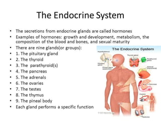
- 2. ’é× The endocrine system includes the endocrine glands and their hormones ’é× The function of the endocrine system is to secrete hormones into the bloodstream. ’é× Hormone: A Chemical messenger which targets a specific group of cells, in order to cause that group of cells do some activity or stop doing an activity.
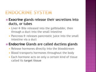
- 3. ’é× Exocrine glands release their secretions into ducts, or tubes ’éĪ Liver’āĀ Bile released into the gallbladder, then through a duct into the small intestine ’éĪ Pancreas’āĀ releases pancreatic juice into the small intestine via a duct ’é× Endocrine Glands are called ductless glands ’éĪ Release hormones directly into the bloodstream ’éĪ Blood transports hormones throughout the body ’éĪ Each hormone acts on only a certain kind of tissue called its target tissue
- 5. ’é× Anatomical Position and Relations ’é× The pituitary gland is a pea-sized oval structure, suspended from the underside of the brain by the pituitary stalk (known as the infundibulum). It sits within a small depression in the sphenoid bone, known as the sella turcica (ŌĆśŌĆÖTurkish saddleŌĆÖŌĆÖ). ’é× The superior surface of the gland is covered by a reflection of the dura mater ŌĆō the diaphragma sellae. This membrane has a central opening which allows passage of the infundibulum.
- 6. ’é× Anatomically, the pituitary gland is a ŌĆśŌĆÖtwo- in-oneŌĆÖŌĆÖ structure consisting of the anterior pituitary and the posterior pituitary. These parts have different embryonic origins and function very differently.
- 7. ’é× Anterior Lobe ’é× The anterior lobe (adenohypophysis) is derived from an outpouching of the roof of the pharynx, called RathkeŌĆÖs pouch. It is composed of glandular epithelium and secretes a number of hormones. The lobe can be further divided into three parts: ’é× Pars anterior ŌĆō the largest part, responsible for hormone secretion. ’é× Pars intermedia ŌĆō a thin epithelial layer that separates the pars anterior from the posterior lobe. ’é× Pars tuberalis ŌĆō an upwards extension of the pars anterior that surrounds the anterolateral aspect of the infundibulum.
- 8. ’é× The release of hormones is under the control of the hypothalamus, which communicates with the gland via neurotransmitters secreted into the hypophyseal portal vessels. These vessels ensure that the hypothalamic hormones remain concentrated, rather than being diluted in the systemic circulation.
- 9. ’é× Posterior Lobe ’é× The posterior lobe (neurohypophysis) consists of nervous tissue. ’é× Upon stimulation, the posterior lobe secretes two hormones ŌĆō ADH (responsible for control of blood osmolarity), and oxytocin (involved in parturition and milk secretion
- 11. ’é× The vasculature of the pituitary gland is complex and unique. Whilst the anterior lobe and posterior lobe have the same venous drainage (anterior and posterior hypophyseal veins), they have an individual arterial supply:
- 12. ’é× Anterior Pituitary ’é× The anterior pituitary gland receives arterial supply from the superior hypophyseal artery (a branch of the internal carotid artery).
- 13. ’é× Posterior Pituitary ’é× The infundibulum and posterior pituitary gland receive a rich blood supply from many arteries. Of these, the major vessels are the superior hypophyseal artery, infundibular artery and inferior hypophyseal artery.
- 14. ’é× The thyroid gland is an endocrine structure located in the neck. It plays a key role in regulating the metabolic rate of the body.
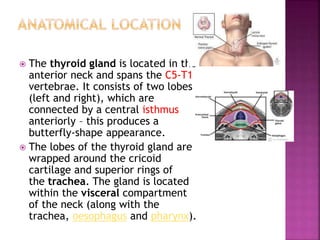
- 15. ’é× The thyroid gland is located in the anterior neck and spans the C5-T1 vertebrae. It consists of two lobes (left and right), which are connected by a central isthmus anteriorly ŌĆō this produces a butterfly-shape appearance. ’é× The lobes of the thyroid gland are wrapped around the cricoid cartilage and superior rings of the trachea. The gland is located within the visceral compartment of the neck (along with the trachea, oesophagus and pharynx).
- 16. ’é× The thyroid gland is closely associated with numerous other structures in the anterior neck: ’é× Anteriorly ŌĆō infrahyoid muscles, namely the sternothyroid, superior belly of the omohyoid and sternohyoid ’é× Laterally ŌĆō carotid sheath, containing the common carotid artey, internal jugular vein and vagus nerve ’é× Medially ŌĆō ’éĪ Organs ŌĆō larynx, pharynx, trachea and oesophagus ’éĪ Nerves ŌĆō external laryngeal and recurrent laryngeal
- 17. ’é× The arterial supply to the thyroid gland is via two main arteries: ’é× Superior thyroid artery ’é× Inferior thyroid artery ’é× In a small proportion of people (around 10%) there is an additional artery present ŌĆō the thyroid ima artery supplies the anterior surface and isthmus of the thyroid gland.
- 18. ’é× Venous Drainage ’é× Venous drainage is carried by the superior, middle, and inferior thyroid veins, which form a venous plexus around the thyroid gland. ’é× The superior and middle veins drain into the internal jugular vein and the inferior empties into the brachiocephalic vein.
- 19. ’é× Innervation ’é× The thyroid gland is innervated by branches derived from the sympathetic trunk. ’é× These nerves do not control the secretory function of the gland ŌĆō the release of thyroid hormones is regulated by the pituitary gland. ’é× Lymphatic Drainage ’é× The lymphatic drainage of the thyroid is to the paratracheal and deep cervical nodes.
- 20. ’é× The parathyroid glands are endocrine glands located in the anterior neck. ’é× They are responsible for the production of parathyroid hormone (PTH), which acts to increase the level of serum calcium.
- 21. ’é× The parathyroid glands are usually located on the posterior aspect of the thyroid gland. They are flattened and oval in shape ŌĆō. ’é× Most individuals have four parathyroid glands, although variation in number (from two to six) is common: ’é× Superior parathyroid glands (x2) ’é× Inferior parathyroid glands (x2)
- 22. ’é× The vascular supply is similar to that of the thyroid gland. ’é× Arterial supply is chiefly via the inferior thyroid artery (as this artery supplies the posterior aspect of the thyroid gland ŌĆō where the parathyroids are located ’é× Venous drainage is into the superior, middle, and inferior thyroid veins.
- 23. ’é× Lymphatics ’é× The lymphatic drainage from the parathyroid glands is to the paratracheal and deep cervical nodes. ’é× Nerves ’é× The parathyroid glands have an extensive supply of sympathetic nerves derived from thyroid branches of the cervical ganglia.
- 24. ’é× The pineal gland is a small endocrine gland located within the brain. Its main secretion is melatonin, which regulates the circadian rhythm of the body.
- 25. ’é× The pineal gland is small glandular body, approximately 6mm long. It is shaped like a pine cone, from which its name is derived. There are two types of cells present within the gland: ’é× Pinealocytes ŌĆō hormone secreting cells. ’é× Glial cells ŌĆō supporting cells.
- 26. ’é× The pineal gland is a midline structure, located between the two cerebral hemispheres. It is attached by a stalk to the posterior wall of third ventricle
- 27. ’é× The arterial supply to the pineal gland is profuse, second only to the kidney. The posterior choroidal arteries are the main supply; they are a set of 10 branches that arise from the posterior cerebral artery. ’é× Venous drainage is via the internal cerebral veins.
- 28. ’é× The adrenal (or suprarenal) glands are paired endocrine glands situated over the medial aspect of the upper poles of each kidney. ’é× They secrete catecholamines harmones directly into blood.
- 29. ’é× The adrenal glands are located in the posterior abdomen, between the superomedial kidney and the diaphragm. They are retroperitoneal, with parietal peritoneum covering their anterior surface only. ’é× The right gland is pyramidal in shape, contrasting with the semi-lunar shape of the left gland.
- 30. ’é× Perinephric (or renal) fascia encloses the adrenal glands and the kidneys. This fascia attaches the glands to the crura of the diaphragm. They are separated from the kidneys by the perirenal fat. ’é× The adrenal glands sit in close proximity to many other structures in the abdomen
- 31. ’é× The adrenal glands consist of an outer connective tissue capsule, a cortex and a medulla. ’é× Veins and lymphatics leave each gland via the hilum, but arteries and nerves enter the glands at numerous sites.
- 32. ’é× The outer cortex and inner medulla are the functional portions of the gland. ’é× Cortex ŌĆō ’é× Medulla ŌĆō ’é× The cortex and medulla synthesise different hormones.
- 33. ’é× Cortex ’é× The cortex is yellowish in colour .Functionally, the cortex can be divided into three regions (superficial to deep): ’é× Zona glomerulosa ŌĆō produces and secretes mineralocorticoids such as aldosterone. ’é× Zona fasciculata ŌĆō produces and secretes corticosteroids such as cortisol. It also secretes a small amount of androgens. ’é× Zona reticularis ŌĆō produces and secretes androgens such as dehydroepiandrosterone (DHES). It also secretes a small amount of corticosteroids.
- 34. ’é× Medulla ’é× The medulla lies in the centre of the gland, and is dark brown in colour. It contains chromaffin cells, which secrete catecholamines (such as adrenaline) into the bloodstream in response to stress. ’é× These hormones produce a ŌĆśflight-or-fightŌĆś response. Chromaffin cells also secrete enkephalins which function in pain control.
- 35. ’é× The adrenal glands have a rich blood supply via three main arteries: ’é× Superior adrenal artery ŌĆō ’é× Middle adrenal artery ’é× Inferior adrenal artery ’é× Right and left adrenal veins drain the glands. The right adrenal vein drains into the inferior vena cava, whereas the left adrenal vein drains into the left renal vein.
- 36. INNERVATION ’é× The adrenal glands are innervated by the coeliac plexus and greater splanchnic nerves. ’é× Sympathetic innervation to the adrenal medulla is via myelinated pre-synaptic fibres, mainly from the T10 to L1 spinal cord segments. LYMPHATICS ’é× Lymph drainage is to the lumbar lymph nodes by adrenal lymphatic vessels. These vessels originate from two lymphatic plexuses ŌĆō one deep to the capsule, and the other in the medulla.
- 37. ’é× The pancreas is an abdominal glandular organ with both digestive (exocrine) and hormonal (endocrine) functions.
- 38. ’é× The pancreas is an oblong-shaped organ positioned at the level of the transpyloric plane (L1). With the exception of the tail of the pancreas, it is a retroperitoneal organ, located deep within the upper abdomen in the epigastrium and left hypochondrium regions.
- 39. ’é× Stomach the stomach and pylorus lie anterior and to the pancreas. ’é× Duodenum ŌĆō The ŌĆ£CŌĆØ shaped duodenum curves around and outlines the head of the pancreas. The first part of the duodenum lies anteriorly whereas the second part of the duodenum including the ampulla of Vater lies laterally to the right of the pancreatic head ’é× Transverse mesocolon ŌĆō Attaches to the anterior surface of the pancreas ’é× Common bile duct ŌĆō Descends behind the head of the pancreas before opening into the second part of the duodenum alongside the major pancreatic duct through the major duodenal papilla ’é× Spleen ŌĆō located posteriorly and laterally.
- 40. ’é× Head ŌĆō the widest part of the pancreas. ’é× Uncinate process ŌĆō a projection arising from the lower part of the head and extending medially to lie beneath the body of the pancreas. ’é× Neck ŌĆō located between the head and the body of the pancreas. ’é× Body ŌĆō centrally located, crossing the midline of the human body to lie behind the stomach ’é× Tail ŌĆō the left end of the pancreas that lies within close proximity to the hilum of the spleen..
- 42. The exocrine pancreas is classified as a lobulated, serous gland which produces digestive enzyme precursors. It is composed of approximately one million ŌĆśberry-likeŌĆÖ clusters of cells called acini, connected by short intercalated ducts. ’é× The intercalated ducts unite with those draining adjacent lobules and drain into a network of intralobular collecting ducts, which in turn drain into the main pancreatic duct. ’é× The pancreatic duct runs the length of the pancreas and unites with the common bile duct, forming the hepatopancreatic ampulla of Vater. This structure then opens into the duodenum via the major duodenal papilla. ’é× Secretions into the duodenum are controlled by a muscular valve ŌĆō the sphincter of Oddi. It surrounds the ampulla of Vater, acting as a valve.
- 44. ’é× The pancreas is supplied by the pancreatic branches of the splenic artery. The head is additionally supplied by the superior and inferior pancreaticoduodenal arteries ’é× Venous drainage of the head of the pancreas is into the superior mesenteric branches of the hepatic portal vein. The pancreatic veins draining the rest of the pancreas do so via the splenic vein.
- 46. ’é× The pancreas is drained by lymphatic vessels that follow the arterial supply. They empty into the pancreaticosplenal nodes and the pyloric nodes, which in turn drain into the superior mesenteric and coeliac lymph nodes.