The document discusses the different tracts of the spinal cord. It describes short tracts that connect parts of the same spinal cord and long tracts that connect the spinal cord to other parts of the central nervous system. The long tracts are further divided into ascending tracts that carry sensory information from the spinal cord to the brain and descending tracts that carry motor information from the brain to the spinal cord. It provides details on major ascending tracts like the posterior, spinothalamic and spinocerebellar tracts. It also describes the descending tracts including the pyramidal and corticospinal tracts, as well as extrapyramidal tracts.
The document provides information on the nervous system, including its main divisions and components. It discusses:
1) The central nervous system (CNS) which includes the brain and spinal cord, and the peripheral nervous system (PNS) which connects the CNS to the rest of the body and is divided into somatic and autonomic systems.
2) The autonomic nervous system has two divisions - the sympathetic and parasympathetic systems which work in opposition to activate the body during stress and restore it to resting state.
3) The brain is the control center of the body and is made up of the cerebrum, cerebellum, and brain stem. It is protected by meninges and
The cerebellum is located in the posterior fossa of the skull below the brain. It has three main lobes - the anterior, middle, and flocculonodular lobes. The cerebellum coordinates voluntary movement through connections with other parts of the brain and spinal cord. It has an outer gray matter cortex containing Purkinje cells and an inner white matter. The cerebellum receives sensory information and regulates motor movements to produce smooth, coordinated activity.
Il diencefalo 竪 una delle 4 componenti maggiori dell'encefalo, quindi 竪 parte integrante del sistema nervoso centrale.
Posto tra cervello e tronco encefalico, alla base della scatola cranica, il diencefalo comprende 6 diverse strutture: il talamo, l'ipotalamo, l'epitalamo (a cui appartiene anche l'epifisi), il subtalamo, la neuroipofisi e il terzo ventricolo.
Il diencefalo agisce da stazione di elaborazione e smistamento di informazioni che intercorrono tra il cervello, il tronco encefalico e il midollo spinale; inoltre, 竪 coinvolto in numerose funzioni fondamentali alla vita, come per esempio il controllo dei sistemi sensoriali, la regolazione dell'ipofisi, l'apprendimento, la memoria e la regolazione del ciclo sonno-veglia.
Encefalo: Cos竪 in breve
L'encefalo 竪, assieme al midollo spinale, una delle due componenti fondamentali del sistema nervoso centrale.
Pesante circa 1,4 chilogrammi e contenente 100 bilioni neuroni (nell'essere umano adulto), l'encefalo 竪 una struttura molto complessa, suddivisibili in 4 grandi regioni, che sono:
Il cervello propriamente detto (o telencefalo o semplicemente cervello),
Il cervelletto,
Il diencefalo e
Il tronco encefalico.
The basal ganglia are a group of nuclei within the central nervous system that include the caudate, putamen, globus pallidus, subthalamic nucleus, and substantia nigra. They predict the effects of actions and execute action plans. The caudate, putamen, and globus pallidus are located within the cerebrum and supplied by the medial cerebral artery. The putamen and globus pallidus form the lentiform nucleus, while the caudate and putamen together are called the striatum.
The basal ganglia are a group of interconnected brain structures that include the caudate nucleus, putamen, globus pallidus, subthalamic nucleus, and substantia nigra. They are involved in regulating movement and certain movement disorders. The basal ganglia receive input from the cortex and thalamus and send output to the thalamus and brainstem areas. Dopamine from the substantia nigra helps modulate input and output signaling in the basal ganglia circuits. Disorders of these circuits can result in dyskinesias like tremors, chorea, athetosis, and ballismus or disturbances of muscle tone.
The pons is the bridge-shaped structure that connects the midbrain to the medulla oblongata. It contains fibers that connect the cerebellum and midbrain. The pons has a ventral surface with sulcus basilaris and lateral continuations with the middle cerebellar peduncle. It attaches to the 6th, 7th and 8th cranial nerves ventrally. Dorsally, it forms the upper part of the floor of the 4th ventricle. Internally, it contains longitudinal and transverse fibers, pontine nuclei in the basilar part, and ascending and descending tracts in the tegmental part such as the medial lemniscus and trigeminal lemn
The brainstem consists of 3 parts - the midbrain, pons, and medulla. It connects the spinal cord to the forebrain and has important functions like transmitting signals between the two, controlling vital reflexes like respiration, and containing nuclei for cranial nerves III-XII. The reticular formation is a network of fibers and neurons that extends through the brainstem and is involved in functions like motor control, sensory processing, visceral control, and neuroendocrine regulation. Other structures in the brainstem include the cerebral peduncles, cerebral aqueduct, inferior and superior colliculi, cranial nerve nuclei, and the pontine nuclei.
Anatomy of ascending and descending tractsMBBS IMS MSU
油
The document summarizes ascending and descending tracts in the spinal cord that transmit sensory and motor information between the spinal cord and brain. It describes the three neuron chain of ascending tracts that transmit exteroceptive, interoceptive, and proprioceptive sensory information from the peripheral receptors to the brain. It outlines the major ascending tracts - lateral and anterior spinothalamic tracts for pain/temperature and touch/pressure, and the posterior white column and cuneate and gracilis fasciculi for proprioception. It also summarizes the descending motor tracts including the corticospinal, rubrospinal, vestibulospinal, reticulospinal and tectospinal tracts that
This document summarizes the structure and types of cells in the nervous system. It describes that the nervous system contains central and peripheral divisions. The central nervous system contains the brain and spinal cord while the peripheral nervous system contains cranial and spinal nerves. There are two main cell types - neurons which are excitable cells that transmit signals and neuroglia which are non-excitable supporting cells. Neurons contain a cell body, dendrites and an axon. Neuroglia include astrocytes, oligodendrocytes and microglia. The document also provides an overview of the spinal nerve, reflex arc and different types of reflex actions.
The cerebellum is located at the back of the brain and is divided into 3 lobes. It coordinates movement and maintains balance and posture. The cerebellum receives input from sensory systems and the motor cortex and sends output through cerebellar nuclei to affect movement. At its core are Purkinje cells that help the cerebellum detect errors and coordinate precise movements through inhibition and excitation of deep nuclear cells and motor areas. Damage results in ataxia and loss of coordinated movement.
The autonomic nervous system regulates involuntary body functions and is divided into the sympathetic and parasympathetic nervous systems. It contains two neurons, with the preganglionic neuron cell body in the CNS and the postganglionic neuron cell body in autonomic ganglia. The sympathetic division is responsible for the fight or flight response and generally opposes the effects of the parasympathetic division, which is responsible for rest and digest functions. The enteric nervous system also helps control the gastrointestinal system independently.
Generaci坦n de Potenciales PostSin叩pticos Potencial de Acci坦nMedical & Gabeents
油
El documento describe la organizaci坦n de la sinapsis y la generaci坦n de potenciales postsin叩pticos. Espec鱈ficamente, explica c坦mo un potencial de acci坦n en la neurona presin叩ptica provoca la liberaci坦n de neurotransmisores que se unen a receptores en la neurona postsin叩ptica, generando potenciales graduados que pueden dar lugar a un potencial de acci坦n si se alcanza el umbral de excitaci坦n. Tambi辿n describe los mecanismos i坦nicos subyacentes a la generaci坦n y conducci坦n de potenciales de acci坦n a lo largo del ax
The thalamus is a structure located in the middle of the brain between the cerebral cortex and midbrain. It is the largest component of the diencephalon. The thalamus acts as a relay station for sensory information (except smell) sending signals to the appropriate areas of the cerebral cortex. It is divided into nuclei that each have distinct functions and connections related to motor control, sensory processing and integration, arousal, memory and cognition. Damage to different thalamic nuclei can disrupt various functions and result in sensory deficits, movement problems or changes to consciousness.
1). The reticular formation is an ill-defined region in the brainstem comprising neurons and fibers that extends from the spinal cord to the thalamus. It is involved in arousal, attention, sleep-wake cycles, and autonomic functions.
2). The ascending reticular activating system projects from the brainstem reticular formation to the thalamus and cortex, promoting wakefulness. The descending pathway projects to the spinal cord and is involved in motor function.
3). The reticular formation receives inputs from sensory systems and projects to the thalamus, hypothalamus, and spinal cord. It regulates functions like muscle tone, respiration, cardiovascular control, and endocrine secretion.
The document summarizes the autonomic nervous system. It describes the central nervous system as consisting of the brain and spinal cord. The peripheral nervous system is divided into the somatic and autonomic systems. The autonomic system is further divided into the sympathetic and parasympathetic systems. The sympathetic system is activated during stress and fights or flight responses. It increases heart rate, blood pressure, and lipolysis to provide energy. The parasympathetic system is active during rest and digestion and maintains normal functions.
This document discusses post-impressionist theories of vision and perception. It references several key figures such as Michel-Eug竪ne Chevreul who studied the interaction of colored light, as well as Charles Blanc, Odgen Rood, and Charles Henry who contributed to understanding how the eye and brain work together in visual processing.
Presentazione della Dott.ssa Eura Quattrone per una lezione su suono ed equilibrio alle classi quinte della scuola primaria Padre Marco d'Aviano del 2属 Circolo di Pordenone
6 cervelletto morfo e organizz interna 2.pdfreaerardr
油
Localizzato alla base del cervello, appena sopra al tronco cerebrale (il punto in cui midollo spinale e cervello si congiungono) e sotto ai lobi occipitali e temporali della corteccia cerebrale, il cervelletto 竪 una struttura formata da due emisferi all'interno dei quali sono presenti pi湛 del 50% dei neuroni contenuti nel cervello, di cui rappresenta il 10% circa del volume.
Storicamente 竪 considerato la sede del controllo dei movimenti. In effetti danni al cervelletto compromettono le capacit motorie e la postura, ma in realt gli impulsi al movimento non nascono qui. Piuttosto, questa struttura riceve informazioni dagli organi di senso, dal midollo spinale e da altre parti del cervello per poi coordinare i movimenti volontari, la postura, l'equilibrio e la parola, rendendoli pi湛 accurati.
Innovazione didattica: collaborative learning
Materiale prodotto dai ragazzi della classe 3属 indirizzo classico del Liceo Statale Vincenzo Monti.
A.s. 2016-2017, prof Gino
2. Funzioni del sistema nervoso
Tre funzioni sovrapposte
I recettori sensoriali eseguono un monitoraggio dei
cambiamenti allinterno e allesterno del corpo
Cambiamento uno stimolo
Guadagno di informazione input sensoriale
Il SNC processa e interpreta gli inputs sensoriali
Prende decisioni integrazione
Il SNC comanda una risposta attivando organi effettori
Risposta output motorio
3. Suddivisione di base del sistema nervoso:
Sistema Nervoso Centrale (SNC)
Sistema Nervoso Periferico SNP)
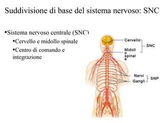
4. Suddivisione di base del sistema nervoso: SNC
Sistema nervoso centrale (SNC)
Cervello e midollo spinale
Centro di comando e
integrazione
SNC
SNP
Cervello
Midoll
o
spinal
e
Nervi
Gangli
5. Suddivisione di base del sistema nervoso: SNP
Al di fuori del SNC
I nervi che si estendono dal
cervello e dal midollo spinale
Nervi cranici
Nervi spinali
Collega tutte le regioni del
corpo al SNC
SNC
SNP
Cervello
Midoll
o
spinal
e
Nervi
Gangli
6. Input sensoriale e output motorio
I segnali sensoriali sono captati dai recettori sensoriali
Sono trasportati da fibre nervose afferenti dal SNP al SNC
I segnali motori partono dal SNC
Sono portati da fibre nervose efferenti del SNP agli effettori
Innervazione dei muscoli e delle ghiandole
7. Suddivisi a seconda della regione innervata
Regione somatica del corpo
Regione viscerale del corpo
Ne risultano quattro suddivisioni principali
Sensibilit somatica
Sensibilit viscerale
Motilit somatica
Motilit viscerale
Inputs sensoriali e outputs motori
8. Sensibilit somatica
Sensibilit somatica
Sensi somatici generali I recettori sono ampiamente
distribuiti
Tatto, dolore, vibrazione, pressione e temperatura
Sensi propriocettivi captano lallungamento dei tendini e
dei muscoli
Sensi corporei posizione e movimento del corpo nello
spazio (recettori articolari e vestibolari)
Sensi somatici speciali
Udito, equilibrio, visione, odorato
9. Sensibilit viscerale
Sensibilit viscerale
I sensi viscerali generici allungamento, dolore,
temperatura, nausea, fame
Ampiamente riscontrabili nei tratti digestivo e urinario
e negli organi riproduttori
Sensi viscerali speciali il gusto
10. Motilit somatica
Motilit somatica
Motilit somatica generale contrazione della
muscolatura scheletrica
Sotto il controllo volontario
Spesso denominato sistema nervoso volontario
11. Motilit viscerale
Motilit viscerale
Regola la contrazione della muscolatura liscia e cardiaca
e della secrezione ghiandolare
Costituisce il sistema nervoso autonomo
Controlla la funzione degli organi viscerali
Spesso definito sistema nervoso involontario
12. Sistema Nervoso Centrale
Cervello
Midollo spinale SNC
SNP
Cervello
Midoll
o
spinal
e
Nervi
Gangli
13. Termini direzionali del SNC
Termini direzionali unici per il SNC
Rostrale verso il naso (anteriore verso lalto)
Caudale verso la coda (posteriore verso il basso)
Dorsale posteriore
Ventrale anteriore
rostrale
caudal
e
14. Le quattro principali regioni del cervello
Emisferi cerebrali
Diencefalo
Tronco encefalico:
Mesencefalo
Ponte
Midollo allungato
(bulbo)
Cervelletto
Tronco
15. Ventricoli
Ripieni di fluido cerebrospinale
Rivestiti di cellule ependimali
In continuit fra di loro
In continuit col canale centrale del midollo spinale
Ventricolo
laterale
Terzo
ventrico
lo
Quarto
ventrico
lo
Ventricol
o
laterale
Terzo
ventrico
lo
Quarto
ventrico
lo
Canale
centrale
Canale
centrale
16. Lobo frontale
Antero-rostrale al solco centrale
Superiore alla fissura trasverso-laterale
Solco centrale
Lobo parietale
Lobo
occipitaleLobo
temporale
Solco
laterale
Fessura
cerebrale
trasversale
Fessura
longitudina
le
Lobo
frontale
Lobo
frontale
Solco
centrale
Lobo
parietale
Lobo
occipitale
anteriore
posteriore
17. Lobo parietale
Posteriore al solco centrale
Superiore alla fissura laterale
Anteriore al solco parieto-occipitale
Fessura
longitudina
le
Solco centrale
Lobo
parietale
Lobo
occipitaleLobo
temporale
Solco
laterale
Fessura
cerebrale
trasversale
Lobo frontale Lobo frontale
Solco
centrale
Lobo
parietaleLobo
occipitale
anteriore
posteriore
18. Lobo temporale
Inferiore alla fissura laterale
Anteriore al lobo occipitale
Solco centrale
Lobo parietale
Lobo
occipitale
Lobo
temporale
Solco
laterale
Fessura
cerebrale
trasversale
Lobo
frontale
Lobo frontale
Solco centrale
Lobo parietale
Lobo
occipita
le
Lobo
temporale
Solco
parieto-
occipital
e
19. Lobo occipitale
Posteriore e inferiore al solco parieto-occipitale
Posteriore al lobo temporale
Fessura
longitudina
le
Solco centrale
Lobo parietale
Lobo
occipitaleLobo
temporale
Solco
laterale
Fessura
cerebrale
trasversale
Lobo frontale Lobo frontale
Solco
centrale
Lobo parietale
Lobo
occipitale
anteriore
posteriore
22. La corteccia cerebrale
Tre tipi di aree funzionali
Aree motorie
Aree sensoriali
Aree associative
Somatosens.
primaria
Somatosens.
di ord. sup.
associative
parietali
motoria
primaria
premotoria
associative
prefrontali
23. Il diencefalo
Circondato dagli emisferi cerebrali
Composto da tre strutture simmetriche:
Talamo, ipotalamo ed epitalamo (comprende lipofisi)
Confina con il terzo ventricolo
Principalmente costituito di materia grigia
diencefal
o
talamo
ipotalamo
ipofis
i
24. Il tronco
Include il mesencefalo, il ponte e il midollo allungato
(bulbo)
Ventrale Laterale Dorsale
Mesencefal
o
Pont
e
Bulb
o
25. Il tronco: funzioni generali
Produce comportamenti automatizzati necessari
per la sopravvivenza
Via di passaggio per tutti i tratti di fibre tra il
cervello e il midollo spinale
Pesantemente coinvolto nellinnervazione della
faccia e del capo
10 delle 12 paia di nervi cranici partono da esso
26. Il cervelletto
Localizzato dorsalmente al ponte e al bulbo
Raffina e coordina i movimenti corporei
Aiuta al mantenimento dellequilibrio
27. Il cervelletto
Consiste di due emisferi cerebellari
La superficie 竪 ripiegata in creste chiamate folia
Separate da fissure
Ciascun emisfero 竪 suddiviso in:
Lobo anteriore
Lobo posteriore
Lobo flocculonodulare
(o verme) Lobo
anterior
e
Lobo
posterior
e
Vermeveduta posteriore
28. Il cervelletto : sezione frontale
Composto da tre regioni
Corteccia sostanza grigia
Sostanza bianca interna
Nuclei profondi materia grigia in profondit
Sostanza
bianca
Mesencefal
o
Corteccia
cerebellare
Nuclei
profondi
Verme
30. Anatomia del midollo spinale
Sostanz
a bianca
Sostanz
a grigia
Nervo
spinale
Radice
dorsale
Radice
ventrale
Dura madre
Pia madre
Canale
centrale
Aracnoide
31. Sostanza grigia del midollo spinale e radici
spinali
A forma di H
Commissura grigia contiene il canale centrale
Corna anteriori contengono i corpi cellulari dei
motoneuroni
Corna posteriori consistono di interneuroni
Sostanza grigia suddivisa tra le regioni viscerali e
somatiche
32. Sostanza grigia del midollo spinale e radici
spinali
Corno posteriore
(interneuroni)
Corno
anteriore
(motoneuroni
)
Radice ventrale
(motoria)
Radice dorsale
(sensoriale)
Ganglio
della radice
dorsale
Nervo
spinale
33. Tratti ascendenti Tratti discendenti
Colonne dorsali
Tratti spino-
cerebellari
Tratti spino-
talamici
Tratti ascendenti
Tratti discendenti
Tratto reticolospin.
lat.
Tratto corticospin.
lat.
Tratto reticolospin.
med.
Tratto corticospin.
ant.
Tratto
vestibolospin.Tratto
tettospinale
Sostanza bianca del midollo spinale
Formata da assoni mielinici e amielinici
Tre tipi di fibre
ascendenti
discendenti
commissurali
34. Vie ascendenti (sensoriali)
Conducono impulsi della sensibilit somatica
Catene di neuroni composti di:
Neuroni del primo, secondo e terzo ordine
Quattro principali vie ascendenti
Via della colonna dorsale
Via spinotalamica
Via spinocerebellare posteriore
Via spinocerebellare anteriore
35. Vie sensoriali ascendenti
Assoni del 3o
ordine di
neuroni
Talamo
Corteccia
cerebrale
Mesencefalo
Cervelletto
Ponte
Tratto del lemnisco mediale
(assoni dei neuroni del 2属 ordine)
Nucleo gracile
Nucleo cuneato
Bulbo Fascicolo cuneato
(assoni dei neuroni del 1属 ordine)
Recettore da stiramento articolare
(propriocettore)
Midollo spinale
toracicoFascicolo gracile
(assoni dei neuroni del 1属 ordine)
Midollo spinale
lombareRecettore tattile
Tratto spinocerebellare post.
(assoni dei neuroni del 2属 ordine)
assoni di neuroni
del 1属 ordine
Fibra intrafusale
(propriocettore)
Via spino- Via delle colonne
36. Vie sensoriali ascendenti
Assoni del 3o
ordine di
neuroni
Talamo
Corteccia
cerebrale
Mesencefalo
Cervelletto
Ponte
Tratto spino-talamico laterale
(assoni dei neuroni del 2属 ordine)Nucleo gracile
Nucleo cuneato
Bulbo
Recettore dolorifico
Midollo spinale
lombare
Recettore termico
Via spino-talamica
assont di neuroni del 1属 ordine
37. Vie discendenti (motorie)
Inviano istruzioni motorie dal cervello al
midollo spinale
Suddivise in due gruppi
Tratti piramidali, o corticospinali
Altre vie motorie
Tratto tettospinali
Tratto vestibulospinale
Tratto rubrospinale
Tratto reticulospinale
38. Vie motorie discendenti
Capsula
interna
Area motoria primaria della
corteccia cerebrale
Mesencefalo
Cervelletto
Ponte
Bulbo
Tratto corticospinale
anteriore
Midollo spinale
cervicale
Midollo spinale
lombareMotoneuroni
Vie piramidali (corticospinali) laterale e
anteriore
Peduncolo
cerebrale
Piramide
Decussazione
delle piramidi
Tratto
corticospinale
laterale
Muscolo scheletrico
44. Il sistema limbico
Il cervello emozionale
Giro del cingolo
Ci permette di viaggiare tra i pensieri
Interpreta il dolore come spiacevole
La formazione dellippocampo
Lippocampo e il giro paraippocampale