The document discusses the lymphatic system. It describes the location and microscopic features of lymphatic organs including lymph nodes, spleen, thymus, and tonsils. It explains the functions of the lymphatic system in reabsorbing excess fluid, transporting lipids, and facilitating immune responses. The components of the lymphatic system include lymph, lymphatic vessels, and lymphatic organs. It provides details on the anatomy and histology of these structures.
This document summarizes the histology of the lymphatic system. It describes the main lymphoid organs as lymph nodes, spleen, thymus, and tonsils. Lymph nodes contain an outer cortex with lymphoid nodules and germinal centers, and an inner medulla with medullary cords and sinuses. The spleen has a capsule dividing it into white pulp with lymphoid nodules and red pulp with splenic cords and sinusoids. The thymus has a dark staining cortex containing immature lymphocytes and a light staining medulla with thymic corpuscles. Tonsils consist of lymphoid nodules in crypts, covered by stratified squ
1. The document summarizes the histology of the central nervous system, including the two main cell types (neurons and neuroglia) and their roles.
2. It describes the structure and cell types of several areas of the CNS in detail, including the cerebral cortex, spinal cord, cerebellum, and sensory ganglia.
3. Neurons are the excitable cells that transmit electrical signals, while neuroglia are the supporting cells that surround neurons and help control their chemical environment.
This document provides information on the histology of cartilage. It discusses the different types of cartilage - hyaline, elastic, and fibrocartilage. It describes their microscopic appearance, including the arrangement of cells and matrix. Key points are highlighted for each cartilage type. Locations of different cartilages in the body are also listed. The document concludes with some clinical applications and references.
09.22.08: Histology of the Peripheral Nervous SystemOpen.Michigan
Ìý
ºÝºÝߣshow is from the University of Michigan Medical School's M1 Cells and Tissues Sequence
View additional course materials on Open.Michigan:
openmi.ch/med-M1CellsTissues
The lymphatic system transports lymph fluid and assists the immune system. It is composed of lymphatic vessels, lymph nodes, the spleen, thymus, and lymphatic cells. Lymph fluid is collected from tissues by lymphatic capillaries and transported through a series of vessels, nodes, and ducts where it is filtered for pathogens. The system's immune cells help fight infection and disease. As the body ages, the thymus and immune function decline, making elderly people more susceptible to illness.
Nervous System is a uniquely designed organ system of our body. This presentation is highlighting over the cellular configuration of this system. Neurons & Neuroglia are the two main players of the system. Neuron is the structural & functional unit of the system, while, Neuroglia are the supporting elements. At the end of this presentation, the young learner would be able to recognize different cell types of the Nervous system & their exclusive function.
The document discusses the endocrine system and its major glands. It provides details on the structure and function of the pituitary gland, thyroid gland, parathyroid glands, adrenal glands, pancreas, and pineal gland. Key points include that the endocrine system is made up of ductless glands that secrete hormones directly into the bloodstream, and that the major glands have distinct zones or areas that secrete different hormones like thyroid hormones or adrenal corticosteroids.
Here are the tissues and structures I see in this slide:
- Simple squamous epithelial tissue - the single layer of flat cells
- Stratified squamous epithelial tissue - the multi-layered epithelium with basal and apical layers. The apical layer contains keratinized cells that will be sloughed off.
Hodgkin's lymphoma is a cancer of the lymphatic system that is characterized by the presence of Reed-Sternberg cells. There are two main types: classical Hodgkin's lymphoma and nodular lymphocyte predominant Hodgkin's lymphoma. Classical Hodgkin's lymphoma is further divided into subtypes based on the cellular background and presence of fibrosis, including nodular sclerosis, mixed cellularity, lymphocyte rich, and lymphocyte depleted. The cause is unknown but Epstein-Barr virus infection plays a role in some cases. Diagnosis involves identifying Reed-Sternberg cells on biopsy.
ºÝºÝߣshow is from the University of Michigan Medical
School's M1 Immunology sequence
View additional course materials on Open.Michigan:
openmi.ch/med-M1Immunology
This document provides an overview of a seminar presentation on blood. It discusses the composition of blood including plasma, red blood cells, white blood cells, and platelets. It also describes the formation and maturation of blood cells through hematopoiesis and the specific processes of erythropoiesis, leukopoiesis, and thrombopoiesis. Additionally, it covers the functions, properties, and role of blood in the body.
The document provides an overview of the urinary system and kidney anatomy and histology. It describes the key functions and components of the kidneys, including the cortex and medulla. It explains the nephron is the functional unit of the kidney, consisting of the renal corpuscle and uriniferous tubule. The renal corpuscle contains the glomerulus and Bowman's capsule. Filtration occurs across the glomerular capillary endothelium, glomerular basement membrane, and podocytes within Bowman's capsule.
The document summarizes the histology of the cardiovascular system. It describes the main components of the system including arteries, arterioles, capillaries, veins and the heart. It explains that arteries bring blood from the heart to tissues, arterioles divide to form capillary networks that pervade tissues, and veins collect blood from capillaries and return it to the heart. It provides details on the layers of arteries and veins as well as cell types in capillaries and sinusoids. In addition, it outlines the layers of the heart and specialized Purkinje fibers for heart conduction. Finally, it briefly discusses atherosclerotic lesions in arteries.
This document discusses the structure and function of nervous tissue. It begins by defining the basic components of nervous tissue: neurons, nerve processes (axons and dendrites), and neuroglia. It then describes in detail the anatomy and roles of neurons, glial cells like astrocytes and oligodendrocytes, myelination of axons, and synaptic transmission of nerve impulses between neurons. In summary, it provides an overview of the key cell types in the nervous system and how they enable neural signaling and communication.
Skeletal muscle tissue functions include movement, posture maintenance, joint stabilization, and heat generation. The main types of muscle tissue are skeletal, cardiac, and smooth muscle. Skeletal muscle is striated and voluntary, attaching to bones and moving the skeleton. Cardiac muscle is only found in the heart walls and has involuntary, rhythmic contractions. Smooth muscle lacks striations and controls involuntary functions like digestion and blood flow. All muscle tissues contain contractile filaments that slide past each other to cause shortening, but the tissues differ in organization, fiber type, and control.
Alveoli are thin-walled air sacs lined with simple squamous epithelium. The walls of larger airways are lined with pseudostratified ciliated columnar epithelium containing goblet cells, smooth muscle, cartilage, and glands. Smaller bronchioles are lined by simple columnar epithelium and lack cartilage, while terminal bronchioles, respiratory bronchioles, and alveolar ducts make up the remaining lung structure, all covered by the pleura.
Histology of lymph node(lymph node histology)pranavguleria2
Ìý
The document summarizes the histology of three lymphoid organs - lymph node, spleen, and thymus. It describes that lymph nodes have an outer cortex containing lymphatic nodules and inner medulla containing medullary cords. The spleen contains white pulp with lymphatic nodules and germinal centers, and red pulp with venous sinuses and splenic cords. The thymus has lobules consisting of a dense cortex and inner medulla containing thymic corpuscles.
The lymphatic system consists of lymph vessels, lymph nodes, spleen, thymus, tonsils, and other lymphoid tissues. The lymph vessels return interstitial fluid to the blood circulation and help protect the body from disease. Primary lymphoid organs like the bone marrow and thymus produce immune cells, while secondary organs like lymph nodes, spleen, and mucosa-associated lymphoid tissue house and transport lymphocytes throughout the body. Histologically, lymphoid tissues contain aggregates of lymphocytes and other immune cells organized into structures like follicles, cords, and sheaths that vary between organs.
Connective tissue is the most abundant tissue in the body and performs many essential functions. It consists of cells and an extracellular matrix of fibers and ground substance. There are several types of connective tissue including loose connective tissue found in skin and around organs, dense connective tissue in tendons and ligaments, cartilage, bone, and blood. The main cell types are fibroblasts, macrophages, mast cells, adipocytes, and leukocytes. Connective tissue disorders are often autoimmune in nature and involve inflammation of connective tissues and blood vessels.
Leukocytes, or white blood cells, are divided into myelocytes and lymphocytes. Myelocytes include granulocytes like neutrophils, eosinophils, and basophils, as well as monocytes. Lymphocytes include B and T lymphocytes. Leukopoiesis is the process by which these cells develop from hematopoietic stem cells in the bone marrow. Morphological abnormalities that can occur include alterations in neutrophil nuclei like the Pelger-Huet anomaly and cytoplasmic changes such as May-Hegglin anomaly. Lymphocytes and monocytes can also demonstrate abnormal morphologies like Reed-Sternberg cells.
This is a powerpoint presentation on the Topic of Diseases of the immune system, part 1 - Chapter 6, based on Robbin's textbook of pathology. Prepared by Dr. Ashish Jawarkar, who is Assistant professor at Parul institute of medical sciences and research, Vadodara. Please subscribe to our youtube channel https://www.youtube.com/channel/UCwjkzK-YnJ-ra4HMOqq3Fkw . Our facebook page: facebook.com/pathologybasics. Instagram handle @pathologybasics
Lymphatic system- Lymph nodes and SpleenKomal Parmar
Ìý
This document discusses the microanatomy of lymphoid organs and immune cells. It describes the structure and function of lymphocytes, macrophages, endothelial cells, lymph nodes, spleen, and lymphatic and circulatory systems. Key cells and tissues discussed include B cells, T cells, macrophages, lymph nodes, spleen, lymphatic vessels, sinusoids, and white and red pulp. The roles of these components in immune function, antigen presentation, and filtration are summarized.
The document provides an overview of the lymphatic system. It describes lymph as fluid that leaves blood vessels and circulates through lymphatic vessels before returning to the bloodstream. Key lymphatic organs include lymph nodes, which filter lymph and add lymphocytes, the spleen, which filters blood and mounts immune responses, and the thymus, which produces T lymphocytes. Together these organs and vessels form a system that drains fluid from tissues, filters pathogens, and supports immune function.
Lymph nodes are oval structures that filter lymph and help the immune system fight infections. They contain a capsule that admits lymph through afferent vessels and releases it through efferent vessels. The lymph circulates through sinuses in the cortex and medulla before exiting. Lymph nodes vary in size and receive blood only at the hilum. They contain lymphatic tissue to combat pathogens by generating antibodies and immune cells that travel via efferent lymph vessels.
LYMPHOID TISSUE AND LYMPHATICS IN OROFACIAL REGIONDr Ramya D
Ìý
The lymphoid tissue and lymphatic system in the orofacial region are integral to immune defense and fluid regulation. Key lymphoid structures include the tonsils (palatine, pharyngeal, and lingual), forming Waldeyer's ring, and mucosa-associated lymphoid tissue (MALT), which provides localized immune responses to pathogens entering the oral and nasal cavities. Lymphatic vessels in this region drain extracellular fluid and immune cells from tissues such as the lips, cheeks, tongue, and gingiva, channeling them toward regional lymph nodes. The submental, submandibular, and deep cervical nodes play pivotal roles in filtering lymph from superficial and deep structures of the face and oral cavity. These nodes eventually drain into the jugular lymphatic trunks, which connect to the thoracic duct or the right lymphatic duct. Clinically, lymphoid tissues and nodes are significant in infections, such as tonsillitis and dental abscesses, and as pathways for cancer metastasis, particularly in oral cancers. Understanding the lymphatics and lymphoid tissues is crucial for diagnosing and managing conditions affecting the orofacial region.
The lymphatic system consists of lymph vessels, lymph nodes, and lymph fluid. Lymph vessels begin as blind-ended lymph capillaries that absorb excess tissue fluid and drain it into larger lymph vessels. These vessels pass through lymph nodes, which filter the lymph and add lymphocytes for immune functions. The largest lymph vessels drain into the subclavian veins, returning lymph to the bloodstream. The lymphatic system helps remove cellular waste and absorbs fatty acids, while the lymph nodes filter the lymph and help fight infection and disease.
Lymphatic System anatomy and physiology pptJRRolfNeuqelet
Ìý
The lymphatic system consists of lymph, lymphatic vessels, lymph nodes, and lymphatic organs that work together to drain excess interstitial fluid, transport lipids and lymphocytes. The lymphatic system includes primary organs like the red bone marrow and thymus that produce lymphocytes and secondary organs like lymph nodes, spleen and lymphatic nodules that filter lymph and initiate immune responses. Lymph flows through lymphatic vessels and is returned to blood circulation through the thoracic duct and right lymphatic duct. Diseases that can affect the lymphatic system include lymphangitis, filariasis, lymphedema, lymphomas and lymphadenopathy.
This document provides an overview of lymph nodes, including their anatomy, components, and function. Lymph nodes are oval shaped structures distributed along lymphatic vessels that filter lymph and help trap foreign substances. They have an outer cortex containing lymphoid follicles and T cells, and an inner medulla containing sinuses that drain lymph. Lymph nodes play an important role in the immune system by filtering lymph and providing an environment for immune cells like lymphocytes and antigen-presenting cells to interact with antigens.
The document summarizes the lymphatic and immune systems. It discusses the organization of the lymphatic system including vessels, lymph nodes, thymus and spleen. It explains how the lymphatic system is related to and works with the circulatory system, and the role of lymphoid tissues and lymphocytes in the body's defense. It provides an introduction to the immune system and its focus on lymphocyte activity.
This document discusses the lymphatic system in the orofacial region. It begins by describing lymphatic tissues and lymph flow from tissues. The lymphatic system consists of lymph, lymphatic vessels, lymph nodes, lymphoid organs, and diffuse lymphoid tissues. Lymph nodes in the head and neck filter lymph and activate lymphocytes. The major lymphoid tissues in the orofacial region are the tonsils, which form Waldeyer's ring to protect the oropharynx. Lymph drainage from different areas of the head and neck flows to specific superficial and deep cervical lymph nodes.
The lymphatic system consists of lymph, lymphatic vessels, lymph nodes, the spleen, thymus, and red bone marrow. It functions to drain excess interstitial fluid, transport lipids and immune cells. Lymph is formed from interstitial fluid drained by lymphatic capillaries. These vessels connect to lymph nodes and eventually drain into the subclavian or thoracic duct. The spleen and red bone marrow are primary lymphoid organs where immune cells develop, while lymph nodes are secondary organs where immune responses occur.
The lymphatic system is a drainage system that works alongside the venous system to remove tissue fluid from interstitial spaces. Lymph capillaries absorb excess tissue fluid and transport it through lymphatic vessels to lymph nodes, where it is filtered. The filtered lymph then drains into either the thoracic duct or right lymphatic duct and returns to the bloodstream. The lymphatic system removes larger particles from tissues and transports mature lymphocytes throughout the body. Key components include lymph vessels, central lymphoid tissues like bone marrow and thymus, peripheral lymphoid organs like lymph nodes and spleen, and circulating lymphocytes in the bloodstream.
The lymphatic system is part of the circulatory system and immune system. It is composed of lymph capillaries that branch throughout the body and collect lymph fluid high in white blood cells. The lymph vessels drain the lymph fluid into the lymph nodes where the fluid is filtered before emptying into the bloodstream via two main lymphatic ducts. The lymphatic system also includes the thymus, spleen, bone marrow, and collections of lymphatic tissue in the respiratory and digestive tracts that help fight infection and produce immune cells.
The lymphatic system consists of lymph and lymphatic vessels that transport lymph throughout the body. Lymph is interstitial fluid that has entered lymphatic capillaries and contains lymphocytes, macrophages, viruses, bacteria, and cellular debris. The main components of the lymphatic system are lymph, lymphatic vessels, lymph nodes, lymphatic trunks and ducts, and lymphatic tissue. Lymph flows from lymphatic capillaries through vessels, nodes, trunks and ducts and eventually returns to the blood circulation via the subclavian veins. The lymphatic system functions to transport fluids and nutrients, support immune responses, and absorb dietary fats.
The lymphatic system returns fluid to the bloodstream, removes antigens from tissues, and exposes antigens to the immune system. It consists of lymph, lymphatic vessels, lymph nodes, diffuse lymphoid tissues like tonsils, and lymphoid organs like the spleen and thymus gland. Lymph passes through a series of vessels and lymph nodes before draining into the subclavian veins near the neck.
The Lymphatic System and Its Functions on Human BodyJervinEleydo1
Ìý
The lymphatic system consists of lymph, lymphatic vessels, lymphatic organs and tissues. It has several functions including draining excess fluid from tissues, transporting dietary lipids, and carrying out immune responses. The main components are lymph, which transports fluid and immune cells; lymphatic vessels and capillaries that collect and transport lymph; lymph nodes that filter lymph; the thymus and bone marrow where immune cells mature; and the spleen and lymphatic nodules where immune responses occur. Together, these components work to maintain fluid balance, absorb fats, and fight infection throughout the body.
Here discussing various cases of Obstructive jaundice namely Choledocholithiassis, Biliary atresia, Carcinoma Pancreas, Periampullary Carcinoma and Cholangiocarcinoma.
Op-eds and commentaries 101: U-M IHPI Elevating Impact seriesKara Gavin
Ìý
A slide set about writing opinion and commentary pieces, created for the University of Michigan Institute for Healthcare Policy and Innovation in Jan. 2025
Unit 1: Introduction to Histological and Cytological techniques
ï‚· Differentiate histology and cytology
ï‚· Overview on tissue types
ï‚· Function and components of the compound light microscope
ï‚· Overview on common Histological Techniques:
o Fixation
o Grossing
o Tissue processing
o Microtomy
o Staining
o Mounting
ï‚· Application of histology and cytology
Flag Screening in Physiotherapy Examination.pptxBALAJI SOMA
Ìý
Flag screening is a crucial part of physiotherapy assessment that helps in identifying medical, psychological, occupational, and social barriers to recovery. Recognizing these flags ensures that physiotherapists make informed decisions, provide holistic care, and refer patients appropriately when necessary. By integrating flag screening into practice, physiotherapists can optimize patient outcomes and prevent chronicity of conditions.
ECZEMA 3rd year notes with images .pptxAyesha Fatima
Ìý
If it’s not Itch It’s not Eczema
Eczema is a group of medical conditions which causes inflammation and irritation to skin.
It is also called as Dermatitis
Eczema is an itchy consisting of ill defined erythremotous patches. The skin surface is usually scaly and As time progress, constant scratching leads to thickened lichenified skin.
Several classifications of eczemas are available based on Etiology, Pattern and chronicity.
According to aetiology Eczema are classified as:
Endogenous eczema: Where constitutional factors predispose the patient to developing an eczema.
Family history (maternal h/o eczema) is often present
Strong genetic predisposition (Filaggrin gene mutations are often present).
Filaggrin is responsible for maintaining moisture in skin (hence all AD patients have dry skin.
Immunilogical factor-Th-2 disease, Type I hypersensitivity (hence serum IgE high)
e.g., Seborrheic dermatitis, Statis dermatitis, Nummular dermatitis, Dyshidrotic Eczema
Exogenous eczema: Where external stimuli trigger development of eczema,
e.g., Irritant dermatitis, Allergic Dermatitis, Neurodermatitis,
Combined eczema: When a combination of constitutional factors and extrinsic triggers are responsible for the development of eczema
e.g., Atopic dermatitis
Extremes of Temperature
Irritants : Soaps, Detergents, Shower gels, Bubble baths and water
Stress
Infection either bacterial or viral,
Bacterial infections caused by Staphylococcus aureus and Streptococcus species.
Viral infections such as Herpes Simplex, Molluscum Contagiosum
Contact allergens
Inhaled allergens
Airborne allergens
Allergens include
Metals eg. Nickle, Cobalt
Neomycin, Topical ointment
Fragrance ingredients such as Balsam of Peru
Rubber compounds
Hair dyes for example p-Phenylediamine
Plants eg. Poison ivy .
Atopic Dermatitis : AD is a chronic, pruritic inflammatory skin disease characterized by itchy inflamed skin.
Allergic Dermatitis: A red itchy weepy reaction where the skin has come in contact with a substance That immune system recognizes as foreign substances.
Ex: Poison envy, Preservatives from creams and lotions.
Contact Irritant Dermatitis: A Localized reaction that include redness, itching and burning where the skin has come In contact with an allergen or with irritant such as acid, cleaning agent or chemical.
Dyshidrotic Eczema: Irritation of skin on the palms and soles by
clear deep blisters that itch and burn.
Clinical Features; Acute Eczema:- Acute eczema is characterized by an erythematous and edematous plaque, which is ill-defined and is surmounted by papules, vesicles, pustules and exudate that dries to form crusts. A subsiding eczematous plaque may be covered with scales.
Chronic Eczema:- Chronic eczema is characterized by lichenification, which is a triad of hyperpigmentation, thickening markings. The lesions are less exudative and more scaly. Flexural lesions may develop fissures.
Pruritus
Characteristic Rash
Chronic or repeatedly occurring symptoms.
Cardiac Arrhythmia definition, classification, normal sinus rhythm, characteristics , types and management with medical ,surgical & nursing, health education and nursing diagnosis for paramedical students.
This presentation provides a detailed exploration of the morphological and microscopic features of pneumonia, covering its histopathology, classification, and clinical significance. Designed for medical students, pathologists, and healthcare professionals, this lecture differentiates bacterial vs. viral pneumonia, explains lobar, bronchopneumonia, and interstitial pneumonia, and discusses diagnostic imaging patterns.
💡 Key Topics Covered:
✅ Normal lung histology vs. pneumonia-affected lung
✅ Morphological changes in lobar, bronchopneumonia, and interstitial pneumonia
✅ Microscopic features: Fibroblastic plugs, alveolar septal thickening, inflammatory cell infiltration
✅ Stages of lobar pneumonia: Congestion, Red hepatization, Gray hepatization, Resolution
✅ Common causative pathogens (Streptococcus pneumoniae, Klebsiella pneumoniae, Mycoplasma, etc.)
✅ Clinical case study with diagnostic approach and differentials
🔬 Who Should Watch?
This is an essential resource for medical students, pathology trainees, and respiratory health professionals looking to enhance their understanding of pneumonia’s morphological aspects.
An X-ray generator is a crucial device used in medical imaging, industry, and research to produce X-rays. It operates by accelerating electrons toward a metal target, generating X-ray radiation. Key components include the X-ray tube, transformer assembly, rectifier system, and high-tension circuits. Various types, such as single-phase, three-phase, constant potential, and high-frequency generators, offer different efficiency levels. High-frequency generators are the most advanced, providing stable, high-quality imaging with minimal radiation exposure. X-ray generators play a vital role in diagnostics, security screening, and industrial testing while requiring strict radiation safety measures.
2. Lymph Nodes in 5 main topics
â–ª General Features
â–ª Microscopic Features
â–ª Lymph and Blood Circulation
â–ª Functions
â–ª Clinical aspects
3. General Features
 Secondary lymphoid organs
 Check-posts of Lymphatic vessels
 Composed of Lymphatic Tissue
 Oval shaped-organ or bean-shaped organ
 1 cm in normal dimensions
 Less than 100 mg in weight
 Concentrated in the Cervical, Axillary, Abdominal and
Inguinal regions
 Each with a specific area of drainage
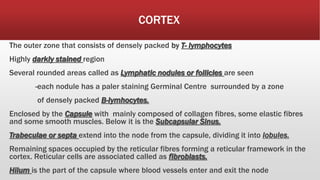
11. CORTEX
The outer zone that consists of densely packed by T- lymphocytes
Highly darkly stained region
Several rounded areas called as Lymphatic nodules or follicles are seen
-each nodule has a paler staining Germinal Centre surrounded by a zone
of densely packed B-lymhocytes.
Enclosed by the Capsule with mainly composed of collagen fibres, some elastic fibres
and some smooth muscles. Below it is the Subcapsular Sinus.
Trabeculae or septa extend into the node from the capsule, dividing it into lobules.
Remaining spaces occupied by the reticular fibres forming a reticular framework in the
cortex. Reticular cells are associated called as fibroblasts.
Hilum is the part of the capsule where blood vessels enter and exit the node
17. MEDULLA
 Surrounded by the Cortex.
 Lighter staining zone.
 Lymphocytes are fewer than the Cortex.
 The lymphocytes present are arranged in the form of anastomosing cords
called Medullary Cords.
 Several blood vessels can be seen.
20. Lymph and Blood Circulation
Lymphatic Circulation
 Apart from the lymphocytes, the reticular fibres and the reticular cells, the
spaces present are modified to form lymph channels through which Lymph
circulates.
 The sinuses are lined by endothelium that permit lymphocyte and
macrophage movement.
1. Lymph enters via the Afferent Lymphatic channels through the convex
surface piercing the capsule entering the subscapular sinus.
2. From this sinus, a number of radial cortical sinuses or trabeculae sinuses
runs to the medulla.
3. When these sinuses reach the medulla, they join to form larger Medullary
Sinuses.
4. These medullary sinuses in turn join to form one or more Efferent Lymph
22. Blood circulation
1. Afferent arteries enter the lymph node at the hilum.
2. They pass through the medulla to reach the cortex where they end in
arterioles and capillaries.
3. These capillaries are arranged in loops that drain into venules.
4. Post -capillary venules in lymph nodes are unusually lined by Cuboidal
epithelium in the place of simple squamous epithelium. These are
therefore called as High Endothelial Venules. This special modification is to
enable the passage of lymphocytes between the bloodstream and the
surrounding tissue due to specialised Tissue Receptors.
24. FUNCTIONS
1. Centres of Lymphocyte production, where both the B–
Lymphocytes and T – Lymphocytes are produced here
from pre-existing lymphocytes. These pass into the
lymph and into the blood stream.
2. Bacteria and particulate matter are filtered from the
lymph by the Macrophages via phagocytosis. These are
then categorised as Antigens which are presented to
the Lymphocytes stimulating their proliferation. THUS,
they act as secondary lymphoid organs.
Thus , they play a very crucial role in Immunity.
25. CLINICAL ASPECTS
1. As they play a key role in immunity, they also tend to get infected
and inflamed. This condition is called as Lymphadenitis.
2. Metastasis {Cancerous cells} spread from the localised regions to
areas which they connect. Pathological examination of the lymph
nodes that are inflamed, can give info about the origin of the
cancer.
3. When the source of cancer is in the lymph node, then it is called
as Lymphoma as it is a cancer of the Lymphoid Tissue
In the above three severe cases, lymph removal or
Lymphadenectomy is performed.