This study investigated the effects of environmental conditions like temperature and UV exposure on the genetic makeup and antibiotic resistance of Pseudomonas aeruginosa and Staphylococcus aureus. The bacteria were exposed to temperatures up to 90°C and UV light for up to 25 minutes. Both bacteria grew under these conditions with P. aeruginosa tolerating 60°C for 30 minutes and UV for 25 minutes. Exposure did not increase resistance to disinfectants. DNA analysis was inconclusive due to degradation or contamination. Further research is needed to analyze genetic changes that could increase antibiotic resistance.
This document describes various culture methods used to isolate and grow bacteria. It discusses streak culture, lawn culture, stroke culture, stab culture, pour plate culture, and liquid culture. It also covers different techniques for culturing anaerobic bacteria, including vacuum production, gas displacement, chemical and biological methods, medium reduction, and use of anaerobic chambers.
1. The document discusses various methods for preserving bacteria cultures, including short-term methods like agar slants and stabs as well as long-term methods like ultra-low temperature freezing and freeze-drying.
2. Freeze-drying, or lyophilization, involves freezing samples, then reducing pressure to allow frozen water to sublimate from the sample without passing through the liquid phase.
3. The freeze-drying process helps preserve bacterial viability for decades by preventing damage from ice crystal formation that can occur with other preservation methods.
Bacterial Culture methods and method of anaerobiosisromagoyal37
Ìý
CULTURE METHODS FOR Medical students
Culture methods are done to:
Isolate bacteria in pure culture from the clinical specimens and their idintification by various methods.
Determination of antibiotic sensitivity.
Prepare antigens for serodiagnosis of infective diseases.
Maintain stock cultures.
Methods to isolate the Bacteria
Streak culture
Stroke
Stab
Pour plate
Liquid culture
Special methods for anaerobic cultures
1. Culture methods are used to isolate bacteria in pure culture, demonstrate their properties, obtain sufficient growth for tests, and maintain stock cultures.
2. Common culture methods include streak culture, lawn culture, stroke culture, stab culture, pour plate culture, and liquid culture.
3. Special methods like anaerobic culture techniques are needed to isolate and grow anaerobic bacteria in the absence of oxygen using methods that generate hydrogen and carbon dioxide gases.
The document discusses antimicrobial susceptibility testing (AST), which determines the susceptibility of bacteria to different antimicrobial agents through in-vitro laboratory procedures. It outlines various guidelines and standardization procedures for AST, including standardized bacterial inoculum, growth medium, incubation conditions, and antimicrobial concentrations. Common AST methods include dilution methods like broth microdilution and agar dilution, which determine minimum inhibitory concentrations (MICs), and diffusion methods like disc diffusion testing, which provide qualitative susceptibility results.
Automated system for bacterial identificationDEEKSHANT KUMAR
Ìý
[DOWNLOAD IT OPEN IT WITH MICROSOFT POWERPOINT THEN YOU WILL BE ABLE TO UNDERSTAND THE TOPIC COVERED.]
1. WHOLE TEXT IS RELIABLE.
2. TEXT HAS BEEN TAKEN FROM STANDARD TEXT BOOK FOR MEDICAL MICROBIOLOGY.
3. SOME PICTURE HAS BEEN TAKEN FROM JOURNAL.
The document discusses methods for identifying bacteria, including phenotypic, immunological, and genetic techniques. Phenotypic methods examine bacterial morphology, staining characteristics, and biochemical reactions. Tests like Gram staining, colony morphology, and catalase can provide initial identification. Further tests of carbohydrate use, enzyme production, and sensitivity to inhibitors allow identification to the species level. Immunological methods detect bacterial antigens, while genetic techniques like PCR and nucleic acid analysis provide accurate identification by examining microbial DNA. Both traditional and molecular methods are used to fully characterize unknown bacteria.
This document discusses capsule staining, which is a technique used to identify the presence of bacterial capsules under a light microscope. It begins by defining bacterial capsules and explaining their functions, which include helping bacteria resist phagocytosis and providing protection. It then discusses the principle of capsule staining, which uses a negative stain to contrast the unstained capsule against stained bacterial cells. The procedure involves smearing a bacterial culture onto a slide with negative stain, staining with a counterstain like crystal violet, and examining under a microscope for unstained capsules surrounding stained cells. Examples of capsule-containing bacteria that can be identified this way include Klebsiella pneumoniae and Bacillus anthracis.
Antimicrobial susceptibility testing – disk diffusion methodsAnn Sam
Ìý
This document provides information on antimicrobial susceptibility testing using disk diffusion methods. It discusses the importance of AST for treating infectious diseases and monitoring antimicrobial resistance. The Kirby-Bauer disk diffusion method is described in detail, including media preparation, inoculum standardization, disk and antibiotic solution preparation, quality control strains, incubation, reading zones of inhibition, and interpreting results according to CLSI guidelines. Special considerations are given to organisms like MRSA, VISA, and inducible clindamycin resistance in Staphylococci.
This document discusses various staining techniques used to visualize bacteria under a microscope. It covers simple staining techniques like Gram staining and acid-fast staining, as well as methods to identify specific structures like volutin granules and bacterial spores. Gram staining uses dyes to differentiate between Gram-positive and Gram-negative bacteria based on their cell wall composition. Acid-fast staining targets bacteria with thick lipid cell walls like Mycobacterium tuberculosis. Specialized techniques employ unique dyes and fixation steps to highlight intracellular inclusions and endospores. Proper staining is crucial for bacterial identification and clinical diagnosis.
This document discusses the fungus Candida. It notes that Candida includes approximately 200 species, about 20 of which can cause infections in humans. C. albicans is the most common cause of infection. Candida is normally found in the gastrointestinal tract but can cause superficial or deep infections. Superficial infections include oral and vaginal candidiasis. Deep infections involve organs and can become disseminated. Virulence factors that enable Candida pathogenesis include adhesion, dimorphism, and production of enzymes. Laboratory diagnosis involves direct microscopic examination of samples and culture isolation followed by identification of Candida species.
This document provides information on the bacteria Bacillus. It discusses two main types of Bacillus - B. anthracis, which causes anthrax, and B. cereus, which can cause two types of food poisoning. For B. anthracis, it describes its morphology, culture characteristics, virulence factors including toxins, clinical manifestations of anthrax in humans and animals, and methods for laboratory diagnosis and treatment. It also provides historical context on the importance of B. anthracis. For B. cereus, it summarizes the two types of food poisoning it can cause and how they differ clinically.
The document describes the macrodilution method for determining the minimal inhibitory concentration (MIC) of antibiotics. Key steps include:
1. Preparing stock solutions of antibiotics at high concentrations like 10 mg/mL and diluting them to make testing concentrations.
2. Creating a standardized bacterial inoculum of around 5x10⁵ CFU/mL.
3. Diluting the antibiotic in a series of tubes containing broth and adding the bacterial inoculum.
4. Incubating the tubes overnight and finding the lowest antibiotic concentration tube that shows no visible growth, which is the MIC. The MBC can also be determined by culturing samples from clear tubes.
This document discusses techniques for cultivating viruses. It explains that viruses require living host cells to replicate and describes three main cultivation methods: animal inoculation, embryonated egg inoculation, and tissue culture. Animal inoculation involves infecting animals like mice to isolate and study viruses, but it is expensive and raises welfare issues. Embryonated egg inoculation is commonly used to grow viruses by inoculating eggs at specific sites, and it is cost-effective but each virus grows at different sites. Tissue culture uses cell monolayers and is versatile but requires specialized facilities and technicians. The document provides details on each technique's advantages, disadvantages and applications.
Sabouraud dextrose agar (SDA) is used to isolate and cultivate fungi and yeasts from clinical specimens. It contains nutrients like dextrose and enzymatic digest of casein to support fungal growth, and antibiotics to inhibit bacteria. The document outlines the materials, composition, and procedure to prepare SDA media. Colonies are examined after incubation and typical morphologies can indicate fungal species present. However, SDA may not promote conidiation in some fungi and antimicrobials could inhibit some pathogens.
This document discusses Salmonella, including its isolation, morphology, cultural characteristics, biochemical activities, epidemiology, virulence factors, pathogenesis, clinical diseases, laboratory diagnosis, and treatment. Salmonella was first isolated in 1885 and causes diseases like typhoid fever, paratyphoid fever, and foodborne illness in humans. It is a gram-negative rod found in the intestines of animals and can contaminate foods. Diagnosis involves culturing specimens from blood, feces, urine or vomit. Treatment includes antibiotics like chloramphenicol, ampicillin, and ciprofloxacin.
Shigella dysenteriae is a gram-negative, non-motile bacillus that causes dysentery. It grows well at 37°C in nutrient broth, forming colorless colonies on MacConkey agar and red colonies without black centers on XLD agar. S. dysenteriae infection is caused by ingesting contaminated food or water and leads to dysentery characterized by bloody mucus in stool. Laboratory diagnosis involves examining stool samples microscopically for pus cells and macrophages, culturing on selective media to isolate non-lactose fermenting colonies, and conducting biochemical tests and slide agglutination with specific antisera. Tetracycline and chloramphenicol are used
This document discusses antifungal susceptibility testing. It provides background on the history of antifungal susceptibility testing and why it is needed. It describes different methods for testing including broth dilution and disk diffusion. It discusses various antifungal agents and their mechanisms of action. The document outlines the procedures for broth microdilution and macrodilution testing according to CLSI guidelines, including preparation of inoculum, drug solutions, reading results, and testing of filamentous fungi.
Vibrio cholerae is the bacteria that causes cholera. It is a facultative anaerobe that grows well between 37°C and pH 7.4-9.6. It can be cultured on ordinary media like nutrient agar as well as special transport and enrichment media. Pathogenesis involves ingesting contaminated food/water, with the bacteria multiplying in the intestines and producing cholera toxin which causes hypersecretion of fluids. Diagnosis involves microscopic examination of rice water stool samples and culturing in selective media. Treatment focuses on fluid replacement therapy and oral rehydration.
This document discusses bacteriocin typing for epidemiological investigations. It defines bacteriocins as bactericidal proteins produced by bacteria that kill closely related bacterial strains. Bacteriocin typing involves determining the bacteriocin production patterns of strains against indicator strains or testing strains for susceptibility to different bacteriocins. This allows differentiation of bacterial isolates and investigation of outbreaks. Specific examples discussed are colicin typing of E. coli and pyocin typing of P. aeruginosa. The document outlines the methods for these typing techniques.
This document summarizes various serological tests used to detect antigens and antibodies. It describes primary, secondary and tertiary serological tests including ELISA, immunofluorescence, radioimmunoassay and more. It also details different types of agglutination tests like qualitative and quantitative tests. Additional tests covered are precipitation tests, complement fixation tests, passive hemagglutination and sandwich ELISA. The document provides information on applications and procedures for many antibody and antigen detection techniques.
This document discusses various bacterial staining techniques used to visualize microorganisms under a microscope. It describes simple staining which uses a single dye, differential staining which allows differentiation using more than one dye, and special staining techniques to highlight specific structures. Gram staining is explained in detail as the most common differential staining method used to classify bacteria as gram-positive or gram-negative. Acid-fast staining and capsule, spore, and flagella staining are also summarized as important special staining methods.
This document presents the results of an experiment to determine the minimum inhibitory concentration (MIC) of samples against various microorganisms. It includes a list of participants and their supervisor, definitions of MIC and methods used to determine it. The experiment involved testing 15 samples against 9 microorganisms by agar dilution. The MIC values were recorded in a table, with most samples having an MIC greater than 200 μg/ml. The agar dilution method was determined to be best suited for this evaluation.
Bacillus is a genus of rod-shaped, Gram-positive bacteria that can form dormant endospores. The document focuses on Bacillus anthracis, which causes anthrax. It describes the morphology, cultural characteristics, virulence factors, and methods of diagnosis and prevention of B. anthracis. Key points include that B. anthracis forms encapsulated, non-motile rods and terminal spores. The anthrax toxins are composed of lethal factor, edema factor, and protective antigen, which combine to cause disease. Diagnosis involves microscopy, culture, and serology. Prevention for humans involves vaccination with anthrax toxoid and occupational hygiene, while animals are vaccinated with attenuated spore
This document discusses Escherichia coli (E. coli), a common bacterium found in the intestines of humans and animals. It describes E. coli's morphology, identification, and ability to cause diseases like urinary tract infections, neonatal meningitis, and intestinal diseases. Prevention methods are outlined, including good hygiene practices like handwashing and thoroughly cooking meats to avoid transmission. Treatment focuses on fluid replacement rather than antibiotics in most cases unless the disease has progressed to a systemic stage.
This study evaluated the effectiveness of a UV keyboard sterilizer against various microorganisms. Methicillin-resistant Staphylococcus aureus (MRSA), Escherichia coli, bacteriophage MS-2, and mold Aspergillus niger were applied to an acrylic surface simulating a keyboard and exposed to 90 seconds of UV light. Results found the sterilizer reduced MRSA and E. coli levels by over 99.999%, MS-2 by over 99.9%, and A. niger by 92%. The sterilizer was highly effective against the bacteria tested but mold spores were more resistant likely due to their ability to withstand drying and UV light.
Presentation based on a research article published in the journal scientific reports in 2017, entitled as "A Novel Pathogen Capturing Device for Removal and Detection"
A biological indicator is a standardized preparation of viable microorganisms, usually bacterial spores, that is carried either directly by some of the items to be sterilized or by carriers such as filter papers, porcelain cylinders, that serve as a challenge to the effectiveness of a given sterilization cycle
This document discusses various methods for assessing the efficacy of disinfectants and sterilization processes, including physical, chemical, and biological indicators. The turbidimetric method uses turbidity measurements after a short incubation period to assess the ability of disinfectants and antibiotics to inhibit bacterial growth. Chemical indicators monitor sterilization processes by undergoing color changes in response to heat, steam, or radiation. Biological indicators use bacterial spores to validate the sterilization of equipment and facilities.
Rotavirus is a viral pathogen that causes gastroenteritis in children. It appears wheel-shaped under microscopy and is transmitted via the fecal-oral route. Symptoms include diarrhea, vomiting, and fever. Diagnosis can be made using ELISA tests or PCR to detect the virus in stool samples. Treatment focuses on rehydration since there are no antiviral drugs available. Vaccines have been developed to help prevent infection.
The Effect of Ultra Violet Radiation on Methicillin Resistant Staphylococcus ...iosrjce
Ìý
IOSR Journal of Dental and Medical Sciences is one of the speciality Journal in Dental Science and Medical Science published by International Organization of Scientific Research (IOSR). The Journal publishes papers of the highest scientific merit and widest possible scope work in all areas related to medical and dental science. The Journal welcome review articles, leading medical and clinical research articles, technical notes, case reports and others.
This document describes the isolation and characterization of a new giant virus called Cedratvirus. Key points:
- Cedratvirus was isolated from an environmental sample in Algeria using Acanthamoeba castellanii.
- It has an ovoid shape with a cork structure at each end, resembling Pithovirus sibericum but with a unique double cork feature.
- The 589kb genome is most closely related to the pithovirus genomes, sharing over 100 genes, but with only 21% of genes involved in best reciprocal hits, indicating genetic distance from known pithoviruses.
5.1.3. Efficacy of antimicrobial preservation (EP 5.0)Guide_Consulting
Ìý
Salah Satu Referensi Yang Digunakan Dalam One Day Seminar "Preservative Effectiveness Validation" 04 Desember 2014.
Detail : info@traininglaboratorium.com
This document discusses various methods of microbial control in healthcare settings. It covers the need for microbial control to prevent infection transmission. Various methods are discussed, including sterilization, disinfection, antisepsis and antimicrobial therapy. Physical sterilization methods like heat, filtration and radiation are outlined. Factors that influence the effectiveness of sterilization and disinfection methods are also summarized.
Inactivation of bacillus cereus spores in liquid food by combination treatmen...Alexander Decker
Ìý
This document summarizes a study that investigated using combination treatments of heat and irradiation to inactivate Bacillus cereus spores in liquid foods like milk and carrot juice. The researchers characterized the heat and radiation resistance of B. cereus spores by determining their D-values (decimal reduction times/doses) and Z-values (temperature increase needed for a 10-fold reduction in D-values) in different media. They found that pre-irradiation at 4 kGy followed by heating reduced D90-values (time to reduce population by 90% at a given temperature) 2.8 to 3.4 times compared to heating alone. The combination treatment was more effective at ensuring safety of foods contaminated with B. cere
Selection and Efficacy Biocontrol Agents in Vitro against Fire Blight (Erwini...IRJET Journal
Ìý
This document summarizes a study that evaluated the effectiveness of biological control agents against the fire blight pathogen Erwinia amylovora in vitro. 114 antagonist bacterial and yeast isolates were tested for their ability to inhibit the growth of E. amylovora. Nine bacterial strains showed satisfactory inhibition (greater than or equal to 20mm) and the best inhibition percentages of 31-41%. These nine strains were generally bacteriolytic, meaning they induced lysis of the pathogen. The study aimed to contribute to fire blight control strategies by selecting effective antagonist microbes that could be developed as biological control agents.
This document discusses various methods of sterilization and disinfection. It begins by describing how early civilizations preserved food using methods like drying and pickling to control microbial growth. In the mid-1800s, Semmelweis and Lister helped develop aseptic techniques to prevent surgical wound infections. The document then defines key terms like sterilization, disinfection, and sanitization. It describes various physical methods of sterilization and disinfection like heat, filtration, and radiation. It provides details on specific heat-based methods like autoclaving, pasteurization, and tyndallization. The document also discusses chemical disinfectants and antiseptics like phenols, iodine, and chlor
Presented by Dr. Brecher at the 40th Annual Symposium "Diagnostic and Clinical Challenges of 20th Century Microbes", held on Nov 18, 2010 in Philadelphia.
To study Prevalence, Pre-disposing factors and Prevention of the following MDRO’s – Klebsiella pneumoniae Carbapenemase Producer, Methicillin Resistant Staphylococcus aureus, Multi Drug Resistant Acinetobacter baumannii, Pseudomonas aeruginosa and Escherichia coli.
Laboratory studies conducted by Bayer Environmental Science found that nets treated with K-O TAB® 1-2-3 insecticide met the WHO criteria for long-lasting insecticidal nets (LLINs) after more than 25 washes. Tests showed the treated nets achieved over 95% knockdown and 80% mortality of Anopheles stephensi mosquitoes after washing, in line with WHO standards. Chemical analysis also confirmed an even distribution of the target dose of deltamethrin insecticide on the nets. Additional washing and testing are underway to evaluate other net materials and detergent amounts.
This study evaluated the effect of reconstitution and storage temperatures (20°C and 30°C) on the growth of Salmonella Typhimurium and Cronobacter sakazakii in powdered infant formula. The study found that reconstituting with water at 70°C had the greatest detrimental impact on bacterial growth, while lower temperatures did not sufficiently reduce risk. Storage at room temperature benefited bacterial growth, so leftover formula must be refrigerated to inhibit growth and minimize outbreak risk.
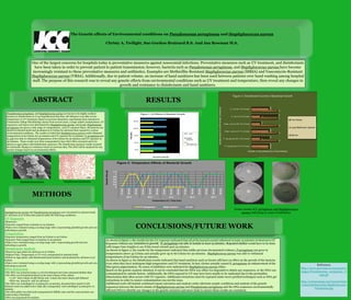
1. The Genetic effects of Environmental conditions on Pseudomonas aeruginosa and Staphylococcus aureus
Christy A. Twilight, Sue Gordon-Brainard B.S. And Jan Bowman M.S.
ABSTRACT
METHODS
RESULTS
Pseudomonas aeruginosa and Staphylococcus aureus are known to be highly resilient
bacteria to disinfectants so it was hypothesized that they will still grow even after severe
temperature or UV treatment. Based on previous laboratory experiments from Jamestown
Community College Microbiology classes from several years, a range relative temperatures, UV
exposures, and times were determined for Staphylococcus aureus and E.coli. Pseudomonas
aeruginosa was given a wide range of temperatures, and UV exposure times. All bacteria was
placed in nutrient broth and incubated at 37 Celsius for 48 hours then exposed to various
environmental conditions. The results revealed that Staphylococcus aureus could withstand
temperatures of 90 Celsius for 30 minutes and UV exposure for 15 minutes. P. aeruginosa and
E. coli were able to withstand temperatures of 60 Celsius for 30 minutes and UV exposure of
25 minutes. These results were then resuspended to have their DNA extracted and to be
placed on agar plates with disinfectant exposures. The disinfectant exposure results revealed
no noticeable changes in resistance based on previous data. The DNA will be analyzed for any
genetic changes based on environmental effects.
One of the largest concerns for hospitals today is preventative measures against nosocomial infections. Preventative measures such as UV treatment, and disinfectants
have been taken in order to prevent patient to patient transmission; however, bacteria such as Pseudomonas aeruginosa, and Staphylococcus aureus have become
increasingly resistant to these preventative measures and antibiotics. Examples are Methicillin-Resistant Staphylococcus aureus (MRSA) and Vancomycin-Resistant
Staphylococcus aureus (VRSA). Additionally, due to patient volume, an increase of hand sanitizers has been used between patients over hand washing among hospital
staff. The purpose of this research was to reveal any genetic effects from environmental conditions such as UV treatment and temperature, then reveal any changes in
growth and resistance to disinfectants and hand sanitizers.
As shown in figure 1, the results for the UV exposure indicated that all of the bacteria tested withstood at least 15 minutes of shortwave UV
Exposure without any inhibition to growth. P. aeruginosa was able to handle at least 25 minutes. Repeated studies would have to be done
with longer time lengths to see if this trend extends past 25 minutes.
As shown in figure 2, the results for the temperature indicated that unlike previous documented evidence, P.aeruginosa can grow in
temperatures above 42 Celsius and actually grow up to 60 Celsius for 30 minutes. Staphylococcus aureus was able to withstand
temperatures of 90 Celsius for 30 minutes.
As shown in figure 3, the disinfectant results indicated that hand sanitizers such as Germx still have no effect on the growth of the bacteria
even when they have undergone high temperature and UV treatment. In fact, Germx actually caused P. aeruginosa an enhancement of the
blue/green pigmentation. No zones of inhibition were indicated for Staphylococcus aureus either.
Based on the genetic analysis obtained, it can be concluded that the DNA was either too degraded to obtain any sequences, or the DNA was
contaminated by outside factors. Additionally, the DNA exposed to UV may have been unable to be replicated due to the pyrimidine
dimerization that often occurs with UV exposure. Additional extractions must be repeated under more purified techniques such as DNA gel
extractions in order to ensure contamination was not the issue.
Additional work will include continued repeat extraction and analysis under alternate aseptic conditions and analysis of the genetic
sequences between the known strains of Staphylococcus aureus and Pseudomonas aeruginosa and the DNA sequences environmentally
affected DNA to identify any changes in the genetic structure and more trials to ensure these results are consistent.
CONCLUSIONS/FUTURE WORK
Staphylococcus aureus and Psuedomonas aeruginosa were inoculated in nutrient broth
for 48 hours at 37 Celsius then placed under the following conditions:
UV Exposure
•Shortwave
•Exposure ranged from 5 minutes to 25 minutes
•Plates were evaluated using a scoring range with 2 representing plentiful growth and zero
indicating no growth .
Temperature
•Exposure temperature ranged from 50 Celsius to 90 Celsius
•Exposure Time ranged from 10 minutes to 30 minutes
•Plates were evaluated using a scoring range with 1 representing growth and zero
indicating no growth.
Disinfectant Analysis
•Placed on Agar plates for 48 hours at 37 Celsius
•Highest Time, Temperature or UV were resuspended in nutrient broth
•Plated on Agar plates with disinfectant/hand sanitizer and incubated for 48 hours at 37
Celsius
•Plates were evaluated using a scoring range with 3 representing plentiful growth and zero
indicating no growth.
DNA Extraction
•The DNA was extracted using 2-3 ml of isobutanol and water saturated diethyl ether
•The DNA was Precipitated based on the total volume of the culture
•1/10th total volume of 3M NaOAc and 2 times the total volume 95% Ethanol
•DNA was placed in -20C for 48 hours
•The DNA was centrifuged at 12,000xg for 15 minutes, decanted then 200ul of 75%
Ethanol wash was added twice while also resuspended and centrifuged 12,000xg for 10
minutes
•The DNA pellet was dried and resuspended in MilliQ water and the concentration was
tested using a Nanodrop
•DNA was sequenced for analysis
Staphylococcus aureus Pseudomonas aeruginosa
0
0.2
0.4
0.6
0.8
1
1.2
10min
20min
30min
10min
20min
30min
10min
20min
30min
10min
20min
30min
10min
20min
30min
50 C 60 C 70 C 80 C 90 C
Growth(Visual)
Temperature (C) Time (min)
Figure 2: Temperature Effects of Bacterial Growth
P. aeruginosa Staph. aureus E. coli
0 1 2 3
8min
10min
15min
20min
25min
Growth (visual)
Time(min)
Figure 1: UV Effects of Bacterial Growth
E. coli
Staph.
aureus
P.
aeruginosa
strain 60 C 70 C 80 C 90 C
10min 20min 30min 10min 20min 30min 10min 20min 30min 10min 20min
#27853 - - - - - - - - - - +
#25923 < < < < < < - - - + -
< < < - - - < < - < <
+
-
<
+ + + + + + + - - + +
+ + + - + + - - - - -
strain 8min 10min 15min 20min 25min Name Germx
Lysol Bathroom
cleaner
P. aeruginosa UV 25
minutes 2 2
#27853 + + + + +
P. aeruginosa 60 C
30 minutes 3 1
#25923 ++ ++ ++ - -
Staph. aureus UV 15
minutes 3 2
++ ++ ++ ++ ++
Staph. aureus 90C
20 minutes 3 1
E. coli UV 25
minutes 3 1
++
E. coli 60C 30
minutes 3 2
2
3
3
3
3
3
2
1
2
1
1
2
2
2
2
1
3
2
P. aeruginosa UV 25 minutes
P. aeruginosa 60 C 30 minutes
Staph. aureus UV 15 minutes
Staph. aureus 90C 20 minutes
E. coli UV 25 minutes
E. coli 60C 30 minutes
Growth in the presence of Disinfectant
BacteriaandEnvironmentalConditions
Figure 3: Disinfectant Control of Bacterial Growth
The Works
Lysol Bathroom cleaner
Germx
Germx results of P. aeruginosa and Staphylococcus
aureus indicating no zones of inhibition
References
http://www.marvistavet.org/assets/i
mages/Pseudomonas_aeruginosa_G
ram.gif
http://entomologia.net/insectos%20
contra%20bacterias/Staphylococcus
%20aureus.jpg