


![INVESTIGATION
EUCr
– Na 142.8, Cl 100.9, K 4.7 ,HCO3 24.7 ,Urea 5.3 ,Cr 84.6
FBC
– PCV – 30%
– WBC differential - Mild neutrophilic leucocytosis.
• TC – 19,000. [N- 78%, L-19%, BEM – 3%]
– Plt – 283,000
Abdominal X-Ray and Abdominal USS
– Not done](https://image.slidesharecdn.com/mmgsteamajune-240919122733-653c10e5/85/Morbidity-and-Mortality-review-Team-A-June-pptx-8-320.jpg)


![1st
Day Post-Op
• Hx
– Fully recovered from anaesthesia.
– Complained of pain at op site and hunger.
• Exam
– Febrile, T – 38.6’C
– Vitals: PR – 102bpm, BP – 130/90mmHg, RR – 24cpm
– Abdominal Exam.
• Drian in situ, draining minimal haemorrhagic effluent.
• Wound dressing clean and dry.
• Nil area of undue tenderness, Abdomen was soft.
– Urine output – 1.3 -1.5mls/kg/hr.
• ASS:
– Severe sepsis on treatment
– KIV Sepsis induced post-op diabetes insipidus
• Plan
– FBC & EUCr [Unable to get results that day]
– Increased Fluid to 4L in 24 hrs
– Ensures antibiotics and analgesia.
Respiratory System.
– RR – 24cpm
– Kussmals breathing
– Chest – CLear
CVS
– PR – 102bpm
– BP – 130/90mmHg
– HS – S1, S2 only](https://image.slidesharecdn.com/mmgsteamajune-240919122733-653c10e5/85/Morbidity-and-Mortality-review-Team-A-June-pptx-14-320.jpg)
![2nd
Day Post-Op
• Hx
– Noted to be restless – necessitating restraint.
– Made incoherent speech.
• Exam
– Febrile, T – 39’C, warm extremities
– Vitals – PR – 132bpm, BP – 110/90mmHg, RR – 30cpm,
– RBS – 20.8mmol/L, repeat 12.9mmol/L
– Abdominal Exam.
• Drian in situ, draining minimal haemorrhagic effluent.
• Wound inspected and seen to be well apposed and clean.
• Nil area of undue tenderness, Abdomen was soft. Not distended.
– Urine output – 2 -2.5mls/kg/hr.
• ASS:
– Sepsis Associated Encephalopathy with Diabetes insipidus.
• Investigations
– FBC
• PCV – 30%, Plt – 620,000.
• WBC – T – 40,200 [N – 86%, L-13%, BEM-1%
– E/U/Cr
• Na – 153, K – 6.1, Cl – 117.2, HCO3- 19, Cr – 133.4 Ur – 6.6
• Hypernatremia, Hyperkalemia, mildly elevated creatinine, and the High anion gap metabolic acidosis.
Respiratory System.
– RR – 30cpm
– Kussmals breathing
– Chest – Clear
CVS
– PR – 132bpm
– BP – 110/90mmHg
– HS – S1, S2 only
CNS
– GCS – 12 [M-5, V-3, E-4]
– Pupils about 3mm, equal reactive to light
bilaterally
qSOFA – 2/3
– RR – 30cpm
– BP – 110/90mmHg
– Altered sensorium](https://image.slidesharecdn.com/mmgsteamajune-240919122733-653c10e5/85/Morbidity-and-Mortality-review-Team-A-June-pptx-15-320.jpg)

![2nd
Day Post-Op
• Hx
– Decrease in consciousness level
• Exam
– GCS – 5 [M-1,V-2,E-2]
– Vitals – PR – 162bpm, BP – 100/60mmHg, RR – 36cpm
– Chest
• RR – 36cpm, SPO2 – 96 on 2L INO2
• Wide spread coarse crepitations in the right Upper and Middle lung zones
– Still pouring urine.
• ASS:
– SAE with Aspiration
• Plan
– Expedite Anesthesiologist review and counselled for ICU care.
– Suction PRN
– Nursed in Left lateral position.](https://image.slidesharecdn.com/mmgsteamajune-240919122733-653c10e5/85/Morbidity-and-Mortality-review-Team-A-June-pptx-17-320.jpg)

![Discussion
• Disease factor
– Disease burden
• 20% Mortality
– Atypical presentation
• Perforated Gastric ulcer.
– 2% as first presentation.
• Uncommon in females
• A concomitant umbilical hernia.
– Atypical course
• Worsening sepsis after source control
– ? Antibiotics [tyonnex brand].
• Sepsis Associated Encephalopathy
– Why? [First organ in the Sequential organ
failure assessment ]
» Rapid deceleration of the CNS with
sparing of other organs.
– 2-3 times greater mortality than Sepsis.
– Devastating out come in a short course.
• Sepsis associated post-op diabetes insipidus.
– Made fluid resuscitation more challenging.
– Masked early diagnosis of organ disfunction.
• Patient and Relative factor
– Domestic violence
• Initial source if the problem.
• Prolonged NSAID use.
– Financial constraint
• Malnourished
– Worsened in the last 2 weeks due to
jaw fracture.
• Subtle delays with investigations
and procurement of medications.
• Hospital factor.
– 12hrs from decision to operate to
knife on skin
• Usual delay
• Ongoing Neurosurgery case.
• Team Factor
– Missed diagnosis
1. Atypical presentation and
course.](https://image.slidesharecdn.com/mmgsteamajune-240919122733-653c10e5/85/Morbidity-and-Mortality-review-Team-A-June-pptx-19-320.jpg)


More Related Content
Similar to Morbidity and Mortality review `Team A June.pptx (20)
Recently uploaded (20)


Morbidity and Mortality review `Team A June.pptx
- 1. Discharge Summary General Surgery Team A June, 2024.
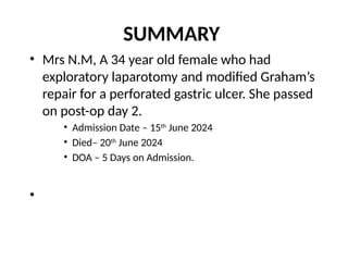
- 2. SUMMARY • Mrs N.M, A 34 year old female who had exploratory laparotomy and modified Graham’s repair for a perforated gastric ulcer. She passed on post-op day 2. • Admission Date – 15th June 2024 • Died– 20th June 2024 • DOA – 5 Days on Admission. •
- 3. Case 1. • Mrs. N.M a 34yr old petty trader who resides at Low cost, Keffi. – Umbilical pain x 3days – Abdominal pain x 1 day • HPC – She was in her usual state of a health until about 3 days prior to presentation she noted an initially painless umbilical budge which failed to resolve spontaneously. • Noted to have become painful, with associated tenderness, but no redness of overlying skin, no colicky abdominal pain, vomiting or constipation.
- 4. Case 1. • HPC – About 12 hours prior to presentation She developed sudden onset burning epigastric pain, radiating to the right upper quadrant with associated hx of non bilious vomiting and passage of loose stool, but no Fever. • No prior hx of colicky abdominal pain, no fever, no abdominal distension. • Nil hx of trauma to the abdomen, nil hx of falls. • Not a known PUD pxt, there was no prior hx of recurrent epigastric pain, nil hx of indiscriminate use of NSAIDs – Had been on NSAIDs for the Jaw fracture and is being managed by the MFU of NHA. • Nil prior hx of abdominal surgeries, • No chronic cough, contact with patient with chronic cough, ingestion of unpasteurized milk, weight loss or drenching night sweats, – For the above she self medicated with some over the counter medications, but with no improvement in symptoms she presented at the AnE of this facility for care.
- 5. Case 1 – She is not a known Hypertensive, Diabetic, asthmatic or epileptic. – No previous surgeries or blood transfusion. – She is married in a monogamous family setting with 2 children. – She does not smoke or take alcohol. No recreational drug of abuse. – No history of drug or food allergy. – Not on any long term medications.
- 6. Examination Young lady, not in mild painful distress, afebrile, anicteric, not pale, not dehydrated, nil pedal edema, nil palpable peripheral lymph nodes. Abdomen – Full, Moved with respiration, • 3 by 2 cm, tender umbilical swelling, • Umbilical defect measuring about 1cm – Mild to moderate tenderness in the epigastrium, R. hypochondrium and lumber region • Nil rebound tenderness. – Rest of abdomen was soft and non tenderness. – Nil palpable organomegaly. – DRE was unremarkable Respiratory System. – RR – 20cpm – Chest – Vesicular breath sounds all over. CVS – PR – 84bpm – BP – 130/80mmHg – HS – S1, S2 only
- 7. Diagnosis Reduced Incarcerated Umbilical hernia with ? Bowel ischemia. Differentials. 1. Uncomplicated umbilical hernia with NSAID induced Gastritis.
- 8. INVESTIGATION EUCr – Na 142.8, Cl 100.9, K 4.7 ,HCO3 24.7 ,Urea 5.3 ,Cr 84.6 FBC – PCV – 30% – WBC differential - Mild neutrophilic leucocytosis. • TC – 19,000. [N- 78%, L-19%, BEM – 3%] – Plt – 283,000 Abdominal X-Ray and Abdominal USS – Not done
- 9. Initial Intervention • NPO • IV Fluids. • IV Antibiotics • IV Omeprazole • IV PCM • Patient admitted for close monitoring and to be properly investigated. • Patient encouraged to do the abdominopelvic scan.
- 10. 2nd Day on Admission • Hx – Worsening of abdominal pain. – 3 episodes of bilious vomiting. • Exam – Febrile, – Vitals – PR – 96bpm, BP – 110/70mmHg, RR – 22cpm – Abdominal Exam. • Moved minimally with respiration, • Marked tenderness with rigidity in the R. hypochondrium and lumber region with guarding. • Rest of abdomen was soft. • ASS: – Localized peritonitis 2’ reduced gangrenous bowel. • Plan – Abandon conservative management and explore.
- 11. INTRA-OPERATIVE 3rd DOA • FINDING 1. Solitary anterior gastric perforation measuring about 5 by 5mm on the distal third of the body. 2. About 250mls of Clear Bilious effluent in the right subhepatic space and extending to the upper right paracolic gutter, walled off by omentum, stomach, and proximal jejunum 3. Rest of the abdomen was clean and grossly normal. • PROCEDURE 1. Bilious effluent suctioned, Gastric perforation identified, and a thorough exploration done. 2. Biopsy of the ulcer edge was taken after stay sutures were applied. 3. A modified grahams repair was done. 4. Copious lavage was done, and the wound closed over an drain in the subhepatic space. Transfused with one unit of blood intra-op
- 12. Post-Op Intervention 1. NG tube maintained – Drained bilious effluent. 2. IV Fluids 3L in 24 hrs. 3. Analgesics – Pentazocine and PCM 4. Antibiotics – Ceftriaxone, Metronidazole 5. Antacids – Omeprazole and Ranitidine. 6. Vitals monitored closely
- 13. 2hrs Post-Op • Hx – Not Fully recovered from anaesthesia. • Exam – Febrile, T – 38.1’C, Sleeping but responded to calls – Vitals – PR – 90bpm, BP – 130/90mmHg, RR – 22cpm – Abdominal Exam. • Drian in situ, draining minimal haemorrhagic effluent. • Wound dressing clean and dry. • Nil area of undue tenderness, Abdomen was soft. – Urine output – 0.5 – 0.8mls/kg/hr. • ASS: – Stable post-op • Plan – Ensured post-op order – Ensured 500mls of fluid every 4hrs
- 14. 1st Day Post-Op • Hx – Fully recovered from anaesthesia. – Complained of pain at op site and hunger. • Exam – Febrile, T – 38.6’C – Vitals: PR – 102bpm, BP – 130/90mmHg, RR – 24cpm – Abdominal Exam. • Drian in situ, draining minimal haemorrhagic effluent. • Wound dressing clean and dry. • Nil area of undue tenderness, Abdomen was soft. – Urine output – 1.3 -1.5mls/kg/hr. • ASS: – Severe sepsis on treatment – KIV Sepsis induced post-op diabetes insipidus • Plan – FBC & EUCr [Unable to get results that day] – Increased Fluid to 4L in 24 hrs – Ensures antibiotics and analgesia. Respiratory System. – RR – 24cpm – Kussmals breathing – Chest – CLear CVS – PR – 102bpm – BP – 130/90mmHg – HS – S1, S2 only
- 15. 2nd Day Post-Op • Hx – Noted to be restless – necessitating restraint. – Made incoherent speech. • Exam – Febrile, T – 39’C, warm extremities – Vitals – PR – 132bpm, BP – 110/90mmHg, RR – 30cpm, – RBS – 20.8mmol/L, repeat 12.9mmol/L – Abdominal Exam. • Drian in situ, draining minimal haemorrhagic effluent. • Wound inspected and seen to be well apposed and clean. • Nil area of undue tenderness, Abdomen was soft. Not distended. – Urine output – 2 -2.5mls/kg/hr. • ASS: – Sepsis Associated Encephalopathy with Diabetes insipidus. • Investigations – FBC • PCV – 30%, Plt – 620,000. • WBC – T – 40,200 [N – 86%, L-13%, BEM-1% – E/U/Cr • Na – 153, K – 6.1, Cl – 117.2, HCO3- 19, Cr – 133.4 Ur – 6.6 • Hypernatremia, Hyperkalemia, mildly elevated creatinine, and the High anion gap metabolic acidosis. Respiratory System. – RR – 30cpm – Kussmals breathing – Chest – Clear CVS – PR – 132bpm – BP – 110/90mmHg – HS – S1, S2 only CNS – GCS – 12 [M-5, V-3, E-4] – Pupils about 3mm, equal reactive to light bilaterally qSOFA – 2/3 – RR – 30cpm – BP – 110/90mmHg – Altered sensorium
- 16. Intervention • Samples for blood culture • Changed antibiotics – Levoflox, Rocephin and Metro. • Commenced on INO2 by nasal prongs • Continuous vitals monitoring. • Internal Medicine review – Severe sepsis in a ? Prediabetic in poly uric phase of AKI. – Monitor RBS. – Maintain other care. • Anaesthesiologist Review – Patient passed on during their review.
- 17. 2nd Day Post-Op • Hx – Decrease in consciousness level • Exam – GCS – 5 [M-1,V-2,E-2] – Vitals – PR – 162bpm, BP – 100/60mmHg, RR – 36cpm – Chest • RR – 36cpm, SPO2 – 96 on 2L INO2 • Wide spread coarse crepitations in the right Upper and Middle lung zones – Still pouring urine. • ASS: – SAE with Aspiration • Plan – Expedite Anesthesiologist review and counselled for ICU care. – Suction PRN – Nursed in Left lateral position.
- 18. 2nd Day Post-Op – Findings • Noted to have stopped making respiratory effort • Nil heart sounds noted. • Commenced CPR – Stat dose of IV Adrenaline given, repeated after 5mins x 3 doses. • Nil ROSC after 30mins • Pupils fixed and dilated • Patient was declared clinically dead at 11:15am – Primary Cause of Death 1. Perforated Peptic Ulcer – Secondary Cause of Death 1. Brain stem failure 2’ Sepsis associated Encephalopathy.
- 19. Discussion • Disease factor – Disease burden • 20% Mortality – Atypical presentation • Perforated Gastric ulcer. – 2% as first presentation. • Uncommon in females • A concomitant umbilical hernia. – Atypical course • Worsening sepsis after source control – ? Antibiotics [tyonnex brand]. • Sepsis Associated Encephalopathy – Why? [First organ in the Sequential organ failure assessment ] » Rapid deceleration of the CNS with sparing of other organs. – 2-3 times greater mortality than Sepsis. – Devastating out come in a short course. • Sepsis associated post-op diabetes insipidus. – Made fluid resuscitation more challenging. – Masked early diagnosis of organ disfunction. • Patient and Relative factor – Domestic violence • Initial source if the problem. • Prolonged NSAID use. – Financial constraint • Malnourished – Worsened in the last 2 weeks due to jaw fracture. • Subtle delays with investigations and procurement of medications. • Hospital factor. – 12hrs from decision to operate to knife on skin • Usual delay • Ongoing Neurosurgery case. • Team Factor – Missed diagnosis 1. Atypical presentation and course.
- 20. •Thank you