neck fascia and spaces
Download as pptx, pdf15 likes1,393 views

The document describes the anatomy of the neck spaces. It is divided into three sections: spaces involving the entire length of the neck, spaces limited to above the hyoid bone, and spaces limited to below the hyoid bone. The key spaces described include the retropharyngeal space, danger space, parapharyngeal space, submandibular space, and anterior visceral space. Various structures form the boundaries of each space and contents are noted.

neck fascia and spaces
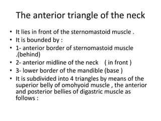
- 2. The anterior triangle of the neck ŌĆó It lies in front of the sternomastoid muscle . ŌĆó It is bounded by : ŌĆó 1- anterior border of sternomastoid muscle .(behind) ŌĆó 2- anterior midline of the neck ( in front ) ŌĆó 3- lower border of the mandible (base ) ŌĆó It is subdivided into 4 triangles by means of the superior belly of omohyoid muscle , the anterior and posterior bellies of digastric muscle as follows :
- 3. ŌĆó 1- Digastric triangle ŌĆó 2- carotid triangle ŌĆó 3- Muscular triangle ŌĆó 4- Submental triangle
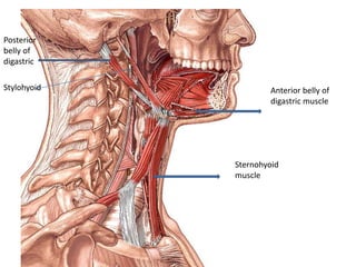
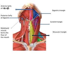
- 4. Anterior belly of digastric muscle Posterior belly of digastric Stylohyoid Sternohyoid muscle
- 5. Digastric triangle Or submandibular triangle ŌĆó Outlines : ŌĆó Above : the lower border of the mandible ŌĆó Below & infront : the anterior belly of Digastric muscle ŌĆó Below & behind : the posterior belly of Digastric and stylohyoid muscles . ŌĆó Floor : ŌĆó Anteriorly : the mylohyoid muscle ŌĆó Posteriorly : part of hyoglossus muscle
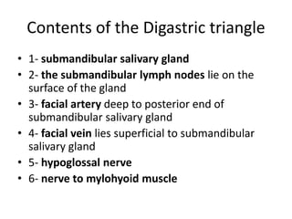
- 6. Contents of the Digastric triangle ŌĆó 1- submandibular salivary gland ŌĆó 2- the submandibular lymph nodes lie on the surface of the gland ŌĆó 3- facial artery deep to posterior end of submandibular salivary gland ŌĆó 4- facial vein lies superficial to submandibular salivary gland ŌĆó 5- hypoglossal nerve ŌĆó 6- nerve to mylohyoid muscle
- 7. ŌĆó d Digastric triangle Carotid triangle Muscular triangle Anterior belly of digastric Posterior belly of digastric Mylohyoid muscle forms the floor of the mouth
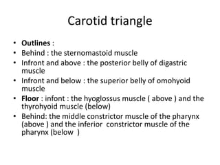
- 8. Carotid triangle ŌĆó Outlines : ŌĆó Behind : the sternomastoid muscle ŌĆó Infront and above : the posterior belly of digastric muscle ŌĆó Infront and below : the superior belly of omohyoid muscle ŌĆó Floor : infont : the hyoglossus muscle ( above ) and the thyrohyoid muscle (below) ŌĆó Behind: the middle constrictor muscle of the pharynx (above ) and the inferior constrictor muscle of the pharynx (below )
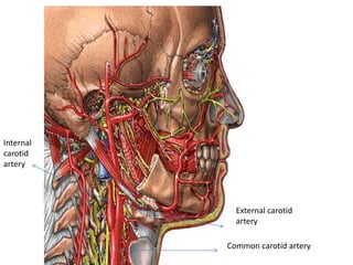
- 9. External carotid artery Common carotid artery Internal carotid artery
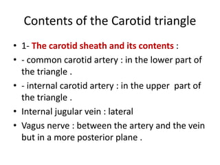
- 10. Contents of the Carotid triangle ŌĆó 1- The carotid sheath and its contents : ŌĆó - common carotid artery : in the lower part of the triangle . ŌĆó - internal carotid artery : in the upper part of the triangle . ŌĆó Internal jugular vein : lateral ŌĆó Vagus nerve : between the artery and the vein but in a more posterior plane .
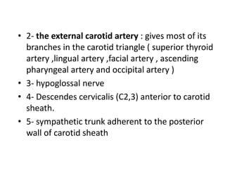
- 11. ŌĆó 2- the external carotid artery : gives most of its branches in the carotid triangle ( superior thyroid artery ,lingual artery ,facial artery , ascending pharyngeal artery and occipital artery ) ŌĆó 3- hypoglossal nerve ŌĆó 4- Descendes cervicalis (C2,3) anterior to carotid sheath. ŌĆó 5- sympathetic trunk adherent to the posterior wall of carotid sheath
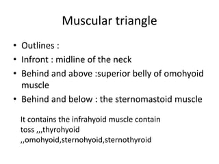
- 12. Muscular triangle ŌĆó Outlines : ŌĆó Infront : midline of the neck ŌĆó Behind and above :superior belly of omohyoid muscle ŌĆó Behind and below : the sternomastoid muscle It contains the infrahyoid muscle contain toss ,,,thyrohyoid ,,omohyoid,sternohyoid,sternothyroid
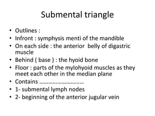
- 13. Submental triangle ŌĆó Outlines : ŌĆó Infront : symphysis menti of the mandible ŌĆó On each side : the anterior belly of digastric muscle ŌĆó Behind ( base ) : the hyoid bone ŌĆó Floor : parts of the mylohyoid muscles as they meet each other in the median plane ŌĆó Contains ŌĆ”ŌĆ”ŌĆ”ŌĆ”ŌĆ”ŌĆ”ŌĆ”ŌĆ”ŌĆ”ŌĆ” ŌĆó 1- submental lymph nodes ŌĆó 2- beginning of the anterior jugular vein
- 14. Deep Neck Space Anatomy ŌĆó Space Involving Entire Length Of Neck ŌĆó Space Limited To Above The Hyoid Bone ŌĆó Space limited To Below The Hyoid Bone
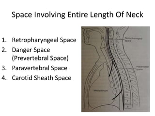
- 15. Space Involving Entire Length Of Neck 1. Retropharyngeal Space 2. Danger Space (Prevertebral Space) 3. Paravertebral Space 4. Carotid Sheath Space
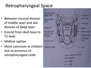
- 16. Retropharyngeal Space ŌĆó Between visceral division of middle layer and alar division of deep layer ŌĆó Extend from skull base to T2 level ŌĆó Midline raphae ŌĆó More commom in children due to presence of retropharyngeal node
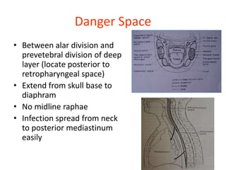
- 17. Danger Space ŌĆó Between alar division and prevetebral division of deep layer (locate posterior to retropharyngeal space) ŌĆó Extend from skull base to diaphram ŌĆó No midline raphae ŌĆó Infection spread from neck to posterior mediastinum easily
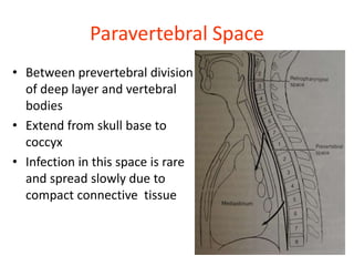
- 18. Paravertebral Space ŌĆó Between prevertebral division of deep layer and vertebral bodies ŌĆó Extend from skull base to coccyx ŌĆó Infection in this space is rare and spread slowly due to compact connective tissue
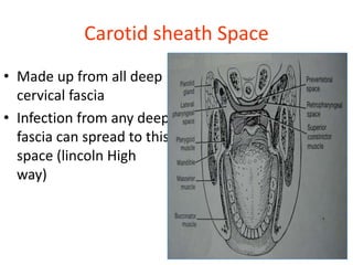
- 19. Carotid sheath Space ŌĆó Made up from all deep cervical fascia ŌĆó Infection from any deep fascia can spread to this space (lincoln High way)
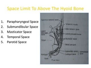
- 20. Space Limit To Above The Hyoid Bone 1. Parapharyngeal Space 2. Submandibular Space 3. Masticator Space 4. Temporal Space 5. Parotid Space
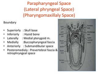
- 21. Parapharyngeal Space (Lateral phryngeal Space) (Pharyngomaxillaly Space) Boundary ŌĆó Superiorly : Skull base ŌĆó Inferiorly : Hyoid bone ŌĆó Laterally : Medial pterygoid m. ŌĆó Medially :Buccopharyngeal fascia ŌĆó Anteriorly : Submandibular space ŌĆó Posteromedialy : Prevertebral fascia & retrophryngeal space
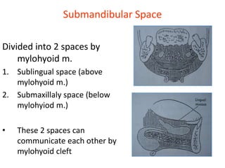
- 22. Submandibular Space Divided into 2 spaces by mylohyoid m. 1. Sublingual space (above mylohyoid m.) 2. Submaxillaly space (below mylohyiod m.) ŌĆó These 2 spaces can communicate each other by mylohyoid cleft
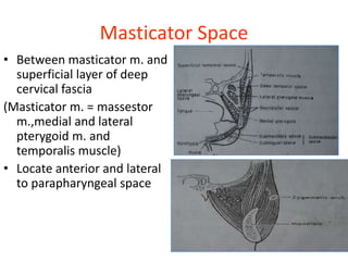
- 23. Masticator Space ŌĆó Between masticator m. and superficial layer of deep cervical fascia (Masticator m. = massestor m.,medial and lateral pterygoid m. and temporalis muscle) ŌĆó Locate anterior and lateral to parapharyngeal space
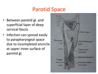
- 24. Parotid Space ŌĆó Between parotid gl. and superficial layer of deep cervical fascia ŌĆó Infection can spread easily to parapharyngeal space due to incompleted encircle at upper inner surface of parotid gl.
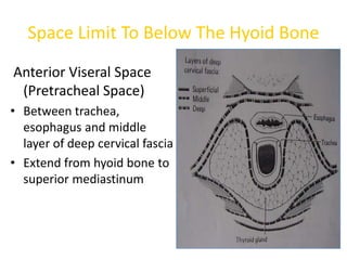
- 25. Space Limit To Below The Hyoid Bone Anterior Viseral Space (Pretracheal Space) ŌĆó Between trachea, esophagus and middle layer of deep cervical fascia ŌĆó Extend from hyoid bone to superior mediastinum