Pulmonary chondroid hamartoma
1 like951 views

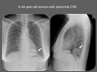
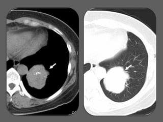
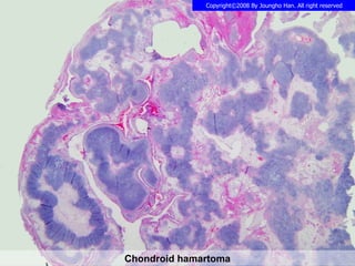
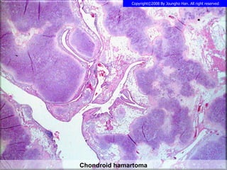
A 44-year-old woman had an abnormal chest x-ray. Further examination found she had a chondroid hamartoma, which is a benign tumor composed of cartilage-like tissue that typically arises in the lungs. A chondroid hamartoma was identified.
1 of 4
Ad
Recommended
Squamous cell carcinoma in WHO B2 thymoma
Squamous cell carcinoma in WHO B2 thymomaJoungho Han
Ìı
A 47-year-old man underwent a chest CT that incidentally discovered a mediastinal mass. Upon examination, doctors found that the man had a B2 thymoma, a type of thymus tumor, that contained squamous cell carcinoma. The squamous cell carcinoma had arisen within the pre-existing B2 thymoma.Sutton 9 limfoma
Sutton 9 limfomaReni Indrastuti
Ìı
Lymphoma is a cancer of the lymphatic system that can arise from either B-cells or T-cells. It is the fourth most common cancer in the UK, with incidence of non-Hodgkin lymphoma increasing. Various viruses and diseases can predispose individuals to developing lymphoma. Clinical manifestations vary between patients but commonly include lymphadenopathy, fever, night sweats, and weight loss. Diagnosis requires a biopsy for examination under microscope. Staging involves CT, PET, or MRI scans to determine spread. The liver, spleen, and lymph nodes are most frequently involved organs. Sonography may reveal hypoechoic lesions in the liver or other organs.Cardiac tumours
Cardiac tumoursAnkur Batra
Ìı
Primary cardiac tumors are rare but can involve the heart directly or through metastasis. Benign tumors like myxomas are the most common primary cardiac tumors and often present with nonspecific symptoms of obstruction, embolism, or constitutional effects. Myxomas typically occur in the left atrium of middle-aged women and can be part of Carney syndrome, characterized by extracardiac tumors and skin pigmentation. Echocardiography is the primary diagnostic tool used to identify location and characteristics of a suspected cardiac tumor.Understanding the thymus
Understanding the thymusAhmed Bahnassy
Ìı
The thymus is a crucial lymphatic organ that plays a key role in immune system development, particularly in T and B cell maturation. It undergoes specific embryological development and can exhibit various abnormalities, including ectopic tissue and neoplasms such as thymoma and thymic carcinoma. The document highlights imaging characteristics, the impact of stress on thymus size, and the distinction between rebound hyperplasia and neoplasia.General pathology lecture 7 neoplasms
General pathology lecture 7 neoplasmsviancksislove
Ìı
This document discusses carcinogenesis and the molecular basis of tumor development. It covers several theories of carcinogenesis, including genetic damage to oncogenes, tumor suppressor genes, genes regulating apoptosis, and DNA repair genes. It also discusses various carcinogenic agents and the multi-step process of carcinogenesis, including initiation, promotion, and progression. Finally, it provides examples of different types of neoplasms and tumors, along with their characteristics.sclerosing hemangioma
sclerosing hemangiomaJoungho Han
Ìı
The document describes a round nodule with a brown tan granular cut surface that is identified as a sclerosing pneumocytoma, also known as a sclerosing hemangioma. This mass is well-circumscribed with partly solid and partly hemorrhagic cut surfaces and a mixed hemorrhagic and solid pattern internally, as well as a mixed papillary, solid and sclerotic pattern.Mucoepidermoid Ca
Mucoepidermoid CaJoungho Han
Ìı
A patient had a mass in their right main bronchus that was removed via rigid bronchoscopy. Upon examination, the mass was found to be a mucoepidermoid carcinoma consisting of many small glands and cysts containing mucin. The lining cells of the mass had round to oval nuclei, abundant mucin-rich cytoplasm, and few mitotic figures.Actinomycosis lung
Actinomycosis lungJoungho Han
Ìı
A 66-year-old man presented with blood tinged sputum and a consolidation in his right middle lung zone. A gun biopsy of the lung tissue showed necrotic tissue, a bacterial colony, and organizing pneumonia. The biopsy revealed actinomycosis, with many filamentous bacteria present.LYMPHANGIOLEIOMYOMATOSIS
LYMPHANGIOLEIOMYOMATOSISJoungho Han
Ìı
A 21-year old woman has recurrent collapsed lungs and many cysts in both lungs. Examinations of the cystic spaces found cells around the edges of the spaces that are characteristic of lymphangioleiomyomatosis, a rare disease where cysts form in the lungs. The woman was diagnosed with lymphangioleiomyomatosis based on her symptoms and the findings of LAM cells around the cystic spaces.Atypical Carcinoid
Atypical CarcinoidJoungho Han
Ìı
A 58-year-old man underwent a routine health check-up where a lung nodule was discovered incidentally in his left lung. Further CT and PET scans revealed a mass in the anterior segment of his left upper lobe. During surgery to remove the mass, a gray tan and lobulated tumor was found growing along the local vessels and lymphatics. Upon microscopic examination, the tumor was found to have an organoid growth pattern, areas of punctate necrosis, and a mitotic rate of 2-10 per 2mm2, consistent with a diagnosis of atypical carcinoid tumor.sclerosing hemangioma
sclerosing hemangiomaJoungho Han
Ìı
A 67-year-old woman had a slowly growing lung nodule that was identified on imaging as a well-defined round mass with calcification and low-attenuation in her left lower lobe. Upon examination, the mass was found to be a round hemorrhagic growth with central irregular sclerosis. The document discusses that this presentation is consistent with a diagnosis of sclerosing hemangioma, with both hemorrhagic and solid patterns present.mucoepidermoid carcinoma lung
mucoepidermoid carcinoma lungJoungho Han
Ìı
A 24-year-old man presented with hemoptysis and was found to have an endobronchial mass obstructing the superior segmental bronchus of his left lower lobe. Further testing revealed a solid and cystic endobronchial mass with focal calcification, which was diagnosed as mucoepidermoid carcinoma, an uncommon tumor containing mucus-secreting, squamous, and intermediate cells often showing glandular formations and calcifications.pulmonary endometriosis
pulmonary endometriosisJoungho Han
Ìı
A 22-year-old woman experienced coughing up blood during her menstrual period for 10 months. A medical scan found abnormalities in her left lung and a biopsy of her lung tissue revealed the presence of endometrial glands and stroma, which are tissues normally found in the uterus. She was diagnosed with endometriosis outside of the uterus that had spread to her lung.Combined small cell carcinoma
Combined small cell carcinomaJoungho Han
Ìı
A 56-year-old man presented with blood tinged sputum for 20 days. Imaging found a nodular lesion and endobronchial mass in the superior segment of his right lower lobe. A biopsy of the mass and bronchial lymph nodes indicated either small cell carcinoma or large cell neuroendocrine carcinoma. Further tests were unable to definitively distinguish between small cell carcinoma and large cell neuroendocrine carcinoma, but suggested a combined diagnosis of both cancers.Cryptococcosis
CryptococcosisJoungho Han
Ìı
A 52-year-old man with a history of hepatitis B and hepatocellular carcinoma underwent evaluation of a new lung nodule seen on chest x-ray. CT and PET scans along with a core biopsy of the lung nodule revealed necrotizing inflammation with granulomas containing many yeast-form organisms. Special stains identified the organisms as Cryptococcus, consistent with a diagnosis of cryptococcosis.Cystic Thymoma
Cystic ThymomaJoungho Han
Ìı
A 63-year-old man underwent VATS excision of an anterior mediastinal mass which was found to be a cystic thymoma of WHO type A. Microscopic examination at low, medium, and high power revealed the tumor cells formed cysts of various sizes consistent with cystic thymoma, WHO type A.lung metastatic adenocarcinoma from colon
lung metastatic adenocarcinoma from colonJoungho Han
Ìı
A 77-year-old man was found to have a lung nodule during follow-up for stage III colon cancer three years prior. Imaging and biopsy of the lung nodule revealed it to be a metastatic adenocarcinoma from the original colon cancer. Microscopic examination showed the metastatic tumor had invaded the mediastinal pleura and consisted of tall columnar tumor cells forming glands, with areas of necrosis and calcification consistent with metastatic colon cancer.small cell carcinoma
small cell carcinomaJoungho Han
Ìı
A 72-year-old man presented with 10 years of hoarseness that worsened over the past year. Imaging found a nodule in his right upper lobe lung and enlarged lymph nodes near his hilar region. A biopsy of the lung nodule revealed small cell carcinoma of the lung that had spread to the lymph nodes.CMV pneumonia
CMV pneumoniaJoungho Han
Ìı
A 6-year-old female with breathing difficulties was previously diagnosed with hemophagocytic lymphohistiocytosis (HLH) based on recurrent infections and enlarged liver and spleen. She received a cord blood transplant one month ago but showed engraftment failure on blood tests. Her respiratory symptoms and enlarged liver and spleen have worsened, suggesting disease progression. A lung biopsy was performed to evaluate the exact cause. The biopsy showed diffuse small nodular and ground-glass opacities in both lungs consistent with CMV pneumonia as shown by cytomegalovirus staining.Adenoid cystic carcinoma trachea
Adenoid cystic carcinoma tracheaJoungho Han
Ìı
A 58-year-old man experienced worsening dyspnea over 4 months. A CT scan showed a multi-lobulated mass in his distal trachea and carina, severely narrowing his airway. During bronchoscopy, the mass was found to obstruct 90% of the distal trachea. After using laser cauterization to remove pieces of the tumor, pathology examination revealed it to be an adenoid cystic carcinoma, shown by a cribriform pattern of tumor cells surrounding pseudocysts.Cryptococcosis
CryptococcosisJoungho Han
Ìı
A 62-year-old man underwent a CT scan and PET scan which found lung nodules incidentally during a work-up for a liver mass. A VATS biopsy of the lung nodules found granulomatous inflammation with many multinucleated giant cells and histiocytes containing many yeast organisms; special stains identified the organisms as Cryptococcus.invasive aspergillosis
invasive aspergillosisJoungho Han
Ìı
A 62-year-old man with a history of hepatitis B, liver cirrhosis, liver transplantation, and recent total gastrectomy presented with cough and sputum. Chest imaging showed multifocal ground-glass opacities and multiple cavitary nodules in both lungs. Biopsy of a cavitary nodule in the left upper lobe revealed fungal organisms consistent with invasive pulmonary aspergillosis.Actinomycosis
ActinomycosisJoungho Han
Ìı
A 46-year-old male presented with cough, sputum, and intermittent chest discomfort. Imaging showed dense consolidation in the right middle lobe. A fine needle aspiration smear revealed a filamentous bacterial organism in a neutrophil-rich background. Biopsy of the right middle lobe showed multiple abscesses with cavities containing necrotic material and inflammatory exudate. The patient was diagnosed with actinomycosis based on the smear and biopsy findings.Sclerosing Hemangioma
Sclerosing HemangiomaJoungho Han
Ìı
A 41-year-old woman had an abnormal chest x-ray and underwent a needle biopsy. The biopsy results indicated a diagnosis of sclerosing hemangioma. Further testing showed the tumor cells were positive for the Ki-67 antigen at the cytoplasmic membrane.Aspergilloma
AspergillomaJoungho Han
Ìı
A 48-year-old man had blood tinged sputum for 8 months and was found to have a 2cm nodular opacity in the posterior portion of his right lower lobe. Biopsies of the area showed inflammation and fibrosis around the bronchus, along with the presence of aspergilloma, a fungus ball caused by the fungus Aspergillus, identified by its branching septated hyphae.How to be and stay healthy: Live Wire Not a Couch Potato
How to be and stay healthy: Live Wire Not a Couch PotatoBiljanaPipovic
Ìı
Live Wire, Not a Couch Potato was an engaging international eTwinning project aimed at promoting physical activity among youth by blending creativity, tradition, and competition. Founded by teachers from Serbia and Portugal, with partners from Turkey and Greece, the project encouraged students to explore the importance of staying active through fun and meaningful challenges. Working within their local contexts, students from all four countries participated in physical activities at school and in their communities. They documented their experiences through photos, videos, and written reflections, which they shared on TwinSpace. The project helped transform exercise from a routine task into an enjoyable, culturally rich experience, while fostering teamwork, creativity, and international exchange.Quality by Design Tools in Pharmaceutical Manufacturing Technology
Quality by Design Tools in Pharmaceutical Manufacturing Technology39KomalZaveri
Ìı
In Qbd why risk assessment needed that explained
Design Space and DoE relationship with each other.
More Related Content
More from Joungho Han (20)
Actinomycosis lung
Actinomycosis lungJoungho Han
Ìı
A 66-year-old man presented with blood tinged sputum and a consolidation in his right middle lung zone. A gun biopsy of the lung tissue showed necrotic tissue, a bacterial colony, and organizing pneumonia. The biopsy revealed actinomycosis, with many filamentous bacteria present.LYMPHANGIOLEIOMYOMATOSIS
LYMPHANGIOLEIOMYOMATOSISJoungho Han
Ìı
A 21-year old woman has recurrent collapsed lungs and many cysts in both lungs. Examinations of the cystic spaces found cells around the edges of the spaces that are characteristic of lymphangioleiomyomatosis, a rare disease where cysts form in the lungs. The woman was diagnosed with lymphangioleiomyomatosis based on her symptoms and the findings of LAM cells around the cystic spaces.Atypical Carcinoid
Atypical CarcinoidJoungho Han
Ìı
A 58-year-old man underwent a routine health check-up where a lung nodule was discovered incidentally in his left lung. Further CT and PET scans revealed a mass in the anterior segment of his left upper lobe. During surgery to remove the mass, a gray tan and lobulated tumor was found growing along the local vessels and lymphatics. Upon microscopic examination, the tumor was found to have an organoid growth pattern, areas of punctate necrosis, and a mitotic rate of 2-10 per 2mm2, consistent with a diagnosis of atypical carcinoid tumor.sclerosing hemangioma
sclerosing hemangiomaJoungho Han
Ìı
A 67-year-old woman had a slowly growing lung nodule that was identified on imaging as a well-defined round mass with calcification and low-attenuation in her left lower lobe. Upon examination, the mass was found to be a round hemorrhagic growth with central irregular sclerosis. The document discusses that this presentation is consistent with a diagnosis of sclerosing hemangioma, with both hemorrhagic and solid patterns present.mucoepidermoid carcinoma lung
mucoepidermoid carcinoma lungJoungho Han
Ìı
A 24-year-old man presented with hemoptysis and was found to have an endobronchial mass obstructing the superior segmental bronchus of his left lower lobe. Further testing revealed a solid and cystic endobronchial mass with focal calcification, which was diagnosed as mucoepidermoid carcinoma, an uncommon tumor containing mucus-secreting, squamous, and intermediate cells often showing glandular formations and calcifications.pulmonary endometriosis
pulmonary endometriosisJoungho Han
Ìı
A 22-year-old woman experienced coughing up blood during her menstrual period for 10 months. A medical scan found abnormalities in her left lung and a biopsy of her lung tissue revealed the presence of endometrial glands and stroma, which are tissues normally found in the uterus. She was diagnosed with endometriosis outside of the uterus that had spread to her lung.Combined small cell carcinoma
Combined small cell carcinomaJoungho Han
Ìı
A 56-year-old man presented with blood tinged sputum for 20 days. Imaging found a nodular lesion and endobronchial mass in the superior segment of his right lower lobe. A biopsy of the mass and bronchial lymph nodes indicated either small cell carcinoma or large cell neuroendocrine carcinoma. Further tests were unable to definitively distinguish between small cell carcinoma and large cell neuroendocrine carcinoma, but suggested a combined diagnosis of both cancers.Cryptococcosis
CryptococcosisJoungho Han
Ìı
A 52-year-old man with a history of hepatitis B and hepatocellular carcinoma underwent evaluation of a new lung nodule seen on chest x-ray. CT and PET scans along with a core biopsy of the lung nodule revealed necrotizing inflammation with granulomas containing many yeast-form organisms. Special stains identified the organisms as Cryptococcus, consistent with a diagnosis of cryptococcosis.Cystic Thymoma
Cystic ThymomaJoungho Han
Ìı
A 63-year-old man underwent VATS excision of an anterior mediastinal mass which was found to be a cystic thymoma of WHO type A. Microscopic examination at low, medium, and high power revealed the tumor cells formed cysts of various sizes consistent with cystic thymoma, WHO type A.lung metastatic adenocarcinoma from colon
lung metastatic adenocarcinoma from colonJoungho Han
Ìı
A 77-year-old man was found to have a lung nodule during follow-up for stage III colon cancer three years prior. Imaging and biopsy of the lung nodule revealed it to be a metastatic adenocarcinoma from the original colon cancer. Microscopic examination showed the metastatic tumor had invaded the mediastinal pleura and consisted of tall columnar tumor cells forming glands, with areas of necrosis and calcification consistent with metastatic colon cancer.small cell carcinoma
small cell carcinomaJoungho Han
Ìı
A 72-year-old man presented with 10 years of hoarseness that worsened over the past year. Imaging found a nodule in his right upper lobe lung and enlarged lymph nodes near his hilar region. A biopsy of the lung nodule revealed small cell carcinoma of the lung that had spread to the lymph nodes.CMV pneumonia
CMV pneumoniaJoungho Han
Ìı
A 6-year-old female with breathing difficulties was previously diagnosed with hemophagocytic lymphohistiocytosis (HLH) based on recurrent infections and enlarged liver and spleen. She received a cord blood transplant one month ago but showed engraftment failure on blood tests. Her respiratory symptoms and enlarged liver and spleen have worsened, suggesting disease progression. A lung biopsy was performed to evaluate the exact cause. The biopsy showed diffuse small nodular and ground-glass opacities in both lungs consistent with CMV pneumonia as shown by cytomegalovirus staining.Adenoid cystic carcinoma trachea
Adenoid cystic carcinoma tracheaJoungho Han
Ìı
A 58-year-old man experienced worsening dyspnea over 4 months. A CT scan showed a multi-lobulated mass in his distal trachea and carina, severely narrowing his airway. During bronchoscopy, the mass was found to obstruct 90% of the distal trachea. After using laser cauterization to remove pieces of the tumor, pathology examination revealed it to be an adenoid cystic carcinoma, shown by a cribriform pattern of tumor cells surrounding pseudocysts.Cryptococcosis
CryptococcosisJoungho Han
Ìı
A 62-year-old man underwent a CT scan and PET scan which found lung nodules incidentally during a work-up for a liver mass. A VATS biopsy of the lung nodules found granulomatous inflammation with many multinucleated giant cells and histiocytes containing many yeast organisms; special stains identified the organisms as Cryptococcus.invasive aspergillosis
invasive aspergillosisJoungho Han
Ìı
A 62-year-old man with a history of hepatitis B, liver cirrhosis, liver transplantation, and recent total gastrectomy presented with cough and sputum. Chest imaging showed multifocal ground-glass opacities and multiple cavitary nodules in both lungs. Biopsy of a cavitary nodule in the left upper lobe revealed fungal organisms consistent with invasive pulmonary aspergillosis.Actinomycosis
ActinomycosisJoungho Han
Ìı
A 46-year-old male presented with cough, sputum, and intermittent chest discomfort. Imaging showed dense consolidation in the right middle lobe. A fine needle aspiration smear revealed a filamentous bacterial organism in a neutrophil-rich background. Biopsy of the right middle lobe showed multiple abscesses with cavities containing necrotic material and inflammatory exudate. The patient was diagnosed with actinomycosis based on the smear and biopsy findings.Sclerosing Hemangioma
Sclerosing HemangiomaJoungho Han
Ìı
A 41-year-old woman had an abnormal chest x-ray and underwent a needle biopsy. The biopsy results indicated a diagnosis of sclerosing hemangioma. Further testing showed the tumor cells were positive for the Ki-67 antigen at the cytoplasmic membrane.Aspergilloma
AspergillomaJoungho Han
Ìı
A 48-year-old man had blood tinged sputum for 8 months and was found to have a 2cm nodular opacity in the posterior portion of his right lower lobe. Biopsies of the area showed inflammation and fibrosis around the bronchus, along with the presence of aspergilloma, a fungus ball caused by the fungus Aspergillus, identified by its branching septated hyphae.Recently uploaded (20)
How to be and stay healthy: Live Wire Not a Couch Potato
How to be and stay healthy: Live Wire Not a Couch PotatoBiljanaPipovic
Ìı
Live Wire, Not a Couch Potato was an engaging international eTwinning project aimed at promoting physical activity among youth by blending creativity, tradition, and competition. Founded by teachers from Serbia and Portugal, with partners from Turkey and Greece, the project encouraged students to explore the importance of staying active through fun and meaningful challenges. Working within their local contexts, students from all four countries participated in physical activities at school and in their communities. They documented their experiences through photos, videos, and written reflections, which they shared on TwinSpace. The project helped transform exercise from a routine task into an enjoyable, culturally rich experience, while fostering teamwork, creativity, and international exchange.Quality by Design Tools in Pharmaceutical Manufacturing Technology
Quality by Design Tools in Pharmaceutical Manufacturing Technology39KomalZaveri
Ìı
In Qbd why risk assessment needed that explained
Design Space and DoE relationship with each other.
Drmohamedaslam_resident_copd2025_fm.pptx
Drmohamedaslam_resident_copd2025_fm.pptxAslam
Ìı
COPD :LATEST GUIDELINES 2025
REFERENCE: Harrisonâs Principles of Internal Medicine
GOLD -2025 Guidelines
It highlights updated diagnostic criteria, pharmacological and non-pharmacological treatment options, and current best practices for resident doctors and healthcare professionals.
Ideal for medical students, residents, and practitioners seeking an up-to-date, evidence-based reference.
ğ Download, share, and feel free to reach out for related study material!nanoparticle and liposomes ppt .(NTDS)pdf
nanoparticle and liposomes ppt .(NTDS)pdfsiddhikalbande
Ìı
Nanoparticles and liposomes are advanced carriers used for targeted drug delivery.
Nanoparticles enhance drug effectiveness by directing treatment to specific sites.
Liposomes are biocompatible vesicles that enable controlled and sustained drug release.Coarse Dispersion, Physical Pharmaceutics
Coarse Dispersion, Physical Pharmaceuticsnishiprakashj
Ìı
Its a compilation of unit 3 as per PCI syllabus of B.Pharm IV sem, Subject Physical Pharmaceutics.Navigating the Open Enrollment Period for Medicare Supplement Insurance in Sa...
Navigating the Open Enrollment Period for Medicare Supplement Insurance in Sa...dfwdirectinsurance
Ìı
Understanding Medicare options can feel like navigating a maze, especially during the open enrollment period. For residents of Sarasota approaching age 65 or already enrolled in Medicare, this window offers a vital opportunity to secure additional health coverage that helps reduce out-of-pocket costs. If you're planning to enhance your Medicare plan, it's essential to understand the rules, deadlines, and options that apply to Medicare supplement insurance in Sarasota.The concept of Druti in Rasashastra- Dr. Aiswarya Babu
The concept of Druti in Rasashastra- Dr. Aiswarya BabuDr. Aiswarya Babu
Ìı
It is a dosage form that is believed to have been utilized for both Deha & Loha siddhi. Druti Kalpana is an important pharmaceutical process of Rasashastra in which metal/minerals are converted into stable liquefied state. reference taken from Rasaratna samuchaya, 8/84.
Viddha karma in Ayurveda-Dr Mahesh Kumar.pdf
Viddha karma in Ayurveda-Dr Mahesh Kumar.pdfCBPACS, Khera Dabar, Najafgarh New Delhi- 73
Ìı
Ayurveda have description of various treatment modalities. Viddhakarma is ayurvedic treatment method described in ancient ayurveda literature. Its actually a Vedhana karma.
Application of Viddha karma in clinical practice is now popular.Severe Acute Respiratory Syndrome (SARS)
Severe Acute Respiratory Syndrome (SARS)Dr. Anu Marhatta
Ìı
This slide is for educational purposes only. Biography and Professional Career of Dr. Seth Eidemiller
Biography and Professional Career of Dr. Seth EidemillerDr. Seth Eidemiller
Ìı
Dr. Seth A. Eidemiller is a board-certified emergency physician whose professional journey began on a fourth-generation dairy farm in Idaho. Early on, he gained experience through farming, wildfire suppression, and construction work, which gave him a strong foundation in practical skills and resilience. After completing degrees in International Studies and Spanish, he returned to Boise to fulfill the prerequisites for medical school and study laboratory sciences. He then attended the University of Nevada, Reno School of Medicine, and continued his training with a residency in emergency medicine in Fresno. Today, he serves as Vice Chair of the Chico Emergency Medicine Physician Group.Update on Anesthesia for Pediatric Ophthalmic Surgery.pptx
Update on Anesthesia for Pediatric Ophthalmic Surgery.pptxDr.Umang Sharma
Ìı
Based on practices on my hospital and 2021 bja articleANATOMY OF LARYNX -Prof.Dr.N.Mugunthan, KMMC.pdf
ANATOMY OF LARYNX -Prof.Dr.N.Mugunthan, KMMC.pdfKanyakumari Medical Mission Research Center, Muttom
Ìı
Anatomy of the Larynx
Larynx is a cartilaginous structure located in the **anterior neck** at the level of **C3âC6 vertebrae**.
* Also called the **voice box**âresponsible for **phonation**, **airway protection**, and **breathing regulation**.
**Cartilages of the Larynx**
**Unpaired Cartilages:**
1. **Thyroid cartilage**:
* Largest cartilage; forms the **Adamâs apple (laryngeal prominence)**.
* Composed of two laminae that fuse anteriorly.
2. **Cricoid cartilage**:
* Ring-shaped; **only complete ring** around the airway.
* Located below thyroid cartilage; articulates with thyroid & arytenoid cartilages.
3. **Epiglottis**:
* Leaf-shaped elastic cartilage.
* Covers the laryngeal inlet during swallowing.
**Paired Cartilages:**
1. **Arytenoid**:
* Pyramid-shaped; sit on top of cricoid.
* Attach vocal cords and muscles.
2. **Corniculate**:
* Small cartilages sitting atop the arytenoids.
3. **Cuneiform**:
* Embedded in the aryepiglottic folds; provide structural support.
**Intrinsic Muscles of the Larynx**
> Responsible for controlling **vocal cord movement**.
| Muscle | Action | Nerve Supply |
| **Cricothyroid** | Tenses vocal cords | External laryngeal nerve |
| **Thyroarytenoid** | Relaxes vocal cords | Recurrent laryngeal nerve |
| **Posterior cricoarytenoid** | Abducts vocal cords (opens glottis) | Recurrent laryngeal nerve |
| **Lateral cricoarytenoid** | Adducts vocal cords | Recurrent laryngeal nerve |
| **Transverse & oblique arytenoids** | Closes the posterior glottis | Recurrent laryngeal nerve |
| **Vocalis** | Fine-tunes pitch | Recurrent laryngeal nerve | **Nerve Supply*
* **Motor**:
* All intrinsic muscles (except cricothyroid): **Recurrent laryngeal nerve** (branch of vagus nerve).
* Cricothyroid: **External branch of superior laryngeal nerve**.
* **Sensory**:
* Above vocal cords: **Internal branch of superior laryngeal nerve**.
* Below vocal cords: **Recurrent laryngeal nerve**.
**Blood Supply**
* **Superior laryngeal artery** (branch of superior thyroid artery).
* **Inferior laryngeal artery** (branch of inferior thyroid artery).
**Laryngeal Cavity & Folds**
Divided into 3 regions:
1. **Vestibule** â from inlet to vestibular folds (false cords).
2. **Ventricle** â between vestibular and vocal folds.
3. **Infraglottic cavity** â below vocal cords to trachea.
**Vocal folds (true cords)** â involved in sound production.INTERPRETATION OF LABORATORY INVESTIGATIONS.pptx
INTERPRETATION OF LABORATORY INVESTIGATIONS.pptxEliLawluvi
Ìı
THE DOCUMENT SUMMARIZES THE KEY COMPONENTS OF INTERPRETING FULL BLOOD CUNTDesign of cosmeceuticals products;sunprotection,sunscreens
Design of cosmeceuticals products;sunprotection,sunscreensSwami ramanand teerth marathwada university
Ìı
Design of cosmeceuticals products and improving the skin to protection sun burning, used sunscreens, sunprotections An interesting case of facial Swelling in an autoimmune rheumatic disease Ahm...
An interesting case of facial Swelling in an autoimmune rheumatic disease Ahm...Internal medicine department, faculty of Medicine Beni-Suef University Egypt
Ìı
An interesting case of facial Swelling in an autoimmune rheumatic disease Ahmed Yehia EGYSIR Conference Air Pollution, air quality index, Nepal air pollution
Air Pollution, air quality index, Nepal air pollutionDr. Anu Marhatta
Ìı
This presentation slides is only for educational purposes. Alzheimerâs Disease Neuroradiology Case Conference: Mastering the New Frontie...
Alzheimerâs Disease Neuroradiology Case Conference: Mastering the New Frontie...PVI, PeerView Institute for Medical Education
Ìı
Co-Chairs, Gloria Chiang, MD, and Ana M. Franceschi, MD, PhD, discuss Alzheimerâs disease in this CME/AAPA activity titled âAlzheimerâs Disease Neuroradiology Case Conference: Mastering the New Frontier in Diagnosis and Treatment.â For the full presentation, downloadable Practice Aids, and complete CME/AAPA information, and to apply for credit, please visit us at https://bit.ly/43CE1XA. CME/AAPA credit will be available until July 3, 2026.From Preservation To Regeneration--The Stem Cell Era of Hair Restoration_DrAl...
From Preservation To Regeneration--The Stem Cell Era of Hair Restoration_DrAl...Alan Bauman
Ìı
How can the latest in Regenerative Medicine help those suffering from hair loss? Cell Surgical Conference 2025 featured Dr Alan Bauman as a faculty member once again to discuss all things related to hair loss, hair transplantation and the use of regenerative stem cell therapies for hair restoration. Navigating the Open Enrollment Period for Medicare Supplement Insurance in Sa...
Navigating the Open Enrollment Period for Medicare Supplement Insurance in Sa...dfwdirectinsurance
Ìı
ANATOMY OF LARYNX -Prof.Dr.N.Mugunthan, KMMC.pdf
ANATOMY OF LARYNX -Prof.Dr.N.Mugunthan, KMMC.pdfKanyakumari Medical Mission Research Center, Muttom
Ìı
Design of cosmeceuticals products;sunprotection,sunscreens
Design of cosmeceuticals products;sunprotection,sunscreensSwami ramanand teerth marathwada university
Ìı
An interesting case of facial Swelling in an autoimmune rheumatic disease Ahm...
An interesting case of facial Swelling in an autoimmune rheumatic disease Ahm...Internal medicine department, faculty of Medicine Beni-Suef University Egypt
Ìı
Alzheimerâs Disease Neuroradiology Case Conference: Mastering the New Frontie...
Alzheimerâs Disease Neuroradiology Case Conference: Mastering the New Frontie...PVI, PeerView Institute for Medical Education
Ìı
Ad
Pulmonary chondroid hamartoma
- 1. A 44-year-old woman with abnormal CXR
- 2. Ìı