Renal system Imaging.pptx
Download as pptx, pdf0 likes111 views

This document discusses two modalities for imaging the renal system: intravenous pyelogram (IVP) and DTPA scan. IVP uses x-rays to anatomically image the urinary tract after injecting contrast media, but exposes patients to higher radiation. DTPA scan uses a radioactive tracer to evaluate renal function and perfusion with gamma cameras, requiring no bowel prep and exposing patients to much less radiation. While IVP is useful for detecting disorders affecting the urinary tract, DTPA scan is the modality of choice as it provides functional information on glomerular filtration rate and kidney blood flow with a significantly lower radiation dose.
1 of 21
Download to read offline
Ad
Recommended
bsc.pptx
bsc.pptxshivambhardwaj399404
Ěý
Percutaneous transhepatic cholangiography (PTC) is a radiographic procedure used to visualize the biliary duct system. It involves inserting a needle through the liver under imaging guidance and injecting contrast dye. PTC can be used diagnostically to distinguish intrahepatic and extrahepatic obstructions or therapeutically to place biliary stents. Potential complications include cholangitis, bile leaks, and hemorrhage. Proper patient preparation and post-procedure monitoring is important to reduce risks.Conventional Radiology In Urology.pptx
Conventional Radiology In Urology.pptxrabi pandit
Ěý
Conventional radiology remains useful in urology for preoperative diagnosis and postoperative evaluation. It includes techniques like intravenous urography to visualize the kidneys and ureters, retrograde pyelography to evaluate the ureters and collecting systems, and retrograde urethrography to assess the urethra. These techniques use iodine-based contrast agents, though complications can occur in 1-5% of cases. The physics of x-rays and factors like radiation dose are also discussed.Intravenous Pyelogram RENAL TESTS ADULTS.pptx
Intravenous Pyelogram RENAL TESTS ADULTS.pptxneeti70
Ěý
Intravenous pyelography (IVP) involves injecting iodinated contrast into a patient's veins and using x-rays to image their urinary tract. It can detect abnormalities in the kidneys, ureters, and bladder. While commonly used in the past, IVP is now less frequent due to superior alternatives like ultrasound, CT, and MRI that have fewer limitations. The test requires contrast material, x-ray equipment, and involves obtaining multiple images over the course of an hour to fully visualize the urinary system.GIT and RENAL SYSTEM NUCLEAR MEDICINE PROCEDURES
GIT and RENAL SYSTEM NUCLEAR MEDICINE PROCEDURESVipin Kumar
Ěý
This document discusses several nuclear medicine procedures related to evaluating the renal system and gastrointestinal tract. It provides information on radiopharmaceuticals, dosages, indications, contraindications, preparation, equipment, procedures, artifacts, and normal/abnormal results for renal cortical imaging, glomerular filtration rate measurement, gastrointestinal bleeding detection, and gastric emptying studies. Key details are given for accurately performing and interpreting these nuclear medicine scans.prospects & constraints in vety Laparoscopy in surgery
prospects & constraints in vety Laparoscopy in surgery shahnawaz ahmad bhat
Ěý
Laparoscopy is a minimally invasive surgical technique that uses small incisions and specialized instruments to visualize the abdominal cavity. It has several advantages over open surgery, including less pain, shorter recovery times, and fewer adhesions. Common veterinary laparoscopic procedures include ovariohysterectomy, hernia repair, and biopsy. While laparoscopy provides benefits, it also faces constraints such as technical challenges, costs, and long learning curves. However, with advances in technology and skills, laparoscopy is projected to replace many traditional open surgeries.Scans.. Dr.Padmesh
Scans.. Dr.PadmeshDr Padmesh Vadakepat
Ěý
Diuretic renal scans use radioactive tracers like DTPA, MAG3, or LLEC to evaluate kidney function and rule out obstruction. DTPA/MAG3 scans provide information on renal blood flow, GFR, tubular function, and excretion. DMSA scans use Technetium99m to visualize renal cortex and assess renal scarring. Bone scans use Technetium99m HDP to detect bone metastases, tumors, and infections. HIDA scans use Technetium99m Hepatolite to evaluate gallbladder function and detect causes of jaundice like cholecystitis. Lung V/Q scans use radioactive gas and injections to detect perfusion mismatches diagnostic of pulmonary embolismCorrelation of CTU with renal scintigraphy in assessing split Renal functio...
Correlation of CTU with renal scintigraphy in assessing split Renal functio...AbiyTadele1
Ěý
This document discusses the correlation of CT urography (CTU) with renal scintigraphy in assessing split renal function. It outlines the methodologies for functional imaging of kidneys, including the use of nuclear renal scans and CTU, and presents findings on their effectiveness in evaluating renal function. The study indicates that CTU can serve as a comprehensive assessment tool for both anatomical and functional characteristics of the kidneys, demonstrating a strong correlation with renal scintigraphy results.Intravenous Urography (IVU)., radiological procedure
Intravenous Urography (IVU)., radiological procedureRitupanta1
Ěý
Intravenous urography (IVU) involves injecting contrast media intravenously and taking x-ray images of the urinary tract over time. It is used to evaluate conditions like hematuria, kidney stones, urinary tract infections, and potential kidney donors. The procedure involves an initial image, then images at 5 minutes, 15 minutes, and after urination to view contrast flow and potential abnormalities in the kidneys, ureters and bladder. Precautions are taken with bowel preparation and creatinine levels, and observations are made for potential side effects of the contrast media.urography.pptxpppppppppppppppppppppppppppppppppppppp
urography.pptxppppppppppppppppppppppppppppppppppppppRitupanta1
Ěý
Intravenous urography (IVU) is a radiographic examination of the urinary tract using contrast media, indicated for conditions like hematuria and nephrolithiasis. Key contraindications include pregnancy and contrast media sensitivity, with specific preparations required for the patient before the procedure. The technique involves multiple stages of imaging post-contrast administration to visualize renal structures and assess urinary system function.Laparoscopic kidney surg
Laparoscopic kidney surgRushabh Shah
Ěý
This document provides an overview of laparoscopic kidney surgery procedures. It begins with a brief history of laparoscopic nephrectomy and then covers kidney anatomy and approaches to laparoscopic surgery including transperitoneal, retroperitoneal, and hand-assisted. It provides detailed descriptions of procedures like simple nephrectomy, radical nephrectomy for renal malignancy, partial nephrectomy, and renal biopsy. Complications of laparoscopic renal surgery and conclusions are also mentioned.ERCP (1).pptx
ERCP (1).pptxSyedFurqan30
Ěý
ERCP (Endoscopic Retrograde Cholangiopancreatography) is an endoscopic procedure that allows visualization of the bile and pancreatic ducts. It has both diagnostic and therapeutic uses. Diagnostically, it is used to evaluate conditions like jaundice, gallstones, and pancreatic diseases. Therapeutically, it can be used to drain the bile ducts, extract stones, dilate strictures, and place stents. The procedure involves passing an endoscope into the duodenum and cannulating the papilla to inject contrast and pass devices into the bile and pancreatic ducts. Complications can include pancreatitis, bleeding, perforation, and infections. Care is taken with techniques and medications to reduceURINARY SYSTEM 1 the formation of urine.pptx
URINARY SYSTEM 1 the formation of urine.pptxkalebomathew
Ěý
Intravenous urography (IVU) is a radiological examination used to visualize the urinary tract, indicated in cases of suspected urinary conditions. The document outlines the anatomy of the urinary system, preparation for the procedure, equipment used, and potential adverse effects of contrast media. It also describes the procedure's steps, including contrast injection, radiographic timing, and assessment of common pathologies related to IVU.INTRAVENOUS UROGRAPHY INDICATIONS CONTRAINDICATIONS
INTRAVENOUS UROGRAPHY INDICATIONS CONTRAINDICATIONSobscmch
Ěý
This document provides an overview of intravenous urography (IVU), including indications, contraindications, risks, procedure details, and imaging techniques. Some key points:
- IVU involves injecting contrast media intravenously to visualize the kidneys, ureters, and bladder. It is used to evaluate the entire urinary tract for various conditions like infections, abnormalities, and potential donors.
- Common indications include screening for hematuria, evaluating obstructive uropathy, and assessing renal anatomy in children with congenital anomalies.
- Risks include allergic reactions. Preparation involves bowel cleansing and fasting prior. The procedure involves serial x-rays over 30-35 minutes to visualize contrastIntravenous Urography lecture detai.pptx
Intravenous Urography lecture detai.pptxssuser504dda
Ěý
Intravenous urography (IVU) involves injecting iodine contrast media intravenously and imaging the urinary tract with x-rays. It was developed in 1929 to noninvasively examine the kidneys, ureters, and bladder. The procedure involves preparing the patient, administering low-osmolar contrast media intravenously, and taking x-ray images over time as the contrast passes through and outlines the urinary system. IVU is used to evaluate urinary obstruction, trauma, anatomy variations, and other conditions. Radiologists examine the images for abnormalities in kidney size and shape, the ureters, and bladder as well as signs of obstruction, tumors, stones,Endoscopic Retrograde Cholangiopancreatography
.pptx
Endoscopic Retrograde Cholangiopancreatography
.pptxNagasai Pelala
Ěý
The document discusses the anatomy and procedure of endoscopic retrograde cholangiopancreatography (ERCP) for examining and intervening in the biliary and pancreatic ducts. It highlights the anatomy of the hepatobiliary system, patient preparation, and equipment used, as well as the steps involved in performing ERCP and its indications, contraindications, and potential complications. The document also addresses the techniques for biliary cannulation, contrast media used, and the significance of imaging during the procedure.Radiographic procedure PTC & PTB..D.pptx
Radiographic procedure PTC & PTB..D.pptxjustinfan550
Ěý
The document provides a comprehensive overview of percutaneous transhepatic cholangiography (PTC) and percutaneous transhepatic biliary drainage (PTBD), detailing the definitions, indications, contraindications, preparation, techniques used, and potential complications associated with these procedures. It covers methods of imaging the biliary tract, essential equipment, and the procedural steps involved in navigating biliary obstruction. The document emphasizes the significance of pre-procedural imaging and patient monitoring to ensure effective management and mitigate risks during and after the interventions.IVP Best presnetation
IVP Best presnetationDR SACHIN SURA
Ěý
The document describes a case presentation of a 10-year-old male child who presented with abdominal pain. An ultrasound and intravenous pyelogram (IVP) were performed. The ultrasound found right pyonephrosis, right and left renal calculi, and left hydroureteronephrosis. The IVP confirmed these findings and also found a right ureteric calculus and question of a left ureteric stricture. The document then provides details on IVP, including how it is performed, indications, advantages, limitations, normal findings, and examples of various abnormalities that can be seen on an IVP.Renal biopsy
Renal biopsyFarragBahbah
Ěý
This document discusses renal biopsy procedures. It provides details on the history, technique, adequacy, contraindications, complications and indications for renal biopsy. Some key points include:
- Renal biopsy has evolved since the 1950s and can now provide a tissue diagnosis in over 95% of patients with a life-threatening complication rate of less than 0.1%.
- An adequate biopsy sample contains 10-15 glomeruli and provides samples for histology, immunofluorescence and electron microscopy.
- Contraindications include bleeding diathesis and inability to comply with instructions. Relative contraindications include hypertension and infection.
- Complications are rare but can include hematuria, pain, and rarely death fromRenal biopsy
Renal biopsyFarragBahbah
Ěý
This document discusses renal biopsy procedures and indications. It provides details on the history and technique of renal biopsy. Key points include that renal biopsy can provide a diagnosis in over 95% of patients with a complication rate of less than 0.1%. An adequate biopsy sample contains 10-15 glomeruli. Tissue is divided for light microscopy, immunofluorescence, and electron microscopy. Complications include hematuria and pain in 3-4% of patients. Indications for biopsy include nephrotic syndrome, acute kidney injury, systemic diseases affecting the kidneys, and unexplained chronic kidney disease.Renal biopsy
Renal biopsyFarragBahbah
Ěý
This document discusses renal biopsy procedures and indications. It provides details on the history and technique of renal biopsy. Key points include that renal biopsy can provide a diagnosis in over 95% of patients with a complication rate of less than 0.1%. An adequate biopsy sample contains 10-15 glomeruli. Tissue is divided for light microscopy, immunofluorescence, and electron microscopy. Complications include hematuria and pain in 3-4% of patients. Indications for biopsy include nephrotic syndrome, acute kidney injury, systemic diseases affecting the kidneys, and unexplained chronic kidney disease.Pre operative preparations of the patients for laparoscopic surgery.pptx
Pre operative preparations of the patients for laparoscopic surgery.pptxQuiyumMdAb
Ěý
The document outlines essential protocols and considerations for laparoscopic surgery, emphasizing patient safety through proper preparation and preoperative assessment. Key factors include patient selection, bowel preparation, and monitoring intraoperative complications, particularly regarding anesthesia and surgical positioning. It stresses the importance of informed consent, DVT prophylaxis, and achieving optimal postoperative management for successful outcomes.Recent surgical updates on pancreatic resections
Recent surgical updates on pancreatic resectionsCancer surgery By Royapettah Oncology Group
Ěý
The document discusses recent surgical updates for pancreatic resections. It introduces novel techniques for pancreatic resections like the Cattell Braasch maneuver, triangle operation, and modified Appleby procedure. It summarizes outcomes from using these techniques on 45 patients, finding no mortality and comparable morbidity. The document also discusses techniques like vein resection without reconstruction that can increase resectability in select cases.Ivu
Ivumr_koky
Ěý
This document provides an overview of the urinary system including the kidneys, ureters, urinary bladder, and urethra. It describes the location and basic functions of the kidneys and notes they filter waste from the blood and help regulate fluid and electrolyte balance. The document also outlines the intravenous urogram procedure used to examine the urinary system including patient preparation, contrast injection, and obtaining radiographic images in multiple projections.Renal cell carcinoma mgt ppt.pptx
Renal cell carcinoma mgt ppt.pptxBari51
Ěý
CT and MRI are the preferred imaging modalities for diagnosing and staging renal cell carcinoma (RCC). Histologically, RCC includes clear cell, papillary, chromophobe, collecting duct, and other subtypes. Treatment depends on tumor stage and includes partial or radical nephrectomy for localized disease. For advanced or metastatic RCC, options include ablation, immunotherapy, targeted therapy, and clinical trials of vaccines or other experimental therapies. Long term monitoring after treatment involves physical exams and imaging to check for recurrence.Ivu
IvuJohn Peter
Ěý
Intravenous urography (IVU) is an x-ray imaging technique used to examine the urinary tract after injecting iodine contrast media intravenously. It was developed in 1929 by American urologist Moses Swick. An IVU allows visualization of the kidneys, ureters, and bladder to detect abnormalities. The procedure involves injecting low-osmolar contrast media intravenously and taking x-ray images over time as the contrast passes through and outlines the urinary system. Radiologists examine the IVU images for any signs of obstruction, masses, stones, or other abnormalities in the kidneys, ureters, or bladder. IVU remains a useful techniqueENDOSCOPIC RETROGRADE CHOLANGIOPACREATOGRAPHY (ERCP).pptx
ENDOSCOPIC RETROGRADE CHOLANGIOPACREATOGRAPHY (ERCP).pptxvigneshsk95
Ěý
Endoscopic retrograde cholangiopancreatography (ERCP) is a diagnostic and therapeutic technique for conditions related to the pancreatobiliary system, evolving from primarily diagnostic to a therapeutic tool. The procedure involves accessing the biliary and pancreatic ducts via the duodenum, using specialized equipment and requiring careful pre-operative preparation and anesthetic management. Complications can include post-ERCP pancreatitis, hemorrhage, and perforation, with various risk factors and methods for reducing complications identified.Great Governors' Send-Off Quiz 2025 Prelims IIT KGP
Great Governors' Send-Off Quiz 2025 Prelims IIT KGPIIT Kharagpur Quiz Club
Ěý
Prelims of the Great Governors' Send-Off Quiz 2025 hosted by the outgoing governors.
QMs: Aarushi, Aatir, Aditya, ArnavHow payment terms are configured in Odoo 18
How payment terms are configured in Odoo 18Celine George
Ěý
Payment terms in Odoo 18 help define the conditions for when invoices are due. This feature can split payments into multiple parts and automate due dates based on specific rules.More Related Content
Similar to Renal system Imaging.pptx (20)
Intravenous Urography (IVU)., radiological procedure
Intravenous Urography (IVU)., radiological procedureRitupanta1
Ěý
Intravenous urography (IVU) involves injecting contrast media intravenously and taking x-ray images of the urinary tract over time. It is used to evaluate conditions like hematuria, kidney stones, urinary tract infections, and potential kidney donors. The procedure involves an initial image, then images at 5 minutes, 15 minutes, and after urination to view contrast flow and potential abnormalities in the kidneys, ureters and bladder. Precautions are taken with bowel preparation and creatinine levels, and observations are made for potential side effects of the contrast media.urography.pptxpppppppppppppppppppppppppppppppppppppp
urography.pptxppppppppppppppppppppppppppppppppppppppRitupanta1
Ěý
Intravenous urography (IVU) is a radiographic examination of the urinary tract using contrast media, indicated for conditions like hematuria and nephrolithiasis. Key contraindications include pregnancy and contrast media sensitivity, with specific preparations required for the patient before the procedure. The technique involves multiple stages of imaging post-contrast administration to visualize renal structures and assess urinary system function.Laparoscopic kidney surg
Laparoscopic kidney surgRushabh Shah
Ěý
This document provides an overview of laparoscopic kidney surgery procedures. It begins with a brief history of laparoscopic nephrectomy and then covers kidney anatomy and approaches to laparoscopic surgery including transperitoneal, retroperitoneal, and hand-assisted. It provides detailed descriptions of procedures like simple nephrectomy, radical nephrectomy for renal malignancy, partial nephrectomy, and renal biopsy. Complications of laparoscopic renal surgery and conclusions are also mentioned.ERCP (1).pptx
ERCP (1).pptxSyedFurqan30
Ěý
ERCP (Endoscopic Retrograde Cholangiopancreatography) is an endoscopic procedure that allows visualization of the bile and pancreatic ducts. It has both diagnostic and therapeutic uses. Diagnostically, it is used to evaluate conditions like jaundice, gallstones, and pancreatic diseases. Therapeutically, it can be used to drain the bile ducts, extract stones, dilate strictures, and place stents. The procedure involves passing an endoscope into the duodenum and cannulating the papilla to inject contrast and pass devices into the bile and pancreatic ducts. Complications can include pancreatitis, bleeding, perforation, and infections. Care is taken with techniques and medications to reduceURINARY SYSTEM 1 the formation of urine.pptx
URINARY SYSTEM 1 the formation of urine.pptxkalebomathew
Ěý
Intravenous urography (IVU) is a radiological examination used to visualize the urinary tract, indicated in cases of suspected urinary conditions. The document outlines the anatomy of the urinary system, preparation for the procedure, equipment used, and potential adverse effects of contrast media. It also describes the procedure's steps, including contrast injection, radiographic timing, and assessment of common pathologies related to IVU.INTRAVENOUS UROGRAPHY INDICATIONS CONTRAINDICATIONS
INTRAVENOUS UROGRAPHY INDICATIONS CONTRAINDICATIONSobscmch
Ěý
This document provides an overview of intravenous urography (IVU), including indications, contraindications, risks, procedure details, and imaging techniques. Some key points:
- IVU involves injecting contrast media intravenously to visualize the kidneys, ureters, and bladder. It is used to evaluate the entire urinary tract for various conditions like infections, abnormalities, and potential donors.
- Common indications include screening for hematuria, evaluating obstructive uropathy, and assessing renal anatomy in children with congenital anomalies.
- Risks include allergic reactions. Preparation involves bowel cleansing and fasting prior. The procedure involves serial x-rays over 30-35 minutes to visualize contrastIntravenous Urography lecture detai.pptx
Intravenous Urography lecture detai.pptxssuser504dda
Ěý
Intravenous urography (IVU) involves injecting iodine contrast media intravenously and imaging the urinary tract with x-rays. It was developed in 1929 to noninvasively examine the kidneys, ureters, and bladder. The procedure involves preparing the patient, administering low-osmolar contrast media intravenously, and taking x-ray images over time as the contrast passes through and outlines the urinary system. IVU is used to evaluate urinary obstruction, trauma, anatomy variations, and other conditions. Radiologists examine the images for abnormalities in kidney size and shape, the ureters, and bladder as well as signs of obstruction, tumors, stones,Endoscopic Retrograde Cholangiopancreatography
.pptx
Endoscopic Retrograde Cholangiopancreatography
.pptxNagasai Pelala
Ěý
The document discusses the anatomy and procedure of endoscopic retrograde cholangiopancreatography (ERCP) for examining and intervening in the biliary and pancreatic ducts. It highlights the anatomy of the hepatobiliary system, patient preparation, and equipment used, as well as the steps involved in performing ERCP and its indications, contraindications, and potential complications. The document also addresses the techniques for biliary cannulation, contrast media used, and the significance of imaging during the procedure.Radiographic procedure PTC & PTB..D.pptx
Radiographic procedure PTC & PTB..D.pptxjustinfan550
Ěý
The document provides a comprehensive overview of percutaneous transhepatic cholangiography (PTC) and percutaneous transhepatic biliary drainage (PTBD), detailing the definitions, indications, contraindications, preparation, techniques used, and potential complications associated with these procedures. It covers methods of imaging the biliary tract, essential equipment, and the procedural steps involved in navigating biliary obstruction. The document emphasizes the significance of pre-procedural imaging and patient monitoring to ensure effective management and mitigate risks during and after the interventions.IVP Best presnetation
IVP Best presnetationDR SACHIN SURA
Ěý
The document describes a case presentation of a 10-year-old male child who presented with abdominal pain. An ultrasound and intravenous pyelogram (IVP) were performed. The ultrasound found right pyonephrosis, right and left renal calculi, and left hydroureteronephrosis. The IVP confirmed these findings and also found a right ureteric calculus and question of a left ureteric stricture. The document then provides details on IVP, including how it is performed, indications, advantages, limitations, normal findings, and examples of various abnormalities that can be seen on an IVP.Renal biopsy
Renal biopsyFarragBahbah
Ěý
This document discusses renal biopsy procedures. It provides details on the history, technique, adequacy, contraindications, complications and indications for renal biopsy. Some key points include:
- Renal biopsy has evolved since the 1950s and can now provide a tissue diagnosis in over 95% of patients with a life-threatening complication rate of less than 0.1%.
- An adequate biopsy sample contains 10-15 glomeruli and provides samples for histology, immunofluorescence and electron microscopy.
- Contraindications include bleeding diathesis and inability to comply with instructions. Relative contraindications include hypertension and infection.
- Complications are rare but can include hematuria, pain, and rarely death fromRenal biopsy
Renal biopsyFarragBahbah
Ěý
This document discusses renal biopsy procedures and indications. It provides details on the history and technique of renal biopsy. Key points include that renal biopsy can provide a diagnosis in over 95% of patients with a complication rate of less than 0.1%. An adequate biopsy sample contains 10-15 glomeruli. Tissue is divided for light microscopy, immunofluorescence, and electron microscopy. Complications include hematuria and pain in 3-4% of patients. Indications for biopsy include nephrotic syndrome, acute kidney injury, systemic diseases affecting the kidneys, and unexplained chronic kidney disease.Renal biopsy
Renal biopsyFarragBahbah
Ěý
This document discusses renal biopsy procedures and indications. It provides details on the history and technique of renal biopsy. Key points include that renal biopsy can provide a diagnosis in over 95% of patients with a complication rate of less than 0.1%. An adequate biopsy sample contains 10-15 glomeruli. Tissue is divided for light microscopy, immunofluorescence, and electron microscopy. Complications include hematuria and pain in 3-4% of patients. Indications for biopsy include nephrotic syndrome, acute kidney injury, systemic diseases affecting the kidneys, and unexplained chronic kidney disease.Pre operative preparations of the patients for laparoscopic surgery.pptx
Pre operative preparations of the patients for laparoscopic surgery.pptxQuiyumMdAb
Ěý
The document outlines essential protocols and considerations for laparoscopic surgery, emphasizing patient safety through proper preparation and preoperative assessment. Key factors include patient selection, bowel preparation, and monitoring intraoperative complications, particularly regarding anesthesia and surgical positioning. It stresses the importance of informed consent, DVT prophylaxis, and achieving optimal postoperative management for successful outcomes.Recent surgical updates on pancreatic resections
Recent surgical updates on pancreatic resectionsCancer surgery By Royapettah Oncology Group
Ěý
The document discusses recent surgical updates for pancreatic resections. It introduces novel techniques for pancreatic resections like the Cattell Braasch maneuver, triangle operation, and modified Appleby procedure. It summarizes outcomes from using these techniques on 45 patients, finding no mortality and comparable morbidity. The document also discusses techniques like vein resection without reconstruction that can increase resectability in select cases.Ivu
Ivumr_koky
Ěý
This document provides an overview of the urinary system including the kidneys, ureters, urinary bladder, and urethra. It describes the location and basic functions of the kidneys and notes they filter waste from the blood and help regulate fluid and electrolyte balance. The document also outlines the intravenous urogram procedure used to examine the urinary system including patient preparation, contrast injection, and obtaining radiographic images in multiple projections.Renal cell carcinoma mgt ppt.pptx
Renal cell carcinoma mgt ppt.pptxBari51
Ěý
CT and MRI are the preferred imaging modalities for diagnosing and staging renal cell carcinoma (RCC). Histologically, RCC includes clear cell, papillary, chromophobe, collecting duct, and other subtypes. Treatment depends on tumor stage and includes partial or radical nephrectomy for localized disease. For advanced or metastatic RCC, options include ablation, immunotherapy, targeted therapy, and clinical trials of vaccines or other experimental therapies. Long term monitoring after treatment involves physical exams and imaging to check for recurrence.Ivu
IvuJohn Peter
Ěý
Intravenous urography (IVU) is an x-ray imaging technique used to examine the urinary tract after injecting iodine contrast media intravenously. It was developed in 1929 by American urologist Moses Swick. An IVU allows visualization of the kidneys, ureters, and bladder to detect abnormalities. The procedure involves injecting low-osmolar contrast media intravenously and taking x-ray images over time as the contrast passes through and outlines the urinary system. Radiologists examine the IVU images for any signs of obstruction, masses, stones, or other abnormalities in the kidneys, ureters, or bladder. IVU remains a useful techniqueENDOSCOPIC RETROGRADE CHOLANGIOPACREATOGRAPHY (ERCP).pptx
ENDOSCOPIC RETROGRADE CHOLANGIOPACREATOGRAPHY (ERCP).pptxvigneshsk95
Ěý
Endoscopic retrograde cholangiopancreatography (ERCP) is a diagnostic and therapeutic technique for conditions related to the pancreatobiliary system, evolving from primarily diagnostic to a therapeutic tool. The procedure involves accessing the biliary and pancreatic ducts via the duodenum, using specialized equipment and requiring careful pre-operative preparation and anesthetic management. Complications can include post-ERCP pancreatitis, hemorrhage, and perforation, with various risk factors and methods for reducing complications identified.Recently uploaded (20)
Great Governors' Send-Off Quiz 2025 Prelims IIT KGP
Great Governors' Send-Off Quiz 2025 Prelims IIT KGPIIT Kharagpur Quiz Club
Ěý
Prelims of the Great Governors' Send-Off Quiz 2025 hosted by the outgoing governors.
QMs: Aarushi, Aatir, Aditya, ArnavHow payment terms are configured in Odoo 18
How payment terms are configured in Odoo 18Celine George
Ěý
Payment terms in Odoo 18 help define the conditions for when invoices are due. This feature can split payments into multiple parts and automate due dates based on specific rules.F-BLOCK ELEMENTS POWER POINT PRESENTATIONS
F-BLOCK ELEMENTS POWER POINT PRESENTATIONSmprpgcwa2024
Ěý
F-block elements are a group of elements in the periodic table that have partially filled f-orbitals. They are also known as inner transition elements. F-block elements are divided into two series:
1.Lanthanides (La- Lu) These elements are also known as rare earth elements.
2.Actinides (Ac- Lr): These elements are radioactive and have complex electronic configurations.
F-block elements exhibit multiple oxidation states due to the availability of f-orbitals.
2. Many f-block compounds are colored due to f-f transitions.
3. F-block elements often exhibit paramagnetic or ferromagnetic behavior.4. Actinides are radioactive.
F-block elements are used as catalysts in various industrial processes.
Actinides are used in nuclear reactors and nuclear medicine.
F-block elements are used in lasers and phosphors due to their luminescent properties.
F-block elements have unique electronic and magnetic properties.Photo chemistry Power Point Presentation
Photo chemistry Power Point Presentationmprpgcwa2024
Ěý
Photochemistry is the branch of chemistry that deals with the study of chemical reactions and processes initiated by light.
Photochemistry involves the interaction of light with molecules, leading to electronic excitation. Energy from light is transferred to molecules, initiating chemical reactions.
Photochemistry is used in solar cells to convert light into electrical energy.
It is used Light-driven chemical reactions for environmental remediation and synthesis. Photocatalysis helps in pollution abatement and environmental cleanup. Photodynamic therapy offers a targeted approach to treating diseases It is used in Light-activated treatment for cancer and other diseases.
Photochemistry is used to synthesize complex organic molecules.
Photochemistry contributes to the development of sustainable energy solutions.Q1_ENGLISH_PPT_WEEK 1 power point grade 3 Quarter 1 week 1
Q1_ENGLISH_PPT_WEEK 1 power point grade 3 Quarter 1 week 1jutaydeonne
Ěý
Grade 3 Quarter 1 Week 1 English part 2How to use search fetch method in Odoo 18
How to use search fetch method in Odoo 18Celine George
Ěý
The search_fetch is a powerful ORM method used in Odoo for some specific addons to combine the functionality of search and read for more efficient data fetching. It might be used to search for records and fetch specific fields in a single call. It stores the result in the cache memory.LDMMIA Yoga S10 Free Workshop Grad Level
LDMMIA Yoga S10 Free Workshop Grad LevelLDM & Mia eStudios
Ěý
This is complete for June 17th. For the weekend of Summer Solstice
June 20th-22nd.
6/17/25: “My now Grads, You’re doing well. I applaud your efforts to continue. We all are shifting to new paradigm realities. Its rough, there’s good and bad days/weeks. However, Reiki with Yoga assistance, does work.”
6/18/25: "For those planning the Training Program Do Welcome. Happy Summer 2k25. You are not ignored and much appreciated. Our updates are ongoing and weekly since Spring. I Hope you Enjoy the Practitioner Grad Level. There's more to come. We will also be wrapping up Level One. So I can work on Levels 2 topics. Please see documents for any news updates. Also visit our websites. Every decade I release a Campus eMap. I will work on that for summer 25. We have 2 old libraries online thats open. https://ldmchapels.weebly.com "
Our Monthly Class Roster is 7,141 for 6/21.
ALL students get privacy naturally. Thx Everyone.
As a Guest Student,
You are now upgraded to Grad Level.
See Uploads for “Student Checkins” & “S9”. Thx.
Happy Summer 25.
These are also timeless.
Thank you for attending our workshops.
If you are new, do welcome.
For visual/Video style learning see our practitioner student status.
This is listed under our new training program. Updates ongoing levels 1-3 this summer. We just started Session 1 for level 1.
These are optional programs. I also would like to redo our library ebooks about Hatha and Money Yoga. THe Money Yoga was very much energy healing without the Reiki Method. An updated ebook/course will be done this year. These Projects are for *all fans, followers, teams, and Readers. TY for being presenting.Paper 108 | Thoreau’s Influence on Gandhi: The Evolution of Civil Disobedience
Paper 108 | Thoreau’s Influence on Gandhi: The Evolution of Civil DisobedienceRajdeep Bavaliya
Ěý
Dive into the powerful journey from Thoreau’s 19th‑century essay to Gandhi’s mass movement, and discover how one man’s moral stand became the backbone of nonviolent resistance worldwide. Learn how conscience met strategy to spark revolutions, and why their legacy still inspires today’s social justice warriors. Uncover the evolution of civil disobedience. Don’t forget to like, share, and follow for more deep dives into the ideas that changed the world.
M.A. Sem - 2 | Presentation
Presentation Season - 2
Paper - 108: The American Literature
Submitted Date: April 2, 2025
Paper Name: The American Literature
Topic: Thoreau’s Influence on Gandhi: The Evolution of Civil Disobedience
[Please copy the link and paste it into any web browser to access the content.]
Video Link: https://youtu.be/HXeq6utg7iQ
For a more in-depth discussion of this presentation, please visit the full blog post at the following link: https://rajdeepbavaliya2.blogspot.com/2025/04/thoreau-s-influence-on-gandhi-the-evolution-of-civil-disobedience.html
Please visit this blog to explore additional presentations from this season:
Hashtags:
#CivilDisobedience #ThoreauToGandhi #NonviolentResistance #Satyagraha #Transcendentalism #SocialJustice #HistoryUncovered #GandhiLegacy #ThoreauInfluence #PeacefulProtest
Keyword Tags:
civil disobedience, Thoreau, Gandhi, Satyagraha, nonviolent protest, transcendentalism, moral resistance, Gandhi Thoreau connection, social change, political philosophyFilipino 9 Maikling Kwento Ang Ama Panitikang Asiyano
Filipino 9 Maikling Kwento Ang Ama Panitikang Asiyanosumadsadjelly121997
Ěý
Filipino 9 Maikling Kwento Ang Ama Panitikang AsiyanoList View Components in Odoo 18 - Odoo şÝşÝߣs
List View Components in Odoo 18 - Odoo şÝşÝߣsCeline George
Ěý
In Odoo, there are many types of views possible like List view, Kanban view, Calendar view, Pivot view, Search view, etc.
The major change that introduced in the Odoo 18 technical part in creating views is the tag <tree> got replaced with the <list> for creating list views. Birnagar High School Platinum Jubilee Quiz.pptx
Birnagar High School Platinum Jubilee Quiz.pptxSourav Kr Podder
Ěý
Birnagar High School Platinum Jubilee Celebration QuizPeer Teaching Observations During School Internship
Peer Teaching Observations During School InternshipAjayaMohanty7
Ěý
FOR B.ED,M.ED,M.A.EDUCATION AND ANY STUDENT OF TEACHER EDUCATIONHow to Manage Different Customer Addresses in Odoo 18 Accounting
How to Manage Different Customer Addresses in Odoo 18 AccountingCeline George
Ěý
A business often have customers with multiple locations such as office, warehouse, home addresses and this feature allows us to associate with different addresses with each customer streamlining the process of creating sales order invoices and delivery orders.YSPH VMOC Special Report - Measles Outbreak Southwest US 6-14-2025.pptx
YSPH VMOC Special Report - Measles Outbreak Southwest US 6-14-2025.pptxYale School of Public Health - The Virtual Medical Operations Center (VMOC)
Ěý
BLUF:
The Texas outbreak has slowed down, but sporadic cases continue to emerge in Kansas, Oklahoma, and New Mexico.
Elsewhere in the US, we continue to see signs of acceleration due to outbreaks outside the Southwest (North Dakota, Montana, and Colorado) and travel-related cases. Measles exposures due to travel are expected to pose a significant challenge throughout the summer.
The U.S. is on track to exceed its 30-year high for measles cases (1,274) within the next two weeks.
Here is the latest update:
CURRENT CASE COUNT: 919
•Texas: 744 (+2) (55% of cases are in Gaines County).
•New Mexico: 81 (83% of cases are from Lea County).
•Oklahoma: 20 (+2)
•Kansas: 74 (+5) (38.89% of the cases are from Gray County).
HOSPITALIZATIONS: 104
• Texas: 96 (+2) – This accounts for 13% of all cases in Texas.
• New Mexico: 7 – This accounts for 9.47% of all cases in New Mexico.
• Kansas: 3 – This accounts for 5.08% of all cases in the state of Kansas.
DEATHS: 3
•Texas: 2 – This is 0.27% of all cases in Texas.
•New Mexico: 1 – This is 1.23% of all cases in New Mexico.
US NATIONAL CASE COUNT: 1,197
INTERNATIONAL SPREAD
•Mexico: 2337 (+257), 5 fatalities
‒Chihuahua, Mexico: 2,179 (+239) cases, 4 fatalities, 7 currently hospitalized.
•Canada: 3,207 (+208), 1 fatality
‒Ontario Outbreak, Canada: 2,115 (+74) cases, 158 hospitalizations, 1 fatality.
‒Alberta, Canada: 879(+118) cases, 5 currently hospitalized.YSPH VMOC Special Report - Measles Outbreak Southwest US 6-14-2025.pptx
YSPH VMOC Special Report - Measles Outbreak Southwest US 6-14-2025.pptxYale School of Public Health - The Virtual Medical Operations Center (VMOC)
Ěý
Ad
Renal system Imaging.pptx
- 1. Renal System Modality of choice for Imaging Nasir Mohiuddin MSc Nuclear Medicine
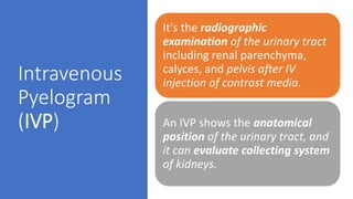
- 3. Intravenous Pyelogram (IVP) It's the radiographic examination of the urinary tract including renal parenchyma, calyces, and pelvis after IV injection of contrast media. An IVP shows the anatomical position of the urinary tract, and it can evaluate collecting system of kidneys.
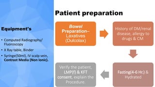
- 4. Patient preparation Equipment's • Computed Radiography/ Fluoroscopy • X Ray table, Binder • Syringe(50ml), IV scalp vein, Contrast Media (Non Ionic). Bowel Preparation– Laxatives (Dulcolax) History of DM/renal disease, allergy to drugs & CM Fasting(4-6 Hr.) & Hydrated Verify the patient, LMP(f) & KFT consent, explain the Procedure.
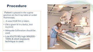
- 5. Procedure Patient is placed in the supine position on the X ray table or under fluoroscopy. • A scout KUB film is taken. • CM is given IV in a bolus, test inj.(1ml) • Adequate Collimation should be used. • Low KV(70-90) high MA(600- 1000) & short exposure technique is used.
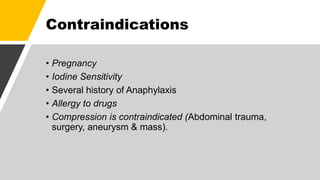
- 7. Contraindications • Pregnancy • Iodine Sensitivity • Several history of Anaphylaxis • Allergy to drugs • Compression is contraindicated (Abdominal trauma, surgery, aneurysm & mass).
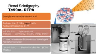
- 8. Renal Scintigraphy Tc99m- DTPA Diethylenetriaminepentaaceticacid Radionuclide- Tc-99m tagged with Radiopharmaceutical-DTPA Half life- 6hrs. Type- generator produced Gamma ray emission, Energy- 140Kev It's used to assess renal function after injecting the Radiopharmaceutical. Dynamic Scan, Mechanism of Action....(100% filtration)
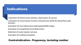
- 9. Indications Evaluation for Renal artery stenosis, obstruction, & trauma, Evaluation of renal tubular function and perfusion (GFR) for blood flow, Split function. Evaluation of renal obstructive nephropathy(HDN study), Evaluation of unexplained renal dysfunction, Detection of acute tubular necrosis, Evaluation of a kidney transplant. Contraindication. Pregnancy, lactating mother
- 10. Patient Preparation • Patient- Well hydrated • History of patient • Check- Height & Weight of patient • Frequent voiding of bladder • Catheterization & PCN Clamping.
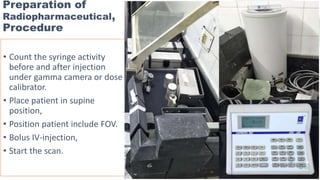
- 11. Preparation of Radiopharmaceutical, Procedure • Count the syringe activity before and after injection under gamma camera or dose calibrator. • Place patient in supine position, • Position patient include FOV. • Bolus IV-injection, • Start the scan.
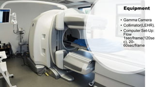
- 12. Equipment • Gamma Camera • Collimator(LEHR), • Computer Set-Up: Flow 1sec/frame(120se c), 20- 60sec/frame
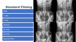
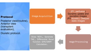
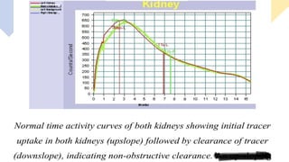
- 13. Protocol Posterior view(routine), Anterior view (transplant evaluation), Diuretic protocol. Image Acquisition (P1, perfusion) Dynamic uptake and excretion images (P2 diuretic). Delayed static images I​mage Processing Draw ROI's, Generate TAC, Differential Renal function GFR/ERPF Calculation.
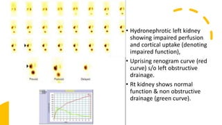
- 18. • Hydronephrotic left kidney showing impaired perfusion and cortical uptake (denoting impaired function), • Uprising renogram curve (red curve) s/o left obstructive drainage. • Rt kidney shows normal function & non obstructive drainage (green curve).
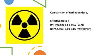
- 19. Comparision of Radiation dose, Effective Dose ! IVP Imaging : 2-5 mSv (01hr) DTPA Scan : 0.01-0.05 mSv(30min)
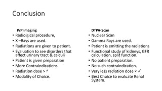
- 20. Conclusion IVP imaging • Radiolgical procedure, • X –Rays are used. • Radiations are given to patient. • Evaluation to see disorders that affect urinary tract & calculi • Patient is given preparation • More Contraindications • Radiation dose > ^ • Modality of Choice. DTPA-Scan • Nuclear Scan • Gamma Rays are used. • Patient is emitting the radiations • Functional study of kidneys, GFR calculation, split function. • No patient preparation. • No such contraindication. • Very less radiation dose × ✓ • Best Choice to evaluate Renal System.
- 21. ThankYou
Editor's Notes
- #5: Patient preparation On the date of Appointment (DOA), On the day of Examination (DOE)