The document summarizes the anatomy of the nose and paranasal sinuses. It describes the external nose, nasal cavity, paranasal sinuses, blood supply, nerve supply, and functions of the nose. The external nose has a bony and cartilaginous framework that provides structure. Internally, the nasal cavity is divided by the nasal septum and contains the paranasal sinuses which include the maxillary, frontal, ethmoid, and sphenoid sinuses. The nose functions include filtration, warming and humidifying air, olfaction, and vocal resonance.
Dentist in pune.(BDS. MDS) - Dr. Amit T. Suryawanshi. Nose & Paranasal sinuses.All Good Things
油
Dentist in pune. (BDS. MDS) - Dr. Amit T. Suryawanshi. Seminar- Nose & Paranasal sinuses.
Email ID- amitsuryawanshi999@gmail.com
Contact -Ph no.-9405622455
Subscribe our channel on youtube - Copy and paste this URL. https://www.youtube.com/channel/UC_gylEXTrjmEbbOTSXjuZ4Q/videos?view_as=public
Follow us on slideshare
The summary of the document is:
1. The nose and paranasal sinuses develop between 4-8 weeks of fetal life from the frontonasal process and maxillary processes.
2. By 5-6 weeks, nasal placodes form and invaginate to form nasal pits which later fuse to form the primitive nasal cavity.
3. Between 7-10 weeks, the paranasal sinuses begin to form from outpocketings of the nasal mucosa. The maxillary sinus is the first to form around 3 months of gestation.
4. The external nose is made up of bones, cartilages and overlying skin and muscle. The internal nasal cavity has
The document summarizes the anatomy of the nose. It describes the external nose, nasal cavity, and structures within including bones, cartilages, walls, openings, blood supply, nerves and lymph drainage. Key points are the pyramidal external nose composed of bones and cartilage, the nasal cavity divided into three regions and outlined by four walls, and various sinuses and structures that open into the nasal cavity.
This document summarizes the anatomy of the nose. It describes the external structures like skin, cartilage, and bones that make up the nose. It then details the internal nasal anatomy including the nasal septum, lateral walls, and turbinates. It discusses the blood supply, nerves, and lymphatic drainage of the nose. The document emphasizes surgical implications for structures like the nasal skin, cartilage, and septum to help surgeons perform reconstructive nasal surgery.
This document outlines the anatomy of the nose, beginning with its external features such as the nasal bones, cartilages, skin and muscles. It then details the internal nasal septum formed by bone and cartilage. The lateral nasal wall contains three turbinates and their air passages. Sensory innervation is provided by the trigeminal and facial nerves. Arterial blood supply originates from the ophthalmic, maxillary and facial arteries, and veins drain to the angular and ophthalmic veins. Precise knowledge of nasal anatomy guides surgical procedures like rhinoplasty and septoplasty.
This document provides an overview of the anatomy of the nose, including both the external nose and nasal cavity. It describes the skin, muscles, bones, and cartilages that make up the external nose. It then details the structures within the nasal cavity, including the nasal septum, lateral wall with its turbinates and meatuses, roof and floor. Throughout, it emphasizes the applied clinical significance of various anatomical structures and relationships as they relate to nasal function, trauma, and surgery.
This document provides an overview of the paranasal air sinuses, including their classification, relationships, and key details about the frontal, maxillary, ethmoidal, and sphenoidal sinuses. It discusses the introduction, drainage, measurements, and applied aspects of each sinus. The sinuses develop as mucosal diverticulae in the bones around the nasal cavity and function to lighten the skull, add resonance to the voice, warm and moisten inspired air, and aid in facial bone growth.
The nose has several important functions including respiration, olfaction, conditioning of inspired air, and protection of the lower airways. It has external and internal structures. The external nose consists of bone and cartilage while the internal nose includes nasal cavities divided by nasal conchae into passages that lead to paranasal sinuses. The nasal mucosa conditions air and supports smell. Olfactory neurons detect smells and relay signals to brain areas involved in processing odors.
The framework of the nose consists of bone and cartilage. Two small nasal bones and extensions of the maxillae form the bridge of the nose, which is the bony portion. The remainder of the framework is cartilage and is the flexible portion. Connective tissue and skin cover the framework.
Air enters the nasal cavity from the outside through two openings: the nostrils or external nares. The openings from the nasal cavity into the pharynx are the internal nares. Nose hairs at the entrance to the nose trap large inhaled particles.
Paranasal sinuses are air-filled cavities in the frontal, maxilae, ethmoid, and sphenoid bones. These sinuses, which have the same names as the bones in which they are located, surround the nasal cavity and open into it. They function to reduce the weight of the skull, to produce mucus, and to influence voice quality by acting as resonating chambers.
This document provides an overview of the paranasal air sinuses, including their classification, functions, drainage pathways, and clinical relevance. It describes the frontal, maxillary, ethmoid, and sphenoid sinuses in detail, covering their locations, openings, relationships to surrounding structures, arterial supply, nerve innervation, and applications. The maxillary sinus is highlighted as the most commonly infected sinus. Sinusitis can potentially lead to complications such as orbital cellulitis if infection spreads.
This document provides an overview of the nasal septum and paranasal sinuses. It describes the anatomy of the external nose, nasal cavity, and paranasal sinuses. Key points include that the nasal septum divides the nose into right and left halves, the nasal cavity has floors, walls and openings that connect to the paranasal sinuses, and the paranasal sinuses include the maxillary, frontal, sphenoid and ethmoid sinuses within facial bones. Clinical correlations regarding examining and treating conditions like sinusitis are also discussed.
nasal_cavity human anatomy lateral wall and venous drainage_1.pptxsiddhimeena3
油
This document provides an overview of the nasal cavity, including its parts, boundaries, blood supply, nerve supply and applied aspects. It discusses the vestibule, nasal cavity proper, roof, floor and septum of the nasal cavity in detail. The septum has bony, cartilaginous and membranous parts. It receives its blood supply from various arteries and drains into facial and pterygoid veins. The septum is innervated by branches of various cranial nerves. Applied aspects of the nasal septum include its role in nasal obstruction and importance of preserving the septal cartilage.
This document provides an outline of the anatomy, examination, and surgical procedures involved in rhinoplasty. It begins with a description of the skin, muscles, blood supply, innervation, and bony and cartilaginous structures of the nose. Assessment of the nasal valves, septum, and turbinates is described. Basic aesthetic parameters and common anatomic variants are discussed. The document concludes with descriptions of routine surgical steps like incision, skin dissection, grafting techniques, and areas commonly augmented like the radix, spreader grafts, and tip grafting.
EAR BASICS SEMINAR.pptx for ENT Pgt in IndiaPrem Nath
油
Introduction to the Ear
The ear is a complex organ responsible for two essential functions: hearing and balance. It receives sound waves from the environment and converts them into electrical signals that the brain interprets. Additionally, the ear plays a crucial role in maintaining equilibrium and spatial orientation. Structurally and functionally, the ear is divided into three major sections: the outer ear, middle ear, and inner ear, each performing distinct roles in the auditory and vestibular systems.
Anatomy of the Ear
1. Outer Ear
The outer ear collects sound waves and directs them toward the middle ear. It consists of:
Pinna (Auricle): The visible part of the ear, composed of cartilage and skin. It captures sound waves and funnels them into the ear canal.
External Auditory Canal: A curved tube that extends from the pinna to the tympanic membrane (eardrum). It amplifies sound and produces cerumen (earwax) to protect the ear from dust and microorganisms.
Tympanic Membrane (Eardrum): A thin, cone-shaped membrane that separates the outer ear from the middle ear. It vibrates in response to sound waves, initiating the process of mechanical sound transmission.
2. Middle Ear
The middle ear is an air-filled cavity that transfers sound vibrations from the eardrum to the inner ear through a series of small bones, collectively called ossicles.
Ossicles: The three tiny bones that amplify sound:
1. Malleus (Hammer): Attached to the eardrum.
2. Incus (Anvil): Connects the malleus and stapes.
3. Stapes (Stirrup): Transmits vibrations to the oval window of the cochlea.
Eustachian Tube: A canal connecting the middle ear to the nasopharynx, which helps equalize pressure between the ear and the atmosphere.
Oval Window and Round Window: Openings in the cochlea that facilitate sound wave transmission to the fluid-filled inner ear.
3. Inner Ear
The inner ear consists of the cochlea (responsible for hearing) and the vestibular system (responsible for balance).
Cochlea: A spiral-shaped, fluid-filled structure that converts mechanical vibrations into electrical signals. It contains:
Organ of Corti: The sensory organ of hearing, lined with hair cells that detect sound vibrations.
Hair Cells: Specialized cells that convert sound vibrations into electrical impulses.
Vestibular Apparatus: A system of three semicircular canals and the utricle and saccule, which detect rotational and linear movements for balance and spatial orientation.
Physiology of Hearing
The process of hearing involves a sequence of steps that convert sound waves into electrical signals understood by the brain.
1. Sound Wave Collection: Sound waves enter the ear through the pinna and travel down the external auditory canal.
2. Vibration of the Eardrum: The tympanic membrane vibrates in response to these sound waves, transmitting the vibrations to the ossicles.
3. Ossicular Chain Amplification: The malleus, incus, and stapes amplify the vibrations and
The nose is the sense organ specialized in olfaction. It has specialized receptors responsible in generating impulses which are transmitted to the brain for interpretation. The presentation summarizes the anatomy and physiology of sense organ of smell.
The nose and paranasal sinuses have complex anatomy. The nose is divided into an external cartilaginous portion and internal bony nasal cavities separated by a nasal septum. The nasal cavities contain turbinates and drain into various meatuses. Several pairs of paranasal sinuses surround the nasal cavities. The sinuses are lined with ciliated respiratory epithelium and drain into the nasal cavities. The nose has roles in breathing, warming, and filtering air as well as detecting smells. Congenital anomalies like choanal atresia can cause nasal obstruction.
The document provides an overview of the anatomy of the nose. It discusses the external and internal structures of the nose, including the nasal cavity and paranasal sinuses. The external nose has bony and cartilaginous parts that provide structure. Internally, the nasal cavity is divided by the nasal septum and lined by various types of epithelium. The document outlines the blood supply, nerve innervation, lymphatic drainage and musculature of the nose. It provides details on the four paired paranasal sinuses within the facial bones that are connected to the nasal cavity.
The document provides an overview of the anatomy of the nose and paranasal sinuses. It describes the external nose, nasal cavity, and paranasal sinuses. The nasal cavity has a floor, roof, medial wall and lateral walls. The lateral walls have three projections called conchae that divide the nasal cavity into inferior, middle, and superior meatuses. The paranasal sinuses include the maxillary, frontal, sphenoid, and ethmoid sinuses. They communicate with the nasal cavity and have functions such as resonating voice and reducing skull weight.
The document discusses the anatomy of the nose and paranasal sinuses. It describes the external nose, nasal cavity, and paranasal sinuses. The nasal cavity is divided by the nasal septum into right and left halves and contains three meatuses - the inferior, middle, and superior. The four paranasal sinuses are the maxillary, frontal, sphenoid, and ethmoid sinuses. Each sinus opens into a meatus of the nasal cavity and drains mucus through small openings. The document outlines the bones, blood supply, nerve supply, and functions of the nose and paranasal sinuses.
The document discusses the anatomy of the nose and paranasal sinuses. It describes the external nose, nasal cavity, and paranasal sinuses. The nasal cavity contains the floor, roof, lateral and medial walls. It is divided by the nasal septum into right and left passages. The paranasal sinuses include the maxillary, frontal, sphenoid, and ethmoid sinuses. They open into the nasal cavity and drain mucus through their openings. The document outlines the blood supply, nerve supply, and lymph drainage of the nose and sinuses.
The document discusses the anatomy and development of the nose and paranasal sinuses. It describes how the nose develops from embryonic structures and the formation of the nasal cavity, septum, and external nose. It then covers the anatomy of the different areas of the nose in detail, including the vestibule, muscles, bones/cartilages, blood supply, nerves and lymphatic drainage. Finally, it discusses the anatomy of each paranasal sinus, including the sphenoid sinus and its variations in pneumatization.
The nasal cavity is divided by the nasal septum into left and right cavities. Each cavity contains 4 passages formed by the nasal conchae: the sphenoethmoidal recess, superior meatus, middle meatus, and inferior meatus. The nasal cavities are lined with mucous membrane and contain paranasal sinuses. Epistaxis or nosebleeds can occur due to various causes like trauma, infections, or anatomical abnormalities. Posterior nosebleeds from Woodruff's plexus are difficult to treat due to its inaccessible location.
Digital Tools with AI for e-Content Development.pptxDr. Sarita Anand
油
This ppt is useful for not only for B.Ed., M.Ed., M.A. (Education) or any other PG level students or Ph.D. scholars but also for the school, college and university teachers who are interested to prepare an e-content with AI for their students and others.
How to attach file using upload button Odoo 18Celine George
油
In this slide, well discuss on how to attach file using upload button Odoo 18. Odoo features a dedicated model, 'ir.attachments,' designed for storing attachments submitted by end users. We can see the process of utilizing the 'ir.attachments' model to enable file uploads through web forms in this slide.
Finals of Rass MELAI : a Music, Entertainment, Literature, Arts and Internet Culture Quiz organized by Conquiztadors, the Quiz society of Sri Venkateswara College under their annual quizzing fest El Dorado 2025.
Prelims of Kaun TALHA : a Travel, Architecture, Lifestyle, Heritage and Activism quiz, organized by Conquiztadors, the Quiz society of Sri Venkateswara College under their annual quizzing fest El Dorado 2025.
APM People Interest Network Conference 2025
- Autonomy, Teams and Tension
- Oliver Randall & David Bovis
- Own Your Autonomy
Oliver Randall
Consultant, Tribe365
Oliver is a career project professional since 2011 and started volunteering with APM in 2016 and has since chaired the People Interest Network and the North East Regional Network. Oliver has been consulting in culture, leadership and behaviours since 2019 and co-developed HPTM速an off the shelf high performance framework for teams and organisations and is currently working with SAS (Stellenbosch Academy for Sport) developing the culture, leadership and behaviours framework for future elite sportspeople whilst also holding down work as a project manager in the NHS at North Tees and Hartlepool Foundation Trust.
David Bovis
Consultant, Duxinaroe
A Leadership and Culture Change expert, David is the originator of BTFA and The Dux Model.
With a Masters in Applied Neuroscience from the Institute of Organisational Neuroscience, he is widely regarded as the Go-To expert in the field, recognised as an inspiring keynote speaker and change strategist.
He has an industrial engineering background, majoring in TPS / Lean. David worked his way up from his apprenticeship to earn his seat at the C-suite table. His career spans several industries, including Automotive, Aerospace, Defence, Space, Heavy Industries and Elec-Mech / polymer contract manufacture.
Published in Londons Evening Standard quarterly business supplement, James Caans Your business Magazine, Quality World, the Lean Management Journal and Cambridge Universities PMA, he works as comfortably with leaders from FTSE and Fortune 100 companies as he does owner-managers in SMEs. He is passionate about helping leaders understand the neurological root cause of a high-performance culture and sustainable change, in business.
Session | Own Your Autonomy The Importance of Autonomy in Project Management
#OwnYourAutonomy is aiming to be a global APM initiative to position everyone to take a more conscious role in their decision making process leading to increased outcomes for everyone and contribute to a world in which all projects succeed.
We want everyone to join the journey.
#OwnYourAutonomy is the culmination of 3 years of collaborative exploration within the Leadership Focus Group which is part of the APM People Interest Network. The work has been pulled together using the 5 HPTM速 Systems and the BTFA neuroscience leadership programme.
https://www.linkedin.com/showcase/apm-people-network/about/
The Constitution, Government and Law making bodies .saanidhyapatel09
油
This PowerPoint presentation provides an insightful overview of the Constitution, covering its key principles, features, and significance. It explains the fundamental rights, duties, structure of government, and the importance of constitutional law in governance. Ideal for students, educators, and anyone interested in understanding the foundation of a nations legal framework.
Information Technology for class X CBSE skill SubjectVEENAKSHI PATHAK
油
These questions are based on cbse booklet for 10th class information technology subject code 402. these questions are sufficient for exam for first lesion. This subject give benefit to students and good marks. if any student weak in one main subject it can replace with these marks.
Database population in Odoo 18 - Odoo slidesCeline George
油
In this slide, well discuss the database population in Odoo 18. In Odoo, performance analysis of the source code is more important. Database population is one of the methods used to analyze the performance of our code.
How to use Init Hooks in Odoo 18 - Odoo 際際滷sCeline George
油
In this slide, well discuss on how to use Init Hooks in Odoo 18. In Odoo, Init Hooks are essential functions specified as strings in the __init__ file of a module.
2. HISTORY
Began in Ancient Egypt and India
Description of nasal reconstruction in Susruta Samhita (500 B.C )
1887 John Orlando Roe performed first intranasal rhinoplasty.
Jacques Joseph Father of Modern facial Plastic surgery published his
Treatise on Rhinoplasty.
4. GLABELLA : Most prominent point of
forehead in mid saggital plane.
NASION : Anterior most point of fronto
nasal suture that joins the nasal part of
frontal bone and nasal bones.
RHINION : Midline point of junction of
nasal bones & upper lateral cartilages.
PHILTRUM : A vertical indentation in the
middle area of the upper lip.
COLUMELLA : Inferior margin of the nasal
septum.
EXTERNAL NOSE
5. TIP DEFINING POINT : Located at the apex of the tip lobule & formed by the
junction of the medial & lateral crura of each Lower lateral cartilage.
6. NASOLABIAL ANGLE : between the columella & the upper lip.
SUBNASALE : Junction of columella & upper lip in mid saggital plane.
8. OSSEOCARTILAGINOUS FRAMEWORK OF NOSE
The upper 1/3rd
is bony & forms bridge of the nose. Lower 2/3 rd is cartilaginous
& forms dorsum of the nose.
BONY PART: The 2 nasal bones meet in the midline and fuse with the nasal
process of the frontal bone. They are held between the frontal process of maxilla.
9. CARTILAGINOUS PART
Upper lateral cartilage : (Paired ) They fuse sideways with the upper border of
septal cartilage & forms dorsal surface of nose. The lower free margin forms limen
vestibuli or nasal valve
Lower lateral cartilage : ( Paired ) This U shaped Alar cartilage has 2 crura lateral
& medial. The lateral crus forms the ala while medial crus lies in columella.
10. Lesser Alar/ Sesamoid
cartilage : (Paired) They may
be 2 or more in number & lie
above & lateral to alar
cartilages.
Septal cartilage : is unpaired.
11. SKIN AND MUSCLES OF EXTERNAL NOSE
Nasal skin in thin & loosely adherent over the dorsum and sides of the nose.
Skin is thicker & more adherant over the nasal tip & alar cartilage where it
contains numerous sebaceous glands.
Muscles are all supplied by branches of facial nerve.
12. The nasal elevators are: Procerus, Leavtor labii- superioris alaeque nasi, &
anamolous nasi muscles
The depressors include Alar nasalis & depressor septi nasi muscles
13. Compressor muscles include transverse nasalis & Compressor nasium minor
The Dilator naris anterior muscle acts as a minor dilator.
14. NASAL CAVITY
1. Median nasal wall : formed by the septum. The Columella forms the most caudal
part of the medial wall
2. Lateral Nasal wall : formed by the turbinates, fibrofatty tissue & cartilages
3. Nasal floor : formed by the floor of the nasal cavity & the nasal vestibule sill.
15. TERMINOLOGIES USED IN NASAL & SEPTAL
ORIENTATION
Red arrow : nasal cavity
Green arrow : nasal septum
16. Nasal septum functions :
1. Separation of the nasal airway into 2 nasal cavities
2. Support of the nasal dorsum
3. Maintenance of the nasal tip
4. Forms part of the nasal valves
Deviation leads to significant nasal airway obstruction and cosmetic deformity.
ANATOMY OF NASAL SEPTUM
17. Nasal septum consists of three parts:
Bony
Cartilaginous
Membranous portion
Anterior septal angle : junction of dorsal &
caudal borders.
Posterior septal angle : junction between caudal
& inferior border anchored to the anterior nasal
spine.
18. The cartilaginous portion consists of
septal or quadrilateral cartilage.
Septal cartilage is continuous with the
upper lateral cartilage towards the bridge
of the nose.
Projection of the septal cartilage known
as Septal tail extends posteriorly between
Vomer and perpendicular plate of
Ethmoid.
Septal tail is used as an additional source
of cartilage to harvest during Revision
Rhinoplasty.
19. NASAL VALVE
Narrowest point of upper airway.
Small changes in nasal septal structure
can have significant effects of airflow
resistance & sensation of obstruction.
Internal Nasal valve : triangular area
bounded by caudal edge of upper lateral
cartilage laterally, septum medially,
nasal cavity floor inferiorlly.
20. LATERAL NASAL WALL
The inferior, middle & superior turbinates are found along the lateral wall.
The space between the lateral nasal wall & the inferior, middle & superior turbinates
are called as inferior, middle & superior meatus respectively
21. BLOOD SUPPLY OF NASAL SEPTUM
External carotid artery branches :
Sphenopalatine & Greater palatine arteries
(branches of the internal maxillary artery )
The Sphenopalatine artery supplies the
posteroinferior septum by a branch called the
Posterior Septal artery. It is the basis for
nasoseptal mucosal flap.
Greater palatine artery supplies the anteroinferior
part.
The septal branch of the superior labial artery
( branch of the facial artery ) supplies caudal septum
& Columella.
22. Internal Carotid artery branches: Anterior & Posterior Ethmoidal arteries
( branches of the ophthalmic artery ).
They supply the anterosuperior & posterosuperior postion of the septum.
The Anterior Ethmoidal & Posterior septal artery form the Kisselbachs plexus.
23. NERVE SUPPLY OF THE NASAL SEPTUM
General sensory nerves derived from the
branches of the trigeminal nerve are distributed
to the whole of the lateral wall:
The anterosuperior quadrant is supplied by the
anterior ethmoidal nerve
The anteroinerior quadrant is supplied by the
anterior superior alveolar nerve, branch of
maxillary nerve.
The posterosuperior quadrant is supplied by the
posterior superior lateral nasal branches from the
pterygoplatine ganglion suspended by the
maxillary nerve.
24. The posteroinferior quadrant is supplied by the anterior or greater palatine branch from the
pterygopalatine ganglion .
Special sensory nerves or olfactory are distributed to the upper part of lateral wall just below the cribriform
plate of the ethmoid up to the superior concha.
25. PTERYGOPALATINE GANGLION
The pterygopalatine (Sphenopalatine) ganglion is the largest parasympathetic
ganglion.
It serves as a relay station for secretomotor fibres to the lacrimal gland & to the
mucous glands of the nose, the paranasal sinuses, the palate & the pharynx.
26. BRANCHES
1. Orbital branches : pass through the inferioir orbital fissure
2. Palatine branches : the greater or anterior palatine nerve descends through the
greater palatine canal & supplies the hard palate & the lateral wall of the nose
inferior concha & adjoining meatuses.The lesser or middle & posterior palatine
nerve supply the soft palate and tonsil
28. 3. Nasal branches enter the nasal cavity through the sphenopaltine foramen
The lateral posterior superior nasal nerves supply the posterior part of superior &
middle cocncha
The medial posterior superior nasal nerves supply the posterior part of roof of the
nose & the septum. The largest of these nerves is known as Nasopalatine nerve
which descend upto the anterior part of the hard palate
29. 4. The Pharyngeal branch supplies the part of the nasopharynx behind the auditory
tube
5. Lacrimal branch : secretomotor fibres to the lacrimal gland
40. Basic principles to be taken care
Be conservative
Should know where to stop
Never promise miraculous results after surgery
Beware of psychotic patients
Consent
47. TIP RECOIL
Tip Recoil is defined as the inherent
strength and support of the nasal tip.
It can be evaluated by depressing the
tip towards the upper lip and
watching for the tip's supportive
structure to spring back into position.
If the recoil is good, and the tip
cartilages resist the deforming
influence, then tip surgery can
usually be performed without fear of
substantial support loss.
48. COLUMELLAR DEFECTS
Type 1 deformities (caudal septum
and/or spine)
Type 2 (medial crura)
Type 3 (soft tissue)
Type 4 (combination)
52. QUALITY OF SKIN
Thick skin
Masks refinement and
definition
Failure to contract
excess soft tissue scar
Does not show small
irregularities
Thin skin
Small irregularities become visible
Early healing
Less oedema
Ensure that all bony,
cartilageneous grafts or
implants are precisely positioned and
smoothly contoured.
54. CLINICAL PHOTOGRAPHS
Blue is chosen as the background
colour as it provides an excellent
contrast to the colour of flesh and hair.
Typically consist of a full-face frontal
view, with oblique and lateral views
on the side of the defect.
If the defect extends to the infra tip
lobule or alar margin, a base
view is also obtained.
Close-up views of the defect may be
obtained when appropriate.
55. TYPES OF RHINOPLASTY
OPEN APPROACH
External rhinoplasty
Trans columellar incision
Closed approach
Endonasal rhinoplasty
Incisions positioned inside the
nostril
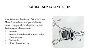
57. CAUDAL SEPTAL INCISION
Also known as hemi-transfixion incision
Made 2 mm above and parallel to the
caudal margin of cartilaginous septum
Incision provides access to-
1. Septum
2. Premaxilla and anterior nasal spine
3. Nasal dorsum
4. Columella
5. Floor of nasal cavity
58. INTERCARTILAGENOUS INCISION
Cut made in the vestibular skin
just cranial to the caudal end of
triangular cartilage
Incision starts halfway along the
lower end of cartilage and continues
past .
Provides access to :
1. Nasal dorsum(cartlaginous and
bony vault)
2. Valve
3. Lobule
59. VESTIBULAR INCISION
Vestibular incision is a slightly curved
cut made in the vestibular skin just
lateral to the margin of pyriform
aperture.
It is used to access:
1. Paranasal area
2. Pyriform aperture
3. Lateral wall of nasal cavity
60. INFRACARTILAGENOUS INCISION
It is an incision at the caudal margin
of the lateral crus, dome and medial
crus of the lobular cartilage.
It gives access to :
1. Lobular cartilages
2. Cartilaginous vault
61. TRANSCOLUMELLAR INVERTED V INCISION
It is a horizontal reversed-v- shaped incision of
the columella at about one-third of the distance
from its base, it is made in combination with
infracartilaginous incision on both sides in the
external approach
Access to
1. Lobular cartilages
2. Cartilaginous dorsum
3. Anterior septum
62. SPECIAL INCISIONS
EXTERNAL
1. Labiogingival
incision
2. Sublabial incision
3. Paranasal incision
4. Lateral columellar
5. Rim incision
6. Alarfacial incision
7. v incision of
columellar base
8. Dorsal incisions
INTERNAL
1. Transfixion incision
2. Transcartilaginous incision
3. Incisions in the turbinate
mucosa
4. Incisions in the septal mucosa
64. INDICATIONS
Extensive revision surgery
Severe nasal trauma
Congenital deformities: cleft lip nose
Marked tip deformities
Elaborate reduction and augmentation procedures
Correction of extreme overprojection
65. PRINCIPLES OF EXTERNAL RHINOPLASTY
Incision- mid-columella incision connected to bilateral marginal incision
Dissection in subperiosteal and subperichondrial planes
Division of medial intercrural tissue offers access to caudal septum and premaxillary
spine
Division of upper lateral cartilages from quadrilateral cartilage offers acccessability
to whole of septum
66. ADVANTAGES
Extensive exposure for both septal and rhinoplasty surgery
Binocular vision
Useof both hands
Control of bleeding and diathermy
Precise placement and suturing of struts,battens and shield grafts
Valve area preserved
68. SURGICAL PROCEDURE
Broken transcolumellar incision
If columella short in case of
cleft lip-V incision
Mid-columella incision situated
above medial crural foot plates
Vertical columellar incision
made 1.5-2mm inside vestibule
Separate lateral incision given
which is joined medially over the
domes
69. Dissection carried in midline just cephalic to dome subperichondrial plane
Dissection of soft tissue of bony pyramid should start above caudal end of nasal bone
Nasal septum- between medial crurae of lower lateral cartilage or hemitransfixion
incision
Strut used to
correct buckled medial crura
strengthen weak medial crura
correct tip asymmetries
stable base for tip graft
71. SPECIFIC APPLICATIONS
The bony pyramid in external rhinoplasty
Allows use of burr or reduction of the soft tissue envelope at nasion to deepen the
nasofrontal angle
Application of soft tissue onlay grafts
Bony dehumping together with lateral, medial, and intermediate osteotomies
72. The middle nasal vault
Placement of cartilaginous
strips or spreader grafts to open
up the nasal valve area and
angles Shaded areas showing placement of
spreader grafts
74. INDICATIONS
Patients with ideal height and position of the nasion associated with
excess dorsal convexity
Oversized alar cartilages producing increase tip and lobule volume
75. AIMS
Aim- strong nasal dorsum in lateral profile-relates to ideal
nasion height
Tip defining point-projecting just above dorsal line-to
create supratip break. In males may be on a straight line
with dorsum
76. CONTRAINDICATIONS
Underprojected tip
Deep radix (deep root) of nose
Short over-rotated nose
TRIAD
PSEUDOHUMP
thin skin
delicate alar side walls
bifidity
79. Tip surgery
Cephalic trim for volume
reduction of lower lateral cartilage
done
Transcartilagenous incision
5mm of continous strip of lateral
crus of lower lateral cartilage is
preserved
For rotation excision of caudal end
of septum
80. Cartilage strip incision for cephalic strip excision of lower lateral cartilage
81. DEHUMP
Nasal hump
1. Bony
2. Cartilagenous
3. Both
Minimal bony hump can be reduced by using endonasal approach with just rasping.
Small cartilagenous humps only require shaving of cartilagenous ridges of the septal
dorsum.
Dorsal hump which involves both cartilagenous and bony vault open approach is
preferred.
82. Cartilagenous dorsum is reduced first.
Blade no.15 is held at the key area in horizontal
plane to incise across left upper lateral cartilage,
quadrilateral cartilage and right upper lateral .
Advanced caudally in the plane of reduction this
transects the upper lateral cartilage and
cartilagenous septum.
Osteotome is then inserted under the
cartilagenous segment removing the
osteocartilagenous hump en bloc
85. MEDIAL OSTEOTOMY
It seperates the nasal bone
from the septum
Made on both side
Nasal bone seperated at
intranasal suture
Short intercartilageneous
incision given
86. 1. Outer peritosteun is pushed to the side
2. Osteotome is placed at about 2mm paramedially
3. Osteotome is worked through the bone slightly below the
level of frontal bone
87. LATERAL OSTEOTOMY
It seperates the lateral bony wall of pyramid
from nasal process of maxilla
Short lateral incision is given
Medial to lateral subperiosteal tunnel is
formed upto level of medial canthus
Osteotome placed across frontal process of
maxilla
Lateral osteotomy done upto the level of
frontal bone
90. TRANSVERSE OSTEOTOMY
Seperates the bony pyramid from frontal
bone and nasal spine of frontal bone
Osteotomy made at a level just below
nasion