Urinary System Anatomy for Med Lab.pptx
- 1. URINARY SYSTEM ANATOMY By: Zekarias B. (MSc. Clinical Anatomy
- 2. An Introduction to the Urinary System 8/3/2022 2
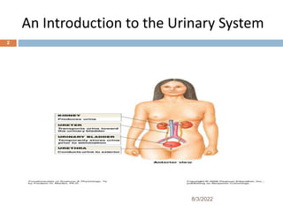
- 3. INTRODUCTION ’é© The urinary system consists of two kidneys, two ureters, one urinary bladder, and one urethra. ’é© After the kidneys filter blood plasma, they return most of the water and solutes to the bloodstream. ’é© The remaining water and solutes constitute urine, which passes through the ureters and is stored in the urinary bladder until it is excreted from the body through the urethra. ’é© Nephrology is the scientific study of the anatomy, physiology, and disorders of the kidneys 8/3/2022 3
- 4. Functions of the Urinary System 1.The kidneys ’ā╝ Regulate blood volume and composition ’ā╝ Help regulate blood pressure(by secreting the enzyme renin, which activates the reninŌĆō angiotensinŌĆō aldosterone pathway) ’ā╝ Regulate blood PH ’ā╝ produce two hormones(Calcitriol & erythropoietin), and ’ā╝ excrete wastes. 8/3/2022 4
- 5. Functions of the Urinary System 2. The ureters transport urine from the kidneys to the urinary bladder. 3. The urinary bladder stores urine and expels it into the urethra. 4. The urethra discharges urine from the body 8/3/2022 5
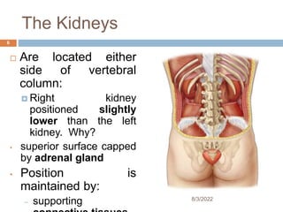
- 6. The Kidneys ’é© Are located either side of vertebral column: ’éż Right kidney positioned slightly lower than the left kidney. Why? ŌĆó superior surface capped by adrenal gland ŌĆó Position is maintained by: ŌĆō supporting 8/3/2022 6
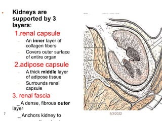
- 7. ’éŚ Kidneys are supported by 3 layers: 1.renal capsule ŌĆō An inner layer of collagen fibers ŌĆō Covers outer surface of entire organ 2.adipose capsule ŌĆō A thick middle layer of adipose tissue ŌĆō Surrounds renal capsule 3. renal fascia _ A dense, fibrous outer layer _ Anchors kidney to 8/3/2022 7
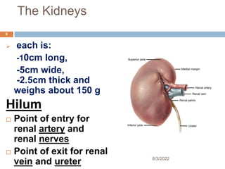
- 8. The Kidneys ’āś each is: -10cm long, -5cm wide, -2.5cm thick and weighs about 150 g Hilum ’é© Point of entry for renal artery and renal nerves ’é© Point of exit for renal vein and ureter 8/3/2022 8
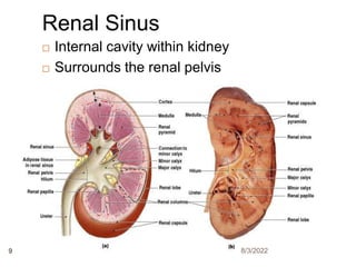
- 9. ’é© Internal cavity within kidney ’é© Surrounds the renal pelvis Renal Sinus 8/3/2022 9
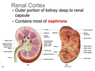
- 10. Renal Cortex ’éŚ Outer portion of kidney deep to renal capsule ’éŚ Contains most of nephrons 8/3/2022 10
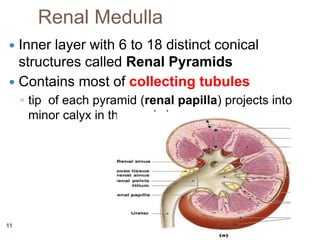
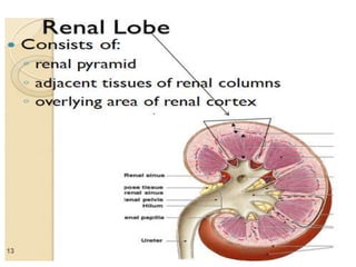
- 11. Renal Medulla ’éŚ Inner layer with 6 to 18 distinct conical structures called Renal Pyramids ’éŚ Contains most of collecting tubules ŌŚ” tip of each pyramid (renal papilla) projects into minor calyx in the renal sinus 8/3/2022 11
- 12. 8/3/2022 12
- 13. 8/3/2022 13
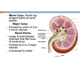
- 14. ’é© Minor Calyx: Small cup shaped drains of renal papilla Major Calyx ’é© Formed by union of 2 to 3 minor calyces Renal Pelvis ’é© Large, funnel-shaped chamber that fills most of renal sinus ’é© Formed by 2 to 3 major calyces ’é© Connected to ureter 8/3/2022 14
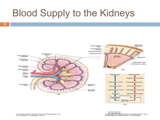
- 15. Blood Supply to the Kidneys ’é© Kidneys receive 20ŌĆō25% of total cardiac output ’é© 1200 ml of blood /minute flows through kidneys ’é© Kidney receives blood through renal artery 8/3/2022 15
- 16. Blood Supply to the Kidneys 8/3/2022 16
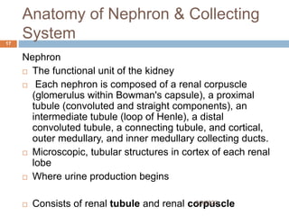
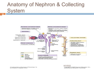
- 17. Anatomy of Nephron & Collecting System Nephron ’é© The functional unit of the kidney ’é© Each nephron is composed of a renal corpuscle (glomerulus within Bowman's capsule), a proximal tubule (convoluted and straight components), an intermediate tubule (loop of Henle), a distal convoluted tubule, a connecting tubule, and cortical, outer medullary, and inner medullary collecting ducts. ’é© Microscopic, tubular structures in cortex of each renal lobe ’é© Where urine production begins ’é© Consists of renal tubule and renal corpuscle 8/3/2022 17
- 18. Anatomy of Nephron & Collecting System 8/3/2022 18
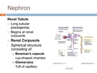
- 19. Nephron Renal Tubule ’é© Long tubular passageway ’é© Begins at renal corpuscle ’é© Renal Corpuscle ŌĆó Spherical structure consisting of: ’éż BowmanŌĆÖs capsule ŌĆō cup-shaped chamber ’éż Glomerulus ŌĆō Tuft of capillary 8/3/2022 19
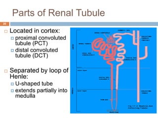
- 20. Parts of Renal Tubule ’é© Located in cortex: ’éż proximal convoluted tubule (PCT) ’éż distal convoluted tubule (DCT) ’é© Separated by loop of Henle: ’éż U-shaped tube ’éż extends partially into medulla 8/3/2022 20
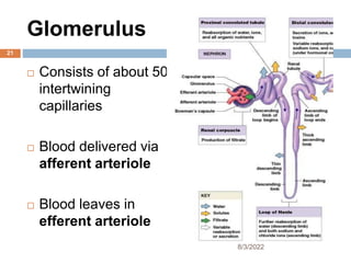
- 21. Glomerulus ’é© Consists of about 50 intertwining capillaries ’é© Blood delivered via afferent arteriole ’é© Blood leaves in efferent arteriole 8/3/2022 21
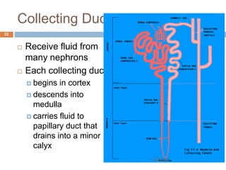
- 22. Collecting Ducts ’é© Receive fluid from many nephrons ’é© Each collecting duct: ’éż begins in cortex ’éż descends into medulla ’éż carries fluid to papillary duct that drains into a minor calyx 8/3/2022 22
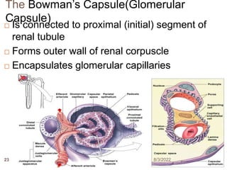
- 23. The BowmanŌĆÖs Capsule(Glomerular Capsule) ’é© Is connected to proximal (initial) segment of renal tubule ’é© Forms outer wall of renal corpuscle ’é© Encapsulates glomerular capillaries 8/3/2022 23
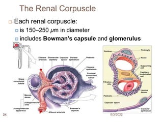
- 24. The Renal Corpuscle ’é© Each renal corpuscle: ’éż is 150ŌĆō250 ┬Ąm in diameter ’éż includes BowmanŌĆÖs capsule and glomerulus 8/3/2022 24
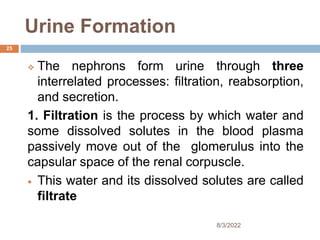
- 25. Urine Formation ’üČ The nephrons form urine through three interrelated processes: filtration, reabsorption, and secretion. 1. Filtration is the process by which water and some dissolved solutes in the blood plasma passively move out of the glomerulus into the capsular space of the renal corpuscle. ’éŚ This water and its dissolved solutes are called filtrate 8/3/2022 25
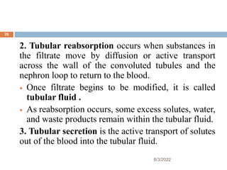
- 26. 2. Tubular reabsorption occurs when substances in the filtrate move by diffusion or active transport across the wall of the convoluted tubules and the nephron loop to return to the blood. ’éŚ Once filtrate begins to be modified, it is called tubular fluid . ’éŚ As reabsorption occurs, some excess solutes, water, and waste products remain within the tubular fluid. 3. Tubular secretion is the active transport of solutes out of the blood into the tubular fluid. 8/3/2022 26
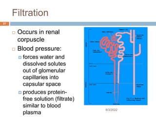
- 27. Filtration ’é© Occurs in renal corpuscle ’é© Blood pressure: ’éż forces water and dissolved solutes out of glomerular capillaries into capsular space ’éż produces protein- free solution (filtrate) similar to blood plasma 8/3/2022 27
- 28. Functions of Renal Tubule 1. Reabsorb useful organic nutrients that enter filtrate 2. Reabsorb more than 90% of water in filtrate 3. Secrete waste products that failed to enter renal corpuscle through filtration at glomerulus 8/3/2022 28
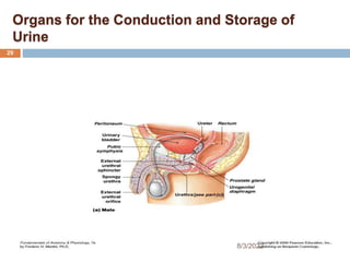
- 29. Organs for the Conduction and Storage of Urine 8/3/2022 29
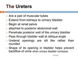
- 30. The Ureters ’é© Are a pair of muscular tubes ’é© Extend from kidneys to urinary bladder ’é© Begin at renal pelvis ’é© attached to posterior abdominal wall ’é© Penetrate posterior wall of the urinary bladder ’é© Pass through bladder wall at oblique angle ’é© Ureteral openings are slit like rather than rounded ’é© Shape of its opening in bladder helps prevent backflow of urine when urinary bladder contracts 8/3/2022 30
- 31. Layers of the Ureter Wall Inner mucosa: ’éż transitional epithelium and lamina propria ’é© Middle muscular layer: ’éż longitudinal and circular bands of smooth muscle ’é© Outer connective-tissue layer (serosa): ’éż continuous with fibrous renal capsule and peritoneum 8/3/2022 31
- 32. Peristaltic Contractions to send urine towards bladder ’é© Begin at renal pelvis ’é© Sweep along ureter ’é© Force urine toward urinary bladder ’é© Every 30 seconds 8/3/2022 32
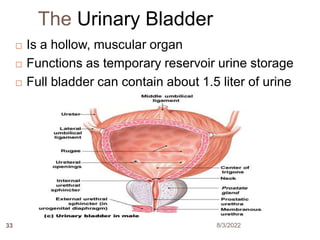
- 33. The Urinary Bladder ’é© Is a hollow, muscular organ ’é© Functions as temporary reservoir urine storage ’é© Full bladder can contain about 1.5 liter of urine 8/3/2022 33
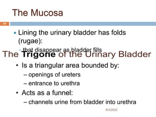
- 34. The Mucosa ’éŚ Lining the urinary bladder has folds (rugae): ŌŚ” that disappear as bladder fills ŌĆó Is a triangular area bounded by: ŌĆō openings of ureters ŌĆō entrance to urethra ŌĆó Acts as a funnel: ŌĆō channels urine from bladder into urethra 8/3/2022 34
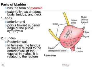
- 35. Parts of bladder ’éż has the form of pyramid ’éż externally has an apex, body, fundus, and neck 1. Apex ’éż anterior end ’éż points toward superior edge of the pubic symphysis 2. Fundus ’éż Posterior wall ’éż In females, the fundus is closely related to the anterior wall of the vagina; in males, it is related to the rectum 8/3/2022 35
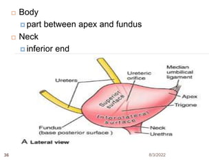
- 36. ’é© Body ’éż part between apex and fundus ’é© Neck ’éż inferior end 8/3/2022 36
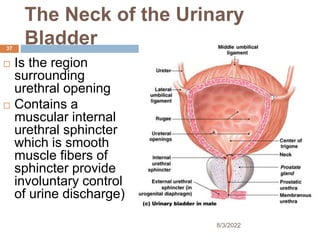
- 37. The Neck of the Urinary Bladder ’é© Is the region surrounding urethral opening ’é© Contains a muscular internal urethral sphincter which is smooth muscle fibers of sphincter provide involuntary control of urine discharge) 8/3/2022 37
- 38. Wall of the Urinary Bladder ’é© Contains mucosa, submucosa, muscularis, and serosa layers: ’éż form powerful detrusor muscle of urinary bladder ’éż contraction compresses urinary bladder and expels urine The Urethra ’é© Extends from neck of urinary bladder ’é© To the exterior of the body The Male Urethra ’é© Extends from neck of urinary bladder ’é© To tip of penis (18ŌĆō20 cm) 8/3/2022 38
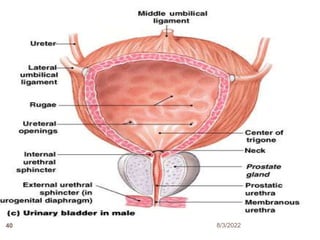
- 39. Parts of the Male Urethra 1. Prostatic urethra: ŌĆō passes through center of prostate gland 2. Membranous urethra: ŌĆō short segment that penetrates the urogenital diaphragm 3. Spongy urethra (penile urethra): ŌŚ” extends from urogenital diaphragm ŌŚ” to external urethral orifice 8/3/2022 39
- 40. 8/3/2022 40
- 41. The Female Urethra ’éŚ Is very short (3ŌĆō5 cm) ’éŚ Extends from bladder to vestibule ’éŚ External urethral orifice is near anterior wall of vagina 8/3/2022 41
- 42. The External Urethral Sphincter ’é© In both sexes: ’éżis a circular band of skeletal muscle ’éżwhere urethra passes through urogenital diaphragm ’é© Acts as a valve ’é© Is under voluntary control: ’é© Voluntarily relaxation permits micturition 8/3/2022 42