


More Related Content
Similar to basalskullfracturesfinale-170707012942.pdf (9)
More from azwararifki1993 (10)
Recently uploaded (20)


basalskullfracturesfinale-170707012942.pdf
- 1. Basal Skull Fractures (Anatomical Base for Signs) M D 1 0 8 : M OT I O N A N D S T R E N G T H P R E S E N T E R : A L I C E M A R I E TA O S D E PA R T M E N T O F M E D I C I N E FA C U LT Y O F M E D I C I N E A N D H E A LT H S C I E N C E S D I V I N E W O R D U N I V E R S I T Y
- 2. Outline Definition of terms Skull Base Anatomy Signs /Clinical Presentations
- 3. Definition Basal Skull – base of the skull Fossa - a depression or hollow (Oxford Concise Medical Dictionary, 2010) Ecchymosis: Passage of blood from ruptured blood vessels into subcutaneous tissue marked by a purple discoloration of the skin
- 4. What is basal skull fracture Basal Skull Fracture - â—¦ Type of skull fracture which occurs in the floor of the skull that is around the eyes, ears, nose or back near the spine (Ellis, 2015) â—¦ Fracture of the base of the skull specifically involves the temporal bone, occipital bone, sphenoid bone, and/or ethmoid bone (Wikipedia, 2016) Morgan, B. (1999, November 19). Overview of Adult Brain Function. Retrieved from https://web.archive.org/web/20080227162001/http://www.orlandoregional.org/pdf%20folder/overview%20adult%20brain% 20injury.pdf
- 5. Morgan, B. (1999, November 19). Overview of Adult Brain Function. Retrieved from https://web.archive.org/web/20080227162001/http://www.orlandoregional.org/pdf%20folder/overview%20adult%20brain% 20injury.pdf
- 6. Skull Base Anatomy The Base of the skull is Made up of 5 bones (Martinez, 2013) â—¦ Frontal Bone â—¦ Cribriform of the last ethmoid â—¦ Sphenoid bone â—¦ Squamous of the temporal bone â—¦ Occipital bone Saladin, K. S. (1998). Anatomy and Physiology: The Unity of Form and Function. Boston: McGraw-Hill.
- 7. Type of Basal Fractures Anterior Skull Base Fracture Middle Skull Base Fracture Posterior Fossa Fracture Martinez, L. (2013, December). Basilar Skull Fractures. Retrieved July 14, 2016, from https://www.utmb.edu/otoref/Grnds/basilar-skull-fx-2013-12/_basilar-skull-fx-pic-2013-12.pdf
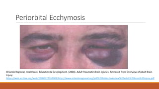
- 9. Signs/Clinical Presentations Diagnosed by clinical findings – Clinical assessment critical Clinical findings consistent with basal skull fracture ◦ Anterior Fossa: rhinorrhea (discharge from the nose), raccoon eyes (periorbital ecchymosis). Anosmia (loss of smell), oculomotor palsies ◦ Middle Fossa: Hemotympanum (blood in the middle ear), otorrhea vertigo, “Battle’s sign (mastoid ecchymosis), unilateral hearing loss. ◦ Posterior Fossa: hypotension, tachycardia, alteration in respirations due to compression of the brainstem. (Orlando Regional, Healthcare, Education & Development, 2004)
- 10. Periorbital Ecchymosis Orlando Regional, Healthcare, Education & Development. (2004). Adult Traumatic Brain Injuries. Retrieved from Overview of Adult Brain Injury: https://web.archive.org/web/20080227162001/http://www.orlandoregional.org/pdf%20folder/overview%20adult%20brain%20injury.pdf
- 14. CSF rhinorrhea
- 17. References 1. Ellis, M. E. (2015, September 29). Skull Fractures. Retrieved July 12, 2016, from Healthline: http://www.healthline.com/health/skull-fracture#Overview1 2. Martinez, L. (2013, December). Basilar Skull Fractures. Retrieved July 14, 2016, from https://www.utmb.edu/otoref/Grnds/basilar-skull-fx-2013-12/_basilar-skull-fx-pic-2013-12.pdf 3. Morgan, B. (1999, November 19). Basal Skull Fractures. Retrieved from London Health Sciences Centre: http://www.lhsc.on.ca/Health_Professionals/CCTC/edubriefs/baseskull.htm 4. Orlando Regional, Healthcare, Education & Development. (2004). Adult Traumatic Brain Injuries. Retrieved from Overview of Adult Brain Injury: https://web.archive.org/web/20080227162001/http://www.orlandoregional.org/pdf%20folder/overvie w%20adult%20brain%20injury.pdf 5. Oxford Concise Medical Dictionary. (2010). New York: Oxford University Press. 6. Saladin, K. S. (1998). Anatomy and Physiology: The Unity of Form and Function. Boston: McGraw-Hill. 7. Wikipedia. (2016, May 31). Retrieved from Basilar skull Fracture: https://en.m.wikipedia.org/wiki/Basilar_skull_fracture