This document provides an overview of fundamental microbiology principles including definitions of microbiology and microorganisms. It describes bacteria and viruses, comparing their structures. Methods for isolating pure cultures, staining bacteria, and different staining techniques like Gram staining and acid fast staining are outlined. Key points covered include how Gram staining is used to classify bacteria as gram positive or negative based on cell wall structure and how acid fast staining identifies Mycobacterium species by their waxy cell walls.
Laboratory diagnosis of bacteria in 5 parts/ dental implant coursesIndian dental academy
油
The Indian Dental Academy is the Leader in continuing dental education , training dentists in all aspects of dentistry and
offering a wide range of dental certified courses in different formats.for more details please visit油
www.indiandentalacademy.com
Outline:
1. Difference between Light microscopy and
electron microscopy
2. Decribe methods for the isolation of
microorganisms in pure culture
3. Techniques for studying live bacteria
4. Distinguish between a simple stain and a
differential stain and give examples
5. Identify steps in the Gram stain procedure
6. List the major categories of microbial characteristics
used to identify microorganisms. Explain why some of
these give more specific info for identification than others
This document provides an overview of microbiology. It discusses that microbiology is the study of microorganisms including their structure, physiology, identification, and relationship to humans and the environment. It describes different types of microorganisms such as bacteria, their structures like cell walls and capsules, and how they are classified. It also discusses laboratory techniques used to study microorganisms like staining, culturing, and biochemical and serological tests.
Identification And Differentiation Of MicroorganismsHaiaykyu
油
Microorganisms can be identified and differentiated using various processes including staining, microscopy, culture techniques, and biochemical analysis. Staining methods like Gram staining and acid-fast staining are used under microscopy to classify bacteria based on cell wall properties and staining patterns. Culture media allow microbes to grow and be distinguished based on colony morphology and biochemical reactions. Together these methods provide essential visual and functional information to identify and classify microorganisms.
Patel college of pharmacy m sandeep mewada.ppt.pptmSANDEEP MEWADA
油
The document discusses various common staining techniques used in microbiology. It begins by explaining the purpose of staining and some key terms like stain, staining, and fixation. It then describes different types of stains including simple stains like methylene blue and differential stains like Gram staining. Gram staining technique and the gram positive and gram negative reactions are explained in detail. Another differential staining method discussed is acid-fast staining using Ziehl-Neelsen stain for tuberculosis diagnosis. Various staining procedures and their applications are outlined.
Bacteria Classification By Gram Staining EssayChristy Hunt
油
Bizzozero staining procedure involves classifying tissues into three categories based on their mitotic activity as seen under the microscope: category I tissues with low mitotic activity, category II tissues with moderate mitotic activity, and category III tissues with high mitotic activity. The staining procedure uses proliferating cell nuclear antigen (PCNA) to label proliferating cells and support Bizzozero's 1894 tissue classification system based on mitotic index determined by examining hematoxylin and eosin stained slides under the microscope. The experiment aims to evaluate if PCNA staining agrees with Bizzozero's original tissue categorization into
The two documents discuss multidrug-resistant organisms and new approaches to treating such infections. The first document aims to inform readers of the growing problem and need for new solutions, targeting healthcare professionals. The second discusses recent findings from a new treatment method, targeting researchers and competitors in the field. Both follow conventions of white papers with background, solutions, conclusions and references to appeal to their respective audiences.
short introduction about microbiology with classification of microorganism, isolation methods, information about staining techniques. those information related to diploma students
1. The document discusses different types of microscopes used to observe microorganisms, including brightfield, darkfield, phase-contrast, fluorescence, confocal, transmission electron, and scanning electron microscopes.
2. It also covers different staining techniques used to prepare specimens for microscopy, including simple stains using a single dye, differential stains like Gram staining and acid-fast staining to classify bacteria, and special stains for structures like capsules, endospores, and flagella.
3. Key points are made about classifying bacteria as gram-positive or gram-negative based on their cell wall composition and how they react to staining, as well as how the acid-fast stain is used to
This document discusses different staining techniques used to identify bacteria under a microscope. It describes simple staining which identifies morphological characteristics using a single dye. Negative staining uses an acidic dye to stain the background while leaving unstained bacteria visible. Gram staining differentiates between gram-positive and gram-negative bacteria using multiple stains. Acid-fast staining identifies bacteria with wax-like cell walls that retain dye after acid treatment. These staining methods enhance contrast and visibility of bacteria for analysis under a microscope.
Staining techniques are used in microbiology to identify bacteria under a microscope. There are several types of staining including simple staining with one dye, Gram staining which differentiates bacteria as Gram-positive or Gram-negative based on cell wall structure, and acid-fast staining used to identify Mycobacterium species. Biochemical tests such as IMViC (Indole, Methyl Red, Voges-Proskauer, Citrate) are also used to identify bacteria based on their metabolic reactions and products.
Lab Report: Isolation of Pure Culture, Gram-staining, and Microscopic Observa...Annisa Hayatunnufus
油
A Lab Report under the subject of Microbiology. Done as a lab session in Josai University, Japan during a twinning program on 2014.
Created by: Annisa Hayatunnufus
Bachelor of Pharmacy
Management & Science University
Isolation and characterization of microbesmeenu sharma
油
This document discusses the isolation and characterization of microbes. It defines key terms like microbes, pure culture, mixed culture, species, and strain. It describes common methods used to isolate pure cultures from mixed populations, including streak plate technique, micromanipulator method, enrichment culture method, and serial dilution method. The document also discusses maintaining and preserving pure cultures through refrigeration, cryopreservation, and lyophilization. It explains how microbes can be characterized based on colony appearance, form, elevation, margins, and optical density.
Microbiology presentation MEDICAL COLLEGEdmfrmicro
油
The document provides an overview of a laboratory presentation on medical microbiology. It discusses several key areas:
1. The introduction defines a laboratory and lists its main departments including clinical chemistry, hematology, microbiology, and blood bank.
2. Microbiology is defined as the study of microorganisms like bacteria, fungi, algae, protozoa and viruses. Medical microbiology deals with infectious disease causative agents.
3. The various fields of medical microbiology covered include bacteriology, virology, parasitology, mycology, and immunology. Common laboratory procedures in each field like microscopy, staining, culture and biochemical testing are summarized.
Microbiology is the study of
living organisms of microscopic
size which includes bacteria ,
Fungi , Algae , Protozoa and Viruses. It is concerned with the forms, structure , reproduction , physiology , metabolism and classification.
Principle Of Microbiology
Medical microbiology deals with the causative agent of the infectious disease of the human , the ways in which they produce disease in the body and essential information for diagnosis and treatment.
Microorganisms can be classified based on their size, shape, and cellular structure. Bacteria are single-celled organisms that can be further classified as cocci, bacilli, or spirochetes depending on their shape. Special stains like Gram stain and acid-fast stain are used to differentiate bacteria and identify medically important types. Fungi have cell walls containing chitin while viruses are protein-coated genes that need host cells. Protozoa are single-celled organisms that move using pseudopods, flagella, or cilia. A variety of staining techniques exist to identify bacteria, fungi, and other microorganisms in clinical samples and tissue sections.
This document provides an overview of the objectives and content covered in the MICI 1100 Health Sciences Microbiology course at QE II HSC, including introductions to microbiology, bacterial structure and classification, growth and metabolism, pathogenicity, and control of microbial growth. Key topics covered include bacterial morphology, staining techniques, taxonomy, requirements for growth, phases of growth, and methods of sterilization and disinfection.
This document discusses different staining techniques used to visualize bacteria under a microscope. It describes simple staining using single dyes like methylene blue, and differential staining techniques like Gram staining and acid-fast staining. Gram staining differentiates bacteria into Gram-positive and Gram-negative groups based on their ability to retain or lose crystal violet dye. Acid-fast staining is used to identify acid-fast bacteria like Mycobacterium that appear bright red after staining. These staining methods allow clear visualization of bacterial morphology and structure.
This document provides an overview of procedures for examining stained microorganisms, including smear preparation and simple and Gram staining. It discusses the steps of inoculation, isolation, incubation, inspection, and identification of microbes in a laboratory setting. Specifically, it outlines the Gram stain procedure, which uses two dyes and a decolorization step to differentiate bacteria based on their cell wall composition into Gram-positive or Gram-negative categories. The Gram stain is a commonly used differential staining technique and first test in bacterial identification.
This document discusses various bacterial staining techniques used to visualize microorganisms under a microscope. It describes simple staining which uses a single dye, differential staining which allows differentiation using more than one dye, and special staining techniques to highlight specific structures. Gram staining is explained in detail as the most common differential staining method used to classify bacteria as gram-positive or gram-negative. Acid-fast staining and capsule, spore, and flagella staining are also summarized as important special staining methods.
The Constitution, Government and Law making bodies .saanidhyapatel09
油
This PowerPoint presentation provides an insightful overview of the Constitution, covering its key principles, features, and significance. It explains the fundamental rights, duties, structure of government, and the importance of constitutional law in governance. Ideal for students, educators, and anyone interested in understanding the foundation of a nations legal framework.
How to Modify Existing Web Pages in Odoo 18Celine George
油
In this slide, well discuss on how to modify existing web pages in Odoo 18. Web pages in Odoo 18 can also gather user data through user-friendly forms, encourage interaction through engaging features.
Database population in Odoo 18 - Odoo slidesCeline George
油
In this slide, well discuss the database population in Odoo 18. In Odoo, performance analysis of the source code is more important. Database population is one of the methods used to analyze the performance of our code.
APM event hosted by the South Wales and West of England Network (SWWE Network)
Speaker: Aalok Sonawala
The SWWE Regional Network were very pleased to welcome Aalok Sonawala, Head of PMO, National Programmes, Rider Levett Bucknall on 26 February, to BAWA for our first face to face event of 2025. Aalok is a member of APMs Thames Valley Regional Network and also speaks to members of APMs PMO Interest Network, which aims to facilitate collaboration and learning, offer unbiased advice and guidance.
Tonight, Aalok planned to discuss the importance of a PMO within project-based organisations, the different types of PMO and their key elements, PMO governance and centres of excellence.
PMOs within an organisation can be centralised, hub and spoke with a central PMO with satellite PMOs globally, or embedded within projects. The appropriate structure will be determined by the specific business needs of the organisation. The PMO sits above PM delivery and the supply chain delivery teams.
For further information about the event please click here.
Blind spots in AI and Formulation Science, IFPAC 2025.pdfAjaz Hussain
油
The intersection of AI and pharmaceutical formulation science highlights significant blind spotssystemic gaps in pharmaceutical development, regulatory oversight, quality assurance, and the ethical use of AIthat could jeopardize patient safety and undermine public trust. To move forward effectively, we must address these normalized blind spots, which may arise from outdated assumptions, errors, gaps in previous knowledge, and biases in language or regulatory inertia. This is essential to ensure that AI and formulation science are developed as tools for patient-centered and ethical healthcare.
SOCIAL CHANGE(a change in the institutional and normative structure of societ...DrNidhiAgarwal
油
This PPT is showing the effect of social changes in human life and it is very understandable to the students with easy language.in this contents are Itroduction, definition,Factors affecting social changes ,Main technological factors, Social change and stress , what is eustress and how social changes give impact of the human's life.
Prelims of Kaun TALHA : a Travel, Architecture, Lifestyle, Heritage and Activism quiz, organized by Conquiztadors, the Quiz society of Sri Venkateswara College under their annual quizzing fest El Dorado 2025.
Finals of Kaun TALHA : a Travel, Architecture, Lifestyle, Heritage and Activism quiz, organized by Conquiztadors, the Quiz society of Sri Venkateswara College under their annual quizzing fest El Dorado 2025.
Prelims of Rass MELAI : a Music, Entertainment, Literature, Arts and Internet Culture Quiz organized by Conquiztadors, the Quiz society of Sri Venkateswara College under their annual quizzing fest El Dorado 2025.
Chapter 3. Social Responsibility and Ethics in Strategic Management.pptxRommel Regala
油
This course provides students with a comprehensive understanding of strategic management principles, frameworks, and applications in business. It explores strategic planning, environmental analysis, corporate governance, business ethics, and sustainability. The course integrates Sustainable Development Goals (SDGs) to enhance global and ethical perspectives in decision-making.
APM People Interest Network Conference 2025
- Autonomy, Teams and Tension
- Oliver Randall & David Bovis
- Own Your Autonomy
Oliver Randall
Consultant, Tribe365
Oliver is a career project professional since 2011 and started volunteering with APM in 2016 and has since chaired the People Interest Network and the North East Regional Network. Oliver has been consulting in culture, leadership and behaviours since 2019 and co-developed HPTM速an off the shelf high performance framework for teams and organisations and is currently working with SAS (Stellenbosch Academy for Sport) developing the culture, leadership and behaviours framework for future elite sportspeople whilst also holding down work as a project manager in the NHS at North Tees and Hartlepool Foundation Trust.
David Bovis
Consultant, Duxinaroe
A Leadership and Culture Change expert, David is the originator of BTFA and The Dux Model.
With a Masters in Applied Neuroscience from the Institute of Organisational Neuroscience, he is widely regarded as the Go-To expert in the field, recognised as an inspiring keynote speaker and change strategist.
He has an industrial engineering background, majoring in TPS / Lean. David worked his way up from his apprenticeship to earn his seat at the C-suite table. His career spans several industries, including Automotive, Aerospace, Defence, Space, Heavy Industries and Elec-Mech / polymer contract manufacture.
Published in Londons Evening Standard quarterly business supplement, James Caans Your business Magazine, Quality World, the Lean Management Journal and Cambridge Universities PMA, he works as comfortably with leaders from FTSE and Fortune 100 companies as he does owner-managers in SMEs. He is passionate about helping leaders understand the neurological root cause of a high-performance culture and sustainable change, in business.
Session | Own Your Autonomy The Importance of Autonomy in Project Management
#OwnYourAutonomy is aiming to be a global APM initiative to position everyone to take a more conscious role in their decision making process leading to increased outcomes for everyone and contribute to a world in which all projects succeed.
We want everyone to join the journey.
#OwnYourAutonomy is the culmination of 3 years of collaborative exploration within the Leadership Focus Group which is part of the APM People Interest Network. The work has been pulled together using the 5 HPTM速 Systems and the BTFA neuroscience leadership programme.
https://www.linkedin.com/showcase/apm-people-network/about/
2. 1 DEFINITION OF MICROIOLOGY
2
3
4
DEFINITION OF MICROORGANISM
CLASSIFICATION OF MICROORGANISM
STRUCTURE OF BACTERIA & VIRUS
5
6
7
ISOLATION OF PURE CULTURE
STAINING OF BACTERIA
TYPES OF STAINING
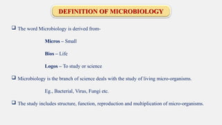
3. DEFINITION OF MICROBIOLOGY
The word Microbiology is derived from-
Micros Small
Bios Life
Logos To study or science
Microbiology is the branch of science deals with the study of living micro-organisms.
Eg., Bacterial, Virus, Fungi etc.
The study includes structure, function, reproduction and multiplication of micro-organisms.
4. DEFINITION OF MICRO-ORGANISMS
These are the small groups of living organisms which can not be seen
by
naked eyes and studied under microscope.
Eg., Bacteria like streptococcal, pneumococcal, salmonella typhi.
Virus like DNA or RNA virus, HIV.
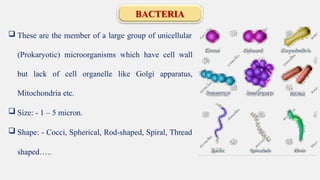
6. BACTERIA
These are the member of a large group of unicellular
(Prokaryotic) microorganisms which have cell wall
but lack of cell organelle like Golgi apparatus,
Mitochondria etc.
Size: - 1 5 micron.
Shape: - Cocci, Spherical, Rod-shaped, Spiral, Thread
shaped..
7. VIRUS
These are non cellular, ultramicroscopic highly infectious agent and posses only one type
of nucleic acid either DNA or RNA surrounded by protein (protective) coat called, Capsid.
Size- 0.02 to 0.2 micron.
Shape As in image
9. Isolation of Pure Culture
Growth of microbes on laboratory medium is known as Culture. A culture which may contains only one
species of microbe is called a pure culture and one which consist of several species is called mixed
culture.
It is very difficult to obtain pure culture of bacterias in nature because they exist as mixed culture. To obtain
organisms in pure culture various techniques are used-
1. Streak plate method
2. Pour plate method
3. Spread plate method
4. Micromanipulator
5. Roll tube method
10. Staining Methods
Stains are the organic dyes used for staining the micro-organisms.
For ex, Crystal violet, methylene blue, safranin etc.
Purpose of Staining: -
1. For greater visualization of cells.
2. For study of their structures.
3. To differentiate the cells.
4. To inhibit the growth of some organism so the others can be
visualized.
12. Simple Staining
It is also called as Monochrome technique. In this method only one stain is used. It used to study morphology i.e.
size, shape and arrangement of microbes.
Prepare a smear and fixed on slide
Add stain for 30 sec to 3 min using
methylene blue
Wash with cool water
Air dry and examine under oil immersion
lens
13. Differential Staining
Grams Staining: -
A differential staining technique used to classify bacteria i.e. gram positive or gram negative and their
specific
structure.
Gram staining was discovered by a Danish Physician Hans Christian Gram while working in Berlin in
1883 and later procedure published in 1884. Hence, it is called Grams staining.
Requirements: - Staining reagents like
1. Crystal violet- Primary Stain
2. Grams Iodine- Mordant - fixative agent
3. Acetone 95% or Alcohol- Decolorizer
4. Saffranine / dilute carbol fuchsin counter stain
14. Grams Staining
Crystal Violet: - All bacteria takes crystal violet so all are appears violet colour.
Iodine: - Crystal violet-iodine (CV-I) complex is formed.
Acetone: - Bacteria with high lipid content loose CV-I complex and appears
colourless but bacteria with less lipid content retains CV-I complex and appears
violet.
Saffranine: - Only colourless bacteria takes saffranine and appears pink
16. Observation
If the bacteria shows violet or
Gram
purple then indicates
Positive.
Eg., E. Coli, Pneumococci
If the bacteria shows pink or red
then indicates Gram Negative.
Eg., S. typhi, H. Influenzae
17. Acid fast staining technique
This technique was discovered by the scientist Zeihl & Neelson,
This technique is used to identify all the separation of Mycobacterium Group members from the others.
It means it is used to identify the acid-fast micro-organism like Mycobacterium
tuberculosis, Mycobacterium Leprae etc.
Acid fast microorganism are characterized by wax like nearly impermeable cell walls means they
contain mycolic acid and large amount of fatty acids, waxes and complex lipids.
Acid-fast organisms are highly resistant to disinfectants because of the cell wall is so resistant to most
compounds. Acid-fast microorganism required a special staining technique i.e. called Acid-fast staining
method.