際際滷

際際滷Share a Scribd company logo
IMPAX PET & SPECT VIEWING 3.0
IMPAX PET &
SPECT Viewing
Agfa HealthCares Advanced Visualization tools that maximize
IMPAX user productivity.
 PET-CT, PET-MRI,
SPECT-CT and SPECT-
MRI viewing and
reporting
 Advanced features
for smart workflows
 Tight integration
with IMPAX for
fast creation and
distribution of results
Follow up with a single click
Advances in imaging have provided radiologists with
a variety of high quality, high resolution imaging
technologies. With IMPAX PET & SPECT Viewing,
Agfa HealthCare provides the physician with an
automated, easy-to-use set of tools for viewing
and comparing studies. Images can come from
independent modalities (CT, MRI, PET, SPECT) or from
hybrid modalities (PET-CT, PET-MRI, SPECT-CT). With
the completely automated application, the radiologist
can initiate registration of two PET/SPECT-CT or PET/
SPECT-MRI studies with a single mouse click.
One system, different modalities
Over the past few years, there has been a strong
increase in the use of combined modalities (e.g.
hybrid PET/SPECT-CT scanners), creating a need for
powerful software tools. IMPAX PET & SPECT Viewing
provides the tools needed to help optimize the value
of combining modalities.
Multi-modality registration and fusion allows the
radiologist to efficiently view images collected
from different imaging devices to provide important
information for follow-up studies.
IMPAX PET & SPECT Viewing allows both side-by-
side viewing and fusion of studies from multiple
modalities. SUVs are automatically calculated for PET
studies and together with volume measurements of
lesions this can provide additional information for
treatment planning.
IMPAX PET & SPECT VIEWING 3.0
2
No specialized staff or extensive training
needed
While IMPAX PET & SPECT Viewing extends IMPAX
functionality, it has been designed to be very easy to use. It
has the same unified look & feel as the IMPAX radiology
workstation, so the reading radiologist feels familiar with the
software from the very first minute. The user interface is very
intuitive: there is no need for specialized staff or extensive
training to be able to use IMPAX PET & SPECT Viewing.
Features
 Registration of PET and SPECT with CT/MRI
 Automatically, based on mutual information
 Semi-automatic, based on user-defined landmarks
 Manual shift and rotate
 Load second PET-CT, PET-MRI, SPECT-CT or SPECT-MRI for
follow-up
 Registration of both data sets based on CT or MRI
images
 Multiple display lay-outs, optimized for dedicated tasks,
a.o.:
 Side-by-side PET/SPECT, CT/MRI and fused view for
comparison
 Simultaneous axial/sagittal/coronal views of PET/
SPECT and CT/MRI per study
 Fusion view with 3D MIP visualization for PET-CT/MRI
and SPECT-CT/MRI
 Create annotations and Regions of Interest
 Region growing algorithm for easy assessment of lesion
shape and volume and calculation of SUVs for PET and
counts for SPECT in the volume of the lesion
 Automatic calculation of SUV for PET
 Save images for further distribution/review within the
PACS.
IMPAX PET & SPECT VIEWING 3.0
3
Technical Specifications
Software prerequisites
 IMPAX Release 6.3.1 SU9 or higher
 IMPAX EE R20 XIII (Germany, Austria and Switzerland
only)
IMPAX PET & SPECT Viewing is available as a concurrent user
license or site license on IMPAX 6 workstations.
Hardware minimum requirements
 On Microsoft Windows XP 32-bit:
 One dual-core CPU (2GHz) or equivalent
 4 GB RAM
 On Microsoft Windows 7 32/64 bit: *
 One dual-core CPU (2GHz) or equivalent
 8 GB RAM for diagnostic workstations
 4 GB RAM for referral viewing stations with IMPAX
Volume Viewing MIP/MPR Enterprise Edition
 On Microsoft Windows 8 32/64-bit: **
 One dual-core CPU (2GHz) or equivalent
 8 GB RAM for diagnostic workstations
 4 GB RAM for referral viewing stations with IMPAX
Volume Viewing MIP/MPR Enterprise Edition
*	 Note: Diagnostic workstations must be 64 bit
	systems.
**	 Note: Microsoft Windows 8 is supported for non-
	 diagnostic workstations starting with IMPAX
	 version 6.5.4.
***	For all configurations 					
	 Minimum number of pixels: 1280 px in horizontal 		
	dimensions
For additional hardware requirements and Windows
compatibility please consult the IMPAX 6 or IMPAX EE
workstation specifications.
 IMPAX PET & SPECT Viewings 3D rendering performance
can be augmented by using a high performance display
controller with a Graphical Processing Unit (GPU). The
following display controllers have been validated for
compatibility: Barco MXRT 5200, MXRT 5400, MXRT
5450, MXRT 7200, MXRT 7300, MXRT 7400
DICOM
IMPAX PET & SPECT Viewing supports display of standard
CT and MRI DICOM objects. The enhanced multiframe CT
and enhanced multiframe MRI DICOM SOP classes are not
supported in this version.
For the most recent edition of Agfa HealthCares DICOM
conformance statements go to Agfa HealthCares web site
and look for the DICOM conformance statement of your
IMPAX solution (6.3, 6.4, 6.5 or EE):
http://www.agfahealthcare.com/global/en/main/products_
services/product-info/interoperability/dicom/conformance_
statements/index.jsp
Language support
Graphical User Interface, Quick Reference Guide and Safety
Instructions are available in Bulgarian, Traditional and
Simplified Chinese, Croatian, Czech, Danish, Dutch, English,
Estonian, Finnish, French, German, Greek, Hungarian, Italian,
Norwegian, Polish, Portuguese, Romanian, Russian, Slovenian,
Spanish and Swedish.
Technical specifications are subject to change without
prior notice.
IMPAX PET & SPECT VIEWING 3.0
息 2013 Agfa HealthCare NV
All rights reserved
Published by Agfa HealthCare NV
B-2640 Mortsel - Belgium
NGOLQ GB 00201405
Agfa and the Agfa rhombus are trademarks of Agfa-Gevaert N.V., Belgium, or its affiliates. IMPAX is
a trademark of Agfa HealthCare NV, Belgium, or its affiliates. All other trademarks are held by their
respective owners and are used in an editorial fashion with no intention of infringement. The data in this
publication are for illustration purposes only and do not necessarily represent standards or specifications,
which must be met by Agfa HealthCare. All information contained herein is intended for guidance purposes
only, and characteristics of the products and services described in this publication can be changed at any
time without notice. Products and services may not be available for your local area. Please contact your
local sales representative for availability information. Agfa HealthCare diligently strives to provide as
accurate information as possible, but shall not be responsible for any typographical error.
Agfa HealthCare is a global leader in the fast growing market of integrated
IT and imaging systems, offering healthcare facilities/hospitals a
seamlessflowofinformationanda360属viewofpatientcare.Thecompany
has a unique, holistic approach, enabling it to provide in-depth clinical
know-how and fully integrated hospital-wide solutions. These specialized
solutions ntegrate IT and imaging systems for Radiology, Cardiology,
Mammography and Orthopaedics. Agfa HealthCares enterprise-wide IT
platformintegratesalladministrativeandclinicaldatawithinahealthcare
facility and is designed to match the unique needs of specific healthcare
professionals.
www.agfahealthcare.com
Insight. Delivered.
Ad

Recommended

IMPAX_Virtual_Colonoscopy_(GB_-_datasheet)
Alexandre Ovtchinnikov
Present existed Driver alert system
krishna495ECE
Analyytikkoesitys Q3 2015 FI
Alma Media
Hidden markov models
Murat im
Alma Media Q4 ja FY 2014
Alma Media
Analyytikkoesitys Q2_2015_FI
Alma Media
Presentation for investors and analysts_Q1_2015
Alma Media
Analyst Presentation Q3 2015 EN
Alma Media
0311
0311
Murat im
Alma Media Q4 and FY 2014
Alma Media
Alma Median taloudellinen tulos Q3 2014
Alma Media
Analyytikkoesitys Q4 2015
Alma Media
Analyytikkoesitys Q1_2015
Alma Media
Analyst presentation Q4 and FY 2015
Alma Media
Ekoloji 02
Ekoloji 02
Murat im
Analyytikkoesitys Q2_2015_ENG
Alma Media
Alma Media's Capital Markets Day 2014
Alma Media
Graph coloring and_applications
mohammad alkhalil
IMPAX_Volume_Viewing_(GB_-_datasheet)
Alexandre Ovtchinnikov
Norway_Oslo_ClinApps
Alexandre Ovtchinnikov
MEDICAL IMAGE PROCESSING
MOUMITA GHOSH
7i PACS Presentation
Hunt Russell
Nsfb 0816 web_opt
Nsfb 0816 web_opt
Robert Farkas
Introduction Frost & Sullivan White Paper: Vision 2027
Marc De Fr辿
The Power of Together
Carestream
Guide Preview: Ensuring your enterprise image-viewer if fully secure
Calgary Scientific Inc.
Carlos Perez Castillo - Imaging biomarkers automated structured
WTHS
Automated image analysis: rescue for diffusion-MRI of threat to radiologists?
Erik R. Ranschaert, MD, PhD
Lec2: Digital Images and Medical Imaging Modalities
Ula Bac脹
spect_student_ppt_spring2012.pdf
AdarshPradeep11

More Related Content

Viewers also liked (10)

0311
0311
Murat im
Alma Media Q4 and FY 2014
Alma Media
Alma Median taloudellinen tulos Q3 2014
Alma Media
Analyytikkoesitys Q4 2015
Alma Media
Analyytikkoesitys Q1_2015
Alma Media
Analyst presentation Q4 and FY 2015
Alma Media
Ekoloji 02
Ekoloji 02
Murat im
Analyytikkoesitys Q2_2015_ENG
Alma Media
Alma Media's Capital Markets Day 2014
Alma Media
Graph coloring and_applications
mohammad alkhalil

Similar to IMPAX_PET_&_SPECT_Viewing_(GB_-_datasheet) (20)

IMPAX_Volume_Viewing_(GB_-_datasheet)
Alexandre Ovtchinnikov
Norway_Oslo_ClinApps
Alexandre Ovtchinnikov
MEDICAL IMAGE PROCESSING
MOUMITA GHOSH
7i PACS Presentation
Hunt Russell
Nsfb 0816 web_opt
Nsfb 0816 web_opt
Robert Farkas
Introduction Frost & Sullivan White Paper: Vision 2027
Marc De Fr辿
The Power of Together
Carestream
Guide Preview: Ensuring your enterprise image-viewer if fully secure
Calgary Scientific Inc.
Carlos Perez Castillo - Imaging biomarkers automated structured
WTHS
Automated image analysis: rescue for diffusion-MRI of threat to radiologists?
Erik R. Ranschaert, MD, PhD
Lec2: Digital Images and Medical Imaging Modalities
Ula Bac脹
spect_student_ppt_spring2012.pdf
AdarshPradeep11
Altaviewer
Altaviewer
odayc01
2013131 webinar slidedeck_ge_universal_viewer_final
Trimed Media Group
Machine Learning in Pathology Diagnostics with Simagis Live
khvatkov
Post proceesing techniques & visualization.pptx
DilshanDillu1
Diagnostic medical imaging
PostDICOM
Hybrid Spectct Imaging In Clinical Practice Ora Israel Stanley J Goldsmith
hajyewhoseop
IMERA Business Plan Sep 2023 Enterprise AI Event.pptx.pdf
brand44
Medical Insight Brochure Nov 2010
ho1234
IMPAX_Volume_Viewing_(GB_-_datasheet)
Alexandre Ovtchinnikov
MEDICAL IMAGE PROCESSING
MOUMITA GHOSH
7i PACS Presentation
Hunt Russell
Nsfb 0816 web_opt
Nsfb 0816 web_opt
Robert Farkas
Introduction Frost & Sullivan White Paper: Vision 2027
Marc De Fr辿
The Power of Together
Carestream
Guide Preview: Ensuring your enterprise image-viewer if fully secure
Calgary Scientific Inc.
Carlos Perez Castillo - Imaging biomarkers automated structured
WTHS
Automated image analysis: rescue for diffusion-MRI of threat to radiologists?
Erik R. Ranschaert, MD, PhD
Lec2: Digital Images and Medical Imaging Modalities
Ula Bac脹
spect_student_ppt_spring2012.pdf
AdarshPradeep11
Altaviewer
Altaviewer
odayc01
2013131 webinar slidedeck_ge_universal_viewer_final
Trimed Media Group
Machine Learning in Pathology Diagnostics with Simagis Live
khvatkov
Post proceesing techniques & visualization.pptx
DilshanDillu1
Diagnostic medical imaging
PostDICOM
Hybrid Spectct Imaging In Clinical Practice Ora Israel Stanley J Goldsmith
hajyewhoseop
IMERA Business Plan Sep 2023 Enterprise AI Event.pptx.pdf
brand44
Medical Insight Brochure Nov 2010
ho1234
Ad

IMPAX_PET_&_SPECT_Viewing_(GB_-_datasheet)

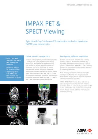
  • 1. IMPAX PET & SPECT VIEWING 3.0 IMPAX PET & SPECT Viewing Agfa HealthCares Advanced Visualization tools that maximize IMPAX user productivity. PET-CT, PET-MRI, SPECT-CT and SPECT- MRI viewing and reporting Advanced features for smart workflows Tight integration with IMPAX for fast creation and distribution of results Follow up with a single click Advances in imaging have provided radiologists with a variety of high quality, high resolution imaging technologies. With IMPAX PET & SPECT Viewing, Agfa HealthCare provides the physician with an automated, easy-to-use set of tools for viewing and comparing studies. Images can come from independent modalities (CT, MRI, PET, SPECT) or from hybrid modalities (PET-CT, PET-MRI, SPECT-CT). With the completely automated application, the radiologist can initiate registration of two PET/SPECT-CT or PET/ SPECT-MRI studies with a single mouse click. One system, different modalities Over the past few years, there has been a strong increase in the use of combined modalities (e.g. hybrid PET/SPECT-CT scanners), creating a need for powerful software tools. IMPAX PET & SPECT Viewing provides the tools needed to help optimize the value of combining modalities. Multi-modality registration and fusion allows the radiologist to efficiently view images collected from different imaging devices to provide important information for follow-up studies. IMPAX PET & SPECT Viewing allows both side-by- side viewing and fusion of studies from multiple modalities. SUVs are automatically calculated for PET studies and together with volume measurements of lesions this can provide additional information for treatment planning.
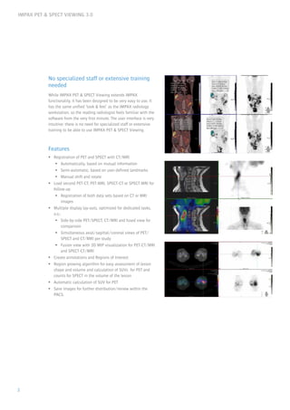
  • 2. IMPAX PET & SPECT VIEWING 3.0 2 No specialized staff or extensive training needed While IMPAX PET & SPECT Viewing extends IMPAX functionality, it has been designed to be very easy to use. It has the same unified look & feel as the IMPAX radiology workstation, so the reading radiologist feels familiar with the software from the very first minute. The user interface is very intuitive: there is no need for specialized staff or extensive training to be able to use IMPAX PET & SPECT Viewing. Features Registration of PET and SPECT with CT/MRI Automatically, based on mutual information Semi-automatic, based on user-defined landmarks Manual shift and rotate Load second PET-CT, PET-MRI, SPECT-CT or SPECT-MRI for follow-up Registration of both data sets based on CT or MRI images Multiple display lay-outs, optimized for dedicated tasks, a.o.: Side-by-side PET/SPECT, CT/MRI and fused view for comparison Simultaneous axial/sagittal/coronal views of PET/ SPECT and CT/MRI per study Fusion view with 3D MIP visualization for PET-CT/MRI and SPECT-CT/MRI Create annotations and Regions of Interest Region growing algorithm for easy assessment of lesion shape and volume and calculation of SUVs for PET and counts for SPECT in the volume of the lesion Automatic calculation of SUV for PET Save images for further distribution/review within the PACS.
  • 3. IMPAX PET & SPECT VIEWING 3.0 3 Technical Specifications Software prerequisites IMPAX Release 6.3.1 SU9 or higher IMPAX EE R20 XIII (Germany, Austria and Switzerland only) IMPAX PET & SPECT Viewing is available as a concurrent user license or site license on IMPAX 6 workstations. Hardware minimum requirements On Microsoft Windows XP 32-bit: One dual-core CPU (2GHz) or equivalent 4 GB RAM On Microsoft Windows 7 32/64 bit: * One dual-core CPU (2GHz) or equivalent 8 GB RAM for diagnostic workstations 4 GB RAM for referral viewing stations with IMPAX Volume Viewing MIP/MPR Enterprise Edition On Microsoft Windows 8 32/64-bit: ** One dual-core CPU (2GHz) or equivalent 8 GB RAM for diagnostic workstations 4 GB RAM for referral viewing stations with IMPAX Volume Viewing MIP/MPR Enterprise Edition * Note: Diagnostic workstations must be 64 bit systems. ** Note: Microsoft Windows 8 is supported for non- diagnostic workstations starting with IMPAX version 6.5.4. *** For all configurations Minimum number of pixels: 1280 px in horizontal dimensions For additional hardware requirements and Windows compatibility please consult the IMPAX 6 or IMPAX EE workstation specifications. IMPAX PET & SPECT Viewings 3D rendering performance can be augmented by using a high performance display controller with a Graphical Processing Unit (GPU). The following display controllers have been validated for compatibility: Barco MXRT 5200, MXRT 5400, MXRT 5450, MXRT 7200, MXRT 7300, MXRT 7400 DICOM IMPAX PET & SPECT Viewing supports display of standard CT and MRI DICOM objects. The enhanced multiframe CT and enhanced multiframe MRI DICOM SOP classes are not supported in this version. For the most recent edition of Agfa HealthCares DICOM conformance statements go to Agfa HealthCares web site and look for the DICOM conformance statement of your IMPAX solution (6.3, 6.4, 6.5 or EE): http://www.agfahealthcare.com/global/en/main/products_ services/product-info/interoperability/dicom/conformance_ statements/index.jsp Language support Graphical User Interface, Quick Reference Guide and Safety Instructions are available in Bulgarian, Traditional and Simplified Chinese, Croatian, Czech, Danish, Dutch, English, Estonian, Finnish, French, German, Greek, Hungarian, Italian, Norwegian, Polish, Portuguese, Romanian, Russian, Slovenian, Spanish and Swedish. Technical specifications are subject to change without prior notice.
  • 4. IMPAX PET & SPECT VIEWING 3.0 息 2013 Agfa HealthCare NV All rights reserved Published by Agfa HealthCare NV B-2640 Mortsel - Belgium NGOLQ GB 00201405 Agfa and the Agfa rhombus are trademarks of Agfa-Gevaert N.V., Belgium, or its affiliates. IMPAX is a trademark of Agfa HealthCare NV, Belgium, or its affiliates. All other trademarks are held by their respective owners and are used in an editorial fashion with no intention of infringement. The data in this publication are for illustration purposes only and do not necessarily represent standards or specifications, which must be met by Agfa HealthCare. All information contained herein is intended for guidance purposes only, and characteristics of the products and services described in this publication can be changed at any time without notice. Products and services may not be available for your local area. Please contact your local sales representative for availability information. Agfa HealthCare diligently strives to provide as accurate information as possible, but shall not be responsible for any typographical error. Agfa HealthCare is a global leader in the fast growing market of integrated IT and imaging systems, offering healthcare facilities/hospitals a seamlessflowofinformationanda360属viewofpatientcare.Thecompany has a unique, holistic approach, enabling it to provide in-depth clinical know-how and fully integrated hospital-wide solutions. These specialized solutions ntegrate IT and imaging systems for Radiology, Cardiology, Mammography and Orthopaedics. Agfa HealthCares enterprise-wide IT platformintegratesalladministrativeandclinicaldatawithinahealthcare facility and is designed to match the unique needs of specific healthcare professionals. www.agfahealthcare.com Insight. Delivered.