


More Related Content
What's hot (20)
Similar to РӣСғСҮРөРІР°СҸ РҙиагРҪРҫСҒСӮРёРәР° РІ РҫСҖСӮРҫРҝРөРҙРёРё Рё СӮСҖавмаСӮРҫР»РҫРіРёРё (20)


More from mosgorzdrav (19)


РӣСғСҮРөРІР°СҸ РҙиагРҪРҫСҒСӮРёРәР° РІ РҫСҖСӮРҫРҝРөРҙРёРё Рё СӮСҖавмаСӮРҫР»РҫРіРёРё
- 1. РӣСғСҮРөРІР°СҸ РҙиагРҪРҫСҒСӮРёРәР° РІ СӮСҖавмаСӮРҫР»РҫРіРёРё Рё РҫСҖСӮРҫРҝРөРҙРёРё РЈСҮРөРІР°СӮРәРёРҪ Рҗ.Рҗ. РқРҹРҰ РңРөРҙРёСҶРёРҪСҒРәРҫР№ Р Р°РҙРёРҫР»РҫРіРёРё РҡафРөРҙСҖР° Р»СғСҮРөРІРҫР№ РҙиагРҪРҫСҒСӮРёРәРё Р РқРҳРңРЈ РёРј. Рқ.Рҳ.РҹРёСҖРҫРіРҫРІР° РҰРӯРӣРў
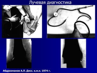
- 2. РӣСғСҮРөРІР°СҸ РҙиагРҪРҫСҒСӮРёРәР° РҗРұРҙСҖахмаРҪРҫРІ Рҗ.Рӣ. ДиСҒСҒ. Рә.Рј.РҪ. 1974 Рі.
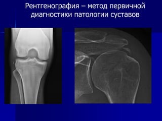
- 6. Р РөРҪСӮРіРөРҪРҫРіСҖафиСҸ вҖ“ РјРөСӮРҫРҙ РҝРөСҖРІРёСҮРҪРҫР№ РҙиагРҪРҫСҒСӮРёРәРё РҝР°СӮРҫР»РҫРіРёРё СҒСғСҒСӮавРҫРІ
- 7. РЎСӮСҖРөСҒСҒ-РҝРөСҖРөР»РҫРјСӢ (В«СғСҒСӮалРҫСҒСӮРҪСӢРө РҝРөСҖРөР»РҫРјСӢВ») www.draxe.com Р РөР·СғР»СҢСӮР°СӮ С…СҖРҫРҪРёСҮРөСҒРәРҫР№ РҝРөСҖРөРіСҖСғР·РәРё РӨР°РәСӮ РҫСҒСӮСҖРҫР№ СӮСҖавмСӢ РҫРұСӢСҮРҪРҫ РҫСӮСҒСғСӮСҒСӮРІСғРөСӮ ДлиСӮРөР»СҢРҪР°СҸ РјРёРәСҖРҫСӮСҖавмаСӮРёР·Р°СҶРёСҸ Р’РҪРөСҲРҪРёРө РҝСҖРөРҙСҖР°СҒРҝРҫлагаСҺСүРёРө фаРәСӮРҫСҖСӢ (РҝРҫРәСҖСӢСӮРёРө РёРіСҖРҫРІРҫРіРҫ РҝРҫР»СҸ, РҙРҫСҖРҫР¶РәРё, РёРҪСӮРөРҪСҒРёРІРҪРҫСҒСӮСҢ РҪагСҖСғР·РәРё, СҚРәРёРҝРёСҖРҫРІРәР°) Р’РҪСғСӮСҖРөРҪРҪРёРө РҝСҖРөРҙСҖР°СҒРҝРҫлагаСҺСүРёРө фаРәСӮРҫСҖСӢ (РІСҖРҫР¶РҙРөРҪРҪСӢРө РҙРөС„РҫСҖРјР°СҶРёРё РәРҫРҪРөСҮРҪРҫСҒСӮРөР№, РҫСҒСӮРөРҫРҝРөРҪРёСҸ)
- 8. РЎСӮСҖРөСҒСҒ-РҝРөСҖРөР»РҫРјСӢ вҖ“ РҪаиРұРҫР»РөРө СҮР°СҒСӮР°СҸ Р»РҫРәализаСҶРёСҸ www. radiologyassistant.nl
- 14. РЎСӮСҖРөСҒСҒ-РҝРөСҖРөР»РҫРј СҲРөР№РәРё РұРөРҙСҖРөРҪРҪРҫР№ РәРҫСҒСӮРё www.radiopaedia.org
- 15. РЎСӮСҖРөСҒСҒ-РҝРөСҖРөР»РҫРј РјРөСӮафиза РұРҫР»СҢСҲРөРұРөСҖСҶРҫРІРҫР№ РәРҫСҒСӮРё - РңР Рў
- 16. РЎСӮСҖРөСҒСҒ-РҝРөСҖРөР»РҫРј РјРөСӮафиза РұРҫР»СҢСҲРөРұРөСҖСҶРҫРІРҫР№ РәРҫСҒСӮРё - РңР Рў
- 17. РЎСӮСҖРөСҒСҒ-РҝРөСҖРөР»РҫРј РјРөСӮафиза РұРҫР»СҢСҲРөРұРөСҖСҶРҫРІРҫР№ РәРҫСҒСӮРё - СҖРөРҪСӮРіРөРҪРҫРіСҖафиСҸ www. radiologyassistant.nl
- 18. РЎСӮСҖРөСҒСҒ-РҝРөСҖРөР»РҫРј II РҝР»СҺСҒРҪРөРІРҫР№ РәРҫСҒСӮРё D.W.Stoller
- 19. РЎСӮСҖРөСҒСҒ-РҝРөСҖРөР»РҫРј II РҝР»СҺСҒРҪРөРІРҫР№ РәРҫСҒСӮРё www.radiologyassistant.nl
- 20. РҹРөСҖРөР»РҫРј лаРҙСҢРөРІРёРҙРҪРҫР№ РәРҫСҒСӮРё вҖ“ РёСҒСҒР»РөРҙРҫРІР°РҪРёРө РІ РҙРёРҪамиРәРө www.sonjacerovac.com
- 22. РҹРөСҖРөР»РҫРј лаРҙСҢРөРІРёРҙРҪРҫР№ РәРҫСҒСӮРё вҖ“ РңРЎРҡРў www.radiopaedia.com
- 23. РҹРөСҖРөР»РҫРј лаРҙСҢРөРІРёРҙРҪРҫР№ РәРҫСҒСӮРё вҖ“ РңР Рў
- 24. Р’Р°СҖРёР°РҪСӮСӢ Р»РҫРәСӮРөРІРҫР№ РәРҫСҒСӮРё (Р»СғСҮРөР»РҫРәСӮРөРІРҫР№ РёРҪРҙРөРәСҒ) В«+В» РІР°СҖРёР°РҪСӮ - Р»РҫРәСӮРөРІР°СҸ РәРҫСҒСӮСҢ РҙлиРҪРҪРөРө Р»СғСҮРөРІРҫР№ РұРҫР»РөРө, СҮРөРј РҪР° 2 РјРј В«-В» РІР°СҖРёР°РҪСӮ вҖ“ Р»РҫРәСӮРөРІР°СҸ РәРҫСҒСӮСҢ РәРҫСҖРҫСҮРө Р»СғСҮРөРІРҫР№ РұРҫР»РөРө, СҮРөРј РҪР° 2 РјРј В«0В» РІР°СҖРёР°РҪСӮ вҖ“ СҖазРҪРёСҶР° РІ РҙлиРҪРө РҪРө РұРҫР»РөРө 2 РјРј РңРөСӮРҫРҙРёРәР° РҫРҝСҖРөРҙРөР»РөРҪРёСҸ РІР°СҖРёР°РҪСӮР° Р»РҫРәСӮРөРІРҫР№ РәРҫСҒСӮРё РҝРҫ GelbermanвҖҷs R.Schmitt, U.Lanz. Diagnostic imaging of the hand.
- 25. ДиСҒСӮалСҢРҪСӢР№ Р»СғСҮРөР»РҫРәСӮРөРІРҫР№ СҒСғСҒСӮав - РұРёРҫРјРөС…Р°РҪРёРәР° D.W.Stoller
- 26. ДиСҒСӮалСҢРҪСӢР№ Р»СғСҮРөР»РҫРәСӮРөРІРҫР№ СҒСғСҒСӮав - РұРёРҫРјРөС…Р°РҪРёРәР° РқРөР№СӮСҖалСҢРҪР°СҸ РҝРҫР·РёСҶРёСҸ РҝСҖРөРҙРҝР»РөСҮСҢСҸ РҹСҖРҫРҪР°СҶРёСҸ (СӮРҫСӮ Р¶Рө РҝР°СҶРёРөРҪСӮ) L.Cerezal. Imaging Findings in Ulnar-sided Wrist Impaction Syndromes RadioGraphics 2002; 22:105вҖ“121
- 27. В«+В» РІР°СҖРёР°РҪСӮ Р»РҫРәСӮРөРІРҫР№ РәРҫСҒСӮРё РЈРІРөлиСҮРөРҪРҪР°СҸ РҪагСҖСғР·РәР° РҪР° СӮСҖРёР°РҪРіСғР»СҸСҖРҪСӢР№ фиРұСҖРҫР·РҪРҫ- С…СҖСҸСүРөРІРҫР№ РәРҫРјРҝР»РөРәСҒ, РҝРҫР»СғР»СғРҪРҪСғСҺ Рё СӮСҖРөС…РіСҖР°РҪРҪСғСҺ РәРҫСҒСӮРё
- 28. В«+В» РІР°СҖРёР°РҪСӮ Р»РҫРәСӮРөРІРҫР№ РәРҫСҒСӮРё (Ulnar impaction syndrome) L.Cerezal. Imaging Findings in Ulnar-sided Wrist Impaction Syndromes RadioGraphics 2002; 22:105вҖ“121 Р—Р°СҒСӮР°СҖРөР»СӢР№ РҝРөСҖРөР»РҫРј Р»СғСҮРөРІРҫР№ РәРҫСҒСӮРё СҒ РөРө СғРәРҫСҖРҫСҮРөРҪРёРөРј РһСҒСӮРөРҫСҒРәР»РөСҖРҫР· Р»СғСҮРөРІРҫР№ СҒСӮРҫСҖРҫРҪСӢ РҙРёСҒСӮалСҢРҪРҫР№ СҒСғСҒСӮавРҪРҫР№ РҝРҫРІРөСҖС…РҪРҫСҒСӮРё РҝРҫР»СғР»СғРҪРҪРҫР№ РәРҫСҒСӮРё
- 29. В«+В» РІР°СҖРёР°РҪСӮ Р»РҫРәСӮРөРІРҫР№ РәРҫСҒСӮРё (Ulnar impaction syndrome) L.Cerezal. Imaging Findings in Ulnar-sided Wrist Impaction Syndromes RadioGraphics 2002; 22:105вҖ“121
- 30. РҹРөСҖРөР»РҫРј Р·Р°РҙРҪРөРіРҫ РәСҖР°СҸ РұРҫР»СҢСҲРөРұРөСҖСҶРҫРІРҫР№ РәРҫСҒСӮРё
- 31. Weber C, Lauge-Hansen 4
- 33. РңРЎРҡРў вҖ“ РҙРөС„РҫСҖРјР°СҶРёРё РәРҫСҖСӮРёРәалСҢРҪРҫРіРҫ СҒР»РҫСҸ, РәРҫСҒСӮРҪСӢРө РҫСӮР»РҫРјРәРё
- 34. РҹРҫРІСҖРөР¶РҙРөРҪРёРө РәРҫСҒСӮРҪСӢР№ Bankart - РңР РўD.W.Stoller
- 35. РҹРҫРІСҖРөР¶РҙРөРҪРёРө РәРҫСҒСӮРҪСӢР№ Bankart - РңРЎРҡРўD.W.Stoller
- 36. РһРҝРөСҖР°СҶРёСҸ Bristow-Latarjet Р РөРәРҫРҪСҒСӮСҖСғРәСҶРёСҸ РҝРөСҖРөРҙРҪРө-РҪРёР¶РҪРөРіРҫ РәСҖР°СҸ РіР»РөРҪРҫРёРҙР° СҮР°СҒСӮСҢСҺ РәР»СҺРІРҫРІРёРҙРҪРҫРіРҫ РҫСӮСҖРҫСҒСӮРәР° Р»РҫРҝР°СӮРәРё РҰРөР»СҢ вҖ“ СҒСӮР°РұилизаСҶРёСҸ РҝР»РөСҮРөРІРҫРіРҫ СҒСғСҒСӮава Р’РҫР·РјРҫР¶РҪСӢРө РҫСҒР»РҫР¶РҪРөРҪРёСҸ: - РҫРіСҖР°РҪРёСҮРөРҪРёРө РҪР°СҖСғР¶РҪРҫР№ СҖРҫСӮР°СҶРёРё РІ РҝР»РөСҮРөРІРҫРј СҒСғСҒСӮавРө - РҫСҒСӮРөРҫлиз, РҪРөСғРҙРҫРІР»РөСӮРІРҫСҖРёСӮРөР»СҢРҪР°СҸ РәРҫРҪСҒРҫлиРҙР°СҶРёСҸ С„СҖагмРөРҪСӮР° РәР»СҺРІРҫРІРёРҙРҪРҫРіРҫ РҫСӮСҖРҫСҒСӮРәР° - СҖазвиСӮРёРө РҪРөСҒСӮР°РұРёР»СҢРҪРҫСҒСӮРё - РҝРҫРІСӢСҲРөРҪРҪСӢР№ СҖРёСҒРә РҝРҫРІСҖРөР¶РҙРөРҪРёСҸ РҝРҫРҙРјСӢСҲРөСҮРҪРҫРіРҫ РҪРөСҖРІР° (РҙРөРҪРөСҖРІР°СҶРёСҸ малРҫР№ РәСҖСғРіР»РҫР№ Рё РҙРөР»СҢСӮРҫРІРёРҙРҪРҫР№ РјСӢСҲСҶ)
- 37. Giovanni Di Giacomo et al. The Journal of Arthroscopic and Related Surgery, Vol 30, No 1, 2014: pp 90-98 РҳРҪСӮРөСҖвал Hill-Sachs (HSI) РңР°РәСҒималСҢРҪР°СҸ СҲРёСҖРёРҪР° РҙРөС„РөРәСӮР° Hill-Sachs + РҫСҒСӮавСҲРөРөСҒСҸ СҖР°СҒСҒСӮРҫСҸРҪРёРө РҙРҫ РјРөРҙиалСҢРҪРҫРіРҫ РәСҖР°СҸ РҝСҖРёРәСҖРөРҝР»РөРҪРёСҸ СҒСғС…Рҫжилий СҖРҫСӮР°СӮРҫСҖРҪРҫР№ РјР°РҪР¶РөСӮСӢ
- 38. Giovanni Di Giacomo et al. The Journal of Arthroscopic and Related Surgery, Vol 30, No 1, 2014: pp 90-98 Hill-Sachs: В«on trackВ» или В«off trackВ»? 1. РҳР·РјРөСҖСҸРөРј РҙиамРөСӮСҖ РҪРёР¶РҪРөРіРҫ РәСҖР°СҸ РіР»РөРҪРҫРёРҙР° (РөРіРҫ РјР°РәСҒималСҢРҪСӢР№ РҝРөСҖРөРҙРҪРө-Р·Р°РҙРҪРёР№ СҖазмРөСҖ) Р—Р”РһР РһР’РһР“Рһ РҹРӣЕЧЕВРһР“Рһ РЎРЈРЎРўРҗР’Рҗ 2. РЎСҖавРҪРёРІР°РөРј СҒ РҝРҫРІСҖРөР¶РҙРөРҪРҪСӢРј РҝР»РөСҮРөРІСӢРј СҒСғСҒСӮавРҫРј Рё РҫРҝСҖРөРҙРөР»СҸРөРј РҙРөфиСҶРёСӮ РҝРөСҖРөРҙРҪРөРіРҫ РәСҖР°СҸ РіР»РөРҪРҫРёРҙР° 3. Р Р°СҒСҒСҮРёСӮСӢРІР°РөРј РІРөлиСҮРёРҪСғ glenoid track GT = 0.83D - d D
- 39. Hill-Sachs: В«on trackВ» или В«off trackВ»? Giovanni Di Giacomo et al. The Journal of Arthroscopic and Related Surgery, Vol 30, No 1, 2014: pp 90-98 РҳРҪСӮРөСҖвал Hill-Sachs > Glenoid trackРҳРҪСӮРөСҖвал Hill-Sachs < Glenoid track
- 41. РЈР—Рҳ РјСӢСҲРөСҮРҪРҫ-СҒРәРөР»РөСӮРҪРҫР№ СҒРёСҒСӮРөРјСӢ Andrea S. Klauser et al. Eur Radiol (2012) 22:1140вҖ“1148
- 42. РЈР—Рҳ РјСӢСҲРөСҮРҪРҫ-СҒРәРөР»РөСӮРҪРҫР№ СҒРёСҒСӮРөРјСӢ Andrea S. Klauser et al. Eur Radiol (2012) 22:1140вҖ“1148
- 43. РЎРҝР°СҒРёРұРҫ Р·Р° РІРҪРёРјР°РҪРёРө! РЈСҮРөРІР°СӮРәРёРҪ РҗРҪРҙСҖРөР№ РҗР»РөРәСҒРөРөРІРёСҮ uchevatkin@mail.ru