Transpedal SFA revascularization using outback re-entry
Download as PPTX, PDF0 likes219 views
SFA revascularization. Transpedal access. CTO. Peripheral artery disease. Peripheral artery disease (PAD).
1 of 8
Download to read offline


Recommended
Instability around elbow -1st part
Instability around elbow -1st partVishnu Raja
?
1) The document discusses various ligaments and structures involved in elbow stability, including the lateral and medial ligamentous complexes.
2) It describes the biomechanics of acute elbow dislocations and different types of elbow instabilities such as posterolateral rotatory instability.
3) Clinical evaluation for elbow instability involves various tests like the lateral pivot shift test and management includes repairing injured ligaments, reconstructing chronic injuries, and addressing any bone deficiencies through procedures like radial head replacement.Spinal immobilization supine
Spinal immobilization supinetimmct
?
This document provides instructions for properly applying a cervical collar during an EMT skills assessment station involving spinal immobilization of a supine patient. Key steps include placing the chin support under the chin and adjusting the size if needed, pulling the back of the collar snug while holding the front in place and fastening it, and for a supine patient, sliding the rear panel behind the neck before placing the chin support. It also notes that cannula hooks are provided for securing supplemental oxygen lines and that the scenario involves immobilizing an unstable spine using a long spine device with the help of an assistant EMT and evaluator.Special tests for knee examination HY
Special tests for knee examination HYRickettsia Rickettsii
?
The document describes several common tests used to examine the knee for injuries. The Valgus stress test and anterior drawer test are used to check for injuries to the medial collateral ligament and anterior cruciate ligament by applying stress and checking for excess laxity in the knee. The Lachman test similarly checks for anterior cruciate ligament injuries. The Thessaly test and McMurray test examine the knee for meniscal tears by having the patient perform rotational motions or applying rotational forces during flexion/extension and checking for pain, clicking or catching.Spinal immobilization seated
Spinal immobilization seatedtimmct
?
1) The document provides instructions for properly applying a cervical collar to immobilize the spine of a seated patient.
2) It describes how to size, position, and secure the collar, including placing the chin support under the chin and pulling the back of the collar snug while holding the front in place.
3) Alternatives to collars for spinal immobilization and the scenario of arriving on the scene of a car crash with one stable patient who requires spinal immobilization using a short device are also outlined.Retronai
Retronaisultanibri
?
This document describes the procedure for intramedullary nailing of a femoral shaft fracture. It involves:
1) Making a small incision over the medial patella tendon to create the entry portal.
2) Advancing a guidewire across the fracture site under fluoroscopy guidance.
3) Measuring the femur to determine the appropriate nail length.
4) Reaming the medullary canal over the guidewire.
5) Inserting the nail and using a jig and fluoroscopy to perform distal and proximal locking of the nail.Agfemnai
Agfemnaisultanibri
?
This document describes the steps for performing antegrade femoral nailing for a femoral shaft fracture. It discusses obtaining imaging to rule out additional injuries, positioning the patient laterally, making an incision over the piriformis fossa, using an awl and guidewire to enter the femoral canal, measuring for nail length, reaming the canal, inserting the nail and locking it proximally and distally with x-ray guidance. Diagrams demonstrate each step of the procedure.Tibnail
Tibnailsultanibri
?
X-rays are taken of the tibial shaft fracture to evaluate the injury. If the knee and ankle are not affected, tibial nailing may be performed. A guidewire is used to identify the optimal portal location, usually just medial to the lateral tibial eminence. An incision is made immediately inferior to the patella on the side aligned with the guidewire. The awl is placed through the incision to start the nail and confirm portal position before reaming and nail insertion. Distal locking is done freehand using a "perfect circle" technique to center the screws.Technique of percutaneous iliosacral screw fixation of sacroiliac disruptions...
Technique of percutaneous iliosacral screw fixation of sacroiliac disruptions...Libin Thomas
?
This document describes a technique for percutaneous iliosacral screw fixation of a sacroiliac disruption using the patient in a supine position. A 39-year-old female presented with lower back pain and inability to move her right hip following a traffic accident. Imaging revealed a right sacroiliac joint disruption and pubic ramus fracture. The technique involves using fluoroscopy to mark the entry point in the safe zone of the sacral ala and advancing a guide wire and cannulated screw under imaging guidance. Intraoperative and postoperative imaging confirmed satisfactory screw placement, and at 35 days post-op the patient had significant pain relief and early healing was seen on x-rays.ANES 1502 - M12 PPT: Positioning (1 of 2)
ANES 1502 - M12 PPT: Positioning (1 of 2)College of DuPage Learning Technologies
?
This document discusses patient positioning for surgery. It notes that positioning usually occurs after anesthesia administration and lists common surgical positions like supine, prone, Trendelenburg, and lithotomy. It also discusses factors that affect positioning sterile team members and equipment, such as the planned procedure, patient position, sizes, and preferences of sterile team members. Finally, it provides images of various patient positioning examples.Lateral Humeral Condyle Fracture
Lateral Humeral Condyle FractureTodd Peterson
?
A 6-year-old male presented to the ED after falling onto his outstretched left hand during a playground accident. He reported immediate throbbing pain and swelling in his right elbow. On examination, he had tenderness over the lateral condyle of the distal humerus with associated bruising but no deformities or nerve issues. An urgent orthopedic consult was requested to evaluate for a possible displaced lateral condyle fracture of the elbow.A case presentation on infected non union
A case presentation on infected non unionAtanu Kayal
?
This document describes a case study of a patient with an infected non-union of the left distal femur fracture treated using the Masquelet technique. The patient underwent debridement, antibiotic cement spacer placement with external fixation, followed by bone grafting and plating. However, the initial plating failed after 8 months, requiring another surgery with fibular grafting and double plating for stability. At the 8-month follow up, the patient had healed with no pain or infection and was walking normally. The conclusion drawn is that Masquelet technique for large distal femur defects should use double plating rather than single plating for sufficient stability.Avellino
Avellinosultanibri
?
The document describes the positioning and procedure for surgically repairing an intertrochanteric hip fracture using a sliding hip screw and side plate. The patient's unaffected leg is scissored to allow lateral imaging of the injured side without risking the other hip. The incision begins at the trochanteric ridge and extends 10cm down the thigh. The iliotibial band and vastus lateralis fascia are incised to expose the femur. Guidewires are placed and the femur is prepared and lag screws inserted to stabilize bone fragments, seen on x-rays. A side plate is slid along the femur and secured with screws. The closure involves suturing the vastus lateralisSupra condylar fractures
Supra condylar fracturesDrzameer
?
Supracondylar fractures of the humerus are very common in children, accounting for around 65% of elbow fractures. They most often occur due to a fall onto an outstretched hand when the elbow is fully extended. Displacement of the distal fragment can place the radial, median or ulnar nerves at risk of injury. Treatment depends on the type of fracture based on the Gartland classification, ranging from splinting for undisplaced fractures to closed or open reduction with pinning for displaced fractures to ensure proper healing. Complications can include loss of reduction, nerve palsies, stiffness and angular deformities like cubitus varus.Shoulder instability
Shoulder instabilityMandeep Singh Brar
?
The document discusses shoulder instability, including its definition, causes, clinical presentation, evaluation, and treatment options. Static factors like bony anatomy and ligaments contribute to stability, while dynamic factors include muscle forces. Pathologies include labral tears and capsular injuries. Treatment depends on factors like age, activity level, and pathology. Non-operative options include immobilization, while surgery repairs tissues like the labrum or tightens the capsule. Rehabilitation progresses from rest to strengthening and sport-specific training.Monteggia fracture-Dislocation reference-appleys,maheshwari,rockwood
Monteggia fracture-Dislocation reference-appleys,maheshwari,rockwoodemillewin
?
The Monteggia fracture-dislocation is a fracture of the upper third of the ulna bone combined with a dislocation of the head of the radius bone in the forearm. It was first described in 1814 and constitutes about 1-2% of forearm fractures. There are four main types classified by Bado based on the direction of radial head dislocation and location of ulna fracture. Treatment involves closed reduction through manipulation and casting, while operative treatment is needed if closed reduction fails or bones fail to stabilize. Complications can include nerve injuries, malunion if bones not properly aligned, and non-union of ulna fracture.Case Review 4: A 58 year old female presented with Scheurmann's Kyphosis
Case Review 4: A 58 year old female presented with Scheurmann's KyphosisRobert Pashman
?
A 58-year-old female with severe rigid thoracolumbar kyphosis due to Scheuermann's disease and degeneration underwent a posterior spinal fusion from T2 to the pelvis. She had a 70 degree curvature that was rigid. The surgery involved segmental spinal instrumentation, multiple osteotomies to correct the curvature, decompression of neural elements, and an 18-level posterior fusion. Post-operatively, the patient's curvature was fully corrected and she gained 2 inches in height.Arthroscopic bankartˇŻs repair
Arthroscopic bankartˇŻs repairorthoprince
?
This document describes the arthroscopic Bankart repair procedure for a 26-year-old male patient with recurrent right shoulder instability. An MRI showed a Bankart lesion and Hill-Sachs lesion. During the arthroscopic procedure, the Bankart lesion extending from the 2 to 5 o'clock position on the glenoid was repaired. Post-operatively, the patient's arm was immobilized for 3 weeks followed by a rehabilitation program. Arthroscopic stabilization allows for a complete inspection of the joint and treatment of all intra-articular lesions while preserving range of motion.Extern conferenceortho-korat-22-12-60
Extern conferenceortho-korat-22-12-60Toey Sutisa
?
1. The patient is a 52-year-old Thai man who fell 3 meters from a wall and landed on his right hip and heel.
2. Examination found tenderness over the right hip and ankle. Imaging showed a right subtrochanteric fracture and comminuted intraarticular fracture of the right calcaneus.
3. The patient underwent surgery for open reduction internal fixation of the right subtrochanteric fracture with a long gamma nail and will have surgery for primary subtalar arthrodesis and open reduction internal fixation of the right calcaneus fracture.Case Review #8: A 29 year old female firefigher presented with Scheurmanns Ky...
Case Review #8: A 29 year old female firefigher presented with Scheurmanns Ky...Robert Pashman
?
A 29 year old female firefighter, diagnosed with progressive Scheurmann's Kyphosis. She presented status post surgery T12-L1 for trauma. Dr. Pashman treated the patient with a Posterior Spinal Fusion from T2 to L2.
Biomechanics
BiomechanicsHamza Ahmad
?
The document describes the manual muscle testing procedure for the tibialis posterior muscle. It details the positioning of the patient and therapist for each grade of testing from 0 to 5. For grades 0 to 2, the therapist palpates the tibialis posterior tendon and provides instructions while the patient attempts to invert their foot. For grades 3 to 5, the therapist provides resistance against foot inversion while the patient performs the motion through their available range.Biomechanics
BiomechanicsHamza Ahmad
?
This document describes the manual muscle testing procedure for the tibialis posterior muscle. It details the positioning, test, and instructions for grades 0 through 5. For each grade, it explains the level of contraction, range of motion, and strength that the patient should be able to demonstrate against the resistance applied by the therapist. The highest grades require the patient to fully invert their foot against strong or maximal resistance through the full available range of motion.Distal end radius, monteggia and galleazi fractures.
Distal end radius, monteggia and galleazi fractures.Dialla Sandouka
?
a simple approach to ditsal end radius, monteggia and galleazi fractures. made by a 5th year medical student at NNUMonteggia fracture dislocation
Monteggia fracture dislocationMd Ashiqur Rahman
?
Simplifying Monteggia fracture-dislocation. All information have been collected from authentic sources.Anchorage control using preadjusted e.w.a /certified fixed orthodontic course...
Anchorage control using preadjusted e.w.a /certified fixed orthodontic course...Indian dental academy
?
The Indian Dental Academy is the Leader in continuing dental education , training dentists in all aspects of dentistry and offering a wide range of dental certified courses in different formats.
Indian dental academy provides dental crown & Bridge,rotary endodontics,fixed orthodontics,
Dental implants courses.for details pls visit www.indiandentalacademy.com ,or call
0091-9248678078Anchorage 1 /certified fixed orthodontic courses by Indian dental academy 
Anchorage 1 /certified fixed orthodontic courses by Indian dental academy Indian dental academy
?
The Indian Dental Academy is the Leader in continuing dental education , training dentists in all aspects of dentistry and offering a wide range of dental certified courses in different formats.
Indian dental academy provides dental crown & Bridge,rotary endodontics,fixed orthodontics,
Dental implants courses.for details pls visit www.indiandentalacademy.com ,or call
0091-9248678078Anchorage in pae technique /certified fixed orthodontic courses by Indian de...
Anchorage in pae technique /certified fixed orthodontic courses by Indian de...Indian dental academy
?
Welcome to Indian Dental Academy
The Indian Dental Academy is the Leader in continuing dental education , training dentists in all aspects of dentistry and offering a wide range of dental certified courses in different formats.
Indian dental academy has a unique training program & curriculum that provides students with exceptional clinical skills and enabling them to return to their office with high level confidence and start treating patients
State of the art comprehensive training-Faculty of world wide repute &Very affordable.After shoulder-replacement - POST-SURGICAL SHOULDER REPLACEMENT REHABILITATIO...
After shoulder-replacement - POST-SURGICAL SHOULDER REPLACEMENT REHABILITATIO...priyaakumarr
?
Shoulder Replacement for arthropathy is an operation for pain relief. These patients often have severe limitations in shoulder function secondary the joint derangement or fracture. Significant gains in function during rehabilitation may be achievable following joint arthroplasty.
To Know more visit - http://www.dramrajani.com/Scaphoid fracture and perilunate dislocation 
Scaphoid fracture and perilunate dislocation Thiyagarajan G
?
This document provides information on scaphoid fractures and perilunate dislocations of the wrist. It begins with an introduction to scaphoid fractures, including their incidence and location. It then describes the anatomy of the scaphoid bone and its articulations. Mechanisms of injury are explained as hyperextension injuries. Classification systems for scaphoid fractures and perilunate dislocations are outlined. Clinical assessment, investigations including imaging, complications, and types of perilunate dislocations are summarized.

More Related Content
What's hot (20)
ANES 1502 - M12 PPT: Positioning (1 of 2)
ANES 1502 - M12 PPT: Positioning (1 of 2)College of DuPage Learning Technologies
?
This document discusses patient positioning for surgery. It notes that positioning usually occurs after anesthesia administration and lists common surgical positions like supine, prone, Trendelenburg, and lithotomy. It also discusses factors that affect positioning sterile team members and equipment, such as the planned procedure, patient position, sizes, and preferences of sterile team members. Finally, it provides images of various patient positioning examples.Lateral Humeral Condyle Fracture
Lateral Humeral Condyle FractureTodd Peterson
?
A 6-year-old male presented to the ED after falling onto his outstretched left hand during a playground accident. He reported immediate throbbing pain and swelling in his right elbow. On examination, he had tenderness over the lateral condyle of the distal humerus with associated bruising but no deformities or nerve issues. An urgent orthopedic consult was requested to evaluate for a possible displaced lateral condyle fracture of the elbow.A case presentation on infected non union
A case presentation on infected non unionAtanu Kayal
?
This document describes a case study of a patient with an infected non-union of the left distal femur fracture treated using the Masquelet technique. The patient underwent debridement, antibiotic cement spacer placement with external fixation, followed by bone grafting and plating. However, the initial plating failed after 8 months, requiring another surgery with fibular grafting and double plating for stability. At the 8-month follow up, the patient had healed with no pain or infection and was walking normally. The conclusion drawn is that Masquelet technique for large distal femur defects should use double plating rather than single plating for sufficient stability.Avellino
Avellinosultanibri
?
The document describes the positioning and procedure for surgically repairing an intertrochanteric hip fracture using a sliding hip screw and side plate. The patient's unaffected leg is scissored to allow lateral imaging of the injured side without risking the other hip. The incision begins at the trochanteric ridge and extends 10cm down the thigh. The iliotibial band and vastus lateralis fascia are incised to expose the femur. Guidewires are placed and the femur is prepared and lag screws inserted to stabilize bone fragments, seen on x-rays. A side plate is slid along the femur and secured with screws. The closure involves suturing the vastus lateralisSupra condylar fractures
Supra condylar fracturesDrzameer
?
Supracondylar fractures of the humerus are very common in children, accounting for around 65% of elbow fractures. They most often occur due to a fall onto an outstretched hand when the elbow is fully extended. Displacement of the distal fragment can place the radial, median or ulnar nerves at risk of injury. Treatment depends on the type of fracture based on the Gartland classification, ranging from splinting for undisplaced fractures to closed or open reduction with pinning for displaced fractures to ensure proper healing. Complications can include loss of reduction, nerve palsies, stiffness and angular deformities like cubitus varus.Shoulder instability
Shoulder instabilityMandeep Singh Brar
?
The document discusses shoulder instability, including its definition, causes, clinical presentation, evaluation, and treatment options. Static factors like bony anatomy and ligaments contribute to stability, while dynamic factors include muscle forces. Pathologies include labral tears and capsular injuries. Treatment depends on factors like age, activity level, and pathology. Non-operative options include immobilization, while surgery repairs tissues like the labrum or tightens the capsule. Rehabilitation progresses from rest to strengthening and sport-specific training.Monteggia fracture-Dislocation reference-appleys,maheshwari,rockwood
Monteggia fracture-Dislocation reference-appleys,maheshwari,rockwoodemillewin
?
The Monteggia fracture-dislocation is a fracture of the upper third of the ulna bone combined with a dislocation of the head of the radius bone in the forearm. It was first described in 1814 and constitutes about 1-2% of forearm fractures. There are four main types classified by Bado based on the direction of radial head dislocation and location of ulna fracture. Treatment involves closed reduction through manipulation and casting, while operative treatment is needed if closed reduction fails or bones fail to stabilize. Complications can include nerve injuries, malunion if bones not properly aligned, and non-union of ulna fracture.Case Review 4: A 58 year old female presented with Scheurmann's Kyphosis
Case Review 4: A 58 year old female presented with Scheurmann's KyphosisRobert Pashman
?
A 58-year-old female with severe rigid thoracolumbar kyphosis due to Scheuermann's disease and degeneration underwent a posterior spinal fusion from T2 to the pelvis. She had a 70 degree curvature that was rigid. The surgery involved segmental spinal instrumentation, multiple osteotomies to correct the curvature, decompression of neural elements, and an 18-level posterior fusion. Post-operatively, the patient's curvature was fully corrected and she gained 2 inches in height.Arthroscopic bankartˇŻs repair
Arthroscopic bankartˇŻs repairorthoprince
?
This document describes the arthroscopic Bankart repair procedure for a 26-year-old male patient with recurrent right shoulder instability. An MRI showed a Bankart lesion and Hill-Sachs lesion. During the arthroscopic procedure, the Bankart lesion extending from the 2 to 5 o'clock position on the glenoid was repaired. Post-operatively, the patient's arm was immobilized for 3 weeks followed by a rehabilitation program. Arthroscopic stabilization allows for a complete inspection of the joint and treatment of all intra-articular lesions while preserving range of motion.Extern conferenceortho-korat-22-12-60
Extern conferenceortho-korat-22-12-60Toey Sutisa
?
1. The patient is a 52-year-old Thai man who fell 3 meters from a wall and landed on his right hip and heel.
2. Examination found tenderness over the right hip and ankle. Imaging showed a right subtrochanteric fracture and comminuted intraarticular fracture of the right calcaneus.
3. The patient underwent surgery for open reduction internal fixation of the right subtrochanteric fracture with a long gamma nail and will have surgery for primary subtalar arthrodesis and open reduction internal fixation of the right calcaneus fracture.Case Review #8: A 29 year old female firefigher presented with Scheurmanns Ky...
Case Review #8: A 29 year old female firefigher presented with Scheurmanns Ky...Robert Pashman
?
A 29 year old female firefighter, diagnosed with progressive Scheurmann's Kyphosis. She presented status post surgery T12-L1 for trauma. Dr. Pashman treated the patient with a Posterior Spinal Fusion from T2 to L2.
Biomechanics
BiomechanicsHamza Ahmad
?
The document describes the manual muscle testing procedure for the tibialis posterior muscle. It details the positioning of the patient and therapist for each grade of testing from 0 to 5. For grades 0 to 2, the therapist palpates the tibialis posterior tendon and provides instructions while the patient attempts to invert their foot. For grades 3 to 5, the therapist provides resistance against foot inversion while the patient performs the motion through their available range.Biomechanics
BiomechanicsHamza Ahmad
?
This document describes the manual muscle testing procedure for the tibialis posterior muscle. It details the positioning, test, and instructions for grades 0 through 5. For each grade, it explains the level of contraction, range of motion, and strength that the patient should be able to demonstrate against the resistance applied by the therapist. The highest grades require the patient to fully invert their foot against strong or maximal resistance through the full available range of motion.Distal end radius, monteggia and galleazi fractures.
Distal end radius, monteggia and galleazi fractures.Dialla Sandouka
?
a simple approach to ditsal end radius, monteggia and galleazi fractures. made by a 5th year medical student at NNUMonteggia fracture dislocation
Monteggia fracture dislocationMd Ashiqur Rahman
?
Simplifying Monteggia fracture-dislocation. All information have been collected from authentic sources.Anchorage control using preadjusted e.w.a /certified fixed orthodontic course...
Anchorage control using preadjusted e.w.a /certified fixed orthodontic course...Indian dental academy
?
The Indian Dental Academy is the Leader in continuing dental education , training dentists in all aspects of dentistry and offering a wide range of dental certified courses in different formats.
Indian dental academy provides dental crown & Bridge,rotary endodontics,fixed orthodontics,
Dental implants courses.for details pls visit www.indiandentalacademy.com ,or call
0091-9248678078Anchorage 1 /certified fixed orthodontic courses by Indian dental academy
Anchorage 1 /certified fixed orthodontic courses by Indian dental academy Indian dental academy
?
The Indian Dental Academy is the Leader in continuing dental education , training dentists in all aspects of dentistry and offering a wide range of dental certified courses in different formats.
Indian dental academy provides dental crown & Bridge,rotary endodontics,fixed orthodontics,
Dental implants courses.for details pls visit www.indiandentalacademy.com ,or call
0091-9248678078Anchorage in pae technique /certified fixed orthodontic courses by Indian de...
Anchorage in pae technique /certified fixed orthodontic courses by Indian de...Indian dental academy
?
Welcome to Indian Dental Academy
The Indian Dental Academy is the Leader in continuing dental education , training dentists in all aspects of dentistry and offering a wide range of dental certified courses in different formats.
Indian dental academy has a unique training program & curriculum that provides students with exceptional clinical skills and enabling them to return to their office with high level confidence and start treating patients
State of the art comprehensive training-Faculty of world wide repute &Very affordable.After shoulder-replacement - POST-SURGICAL SHOULDER REPLACEMENT REHABILITATIO...
After shoulder-replacement - POST-SURGICAL SHOULDER REPLACEMENT REHABILITATIO...priyaakumarr
?
Shoulder Replacement for arthropathy is an operation for pain relief. These patients often have severe limitations in shoulder function secondary the joint derangement or fracture. Significant gains in function during rehabilitation may be achievable following joint arthroplasty.
To Know more visit - http://www.dramrajani.com/Scaphoid fracture and perilunate dislocation
Scaphoid fracture and perilunate dislocation Thiyagarajan G
?
This document provides information on scaphoid fractures and perilunate dislocations of the wrist. It begins with an introduction to scaphoid fractures, including their incidence and location. It then describes the anatomy of the scaphoid bone and its articulations. Mechanisms of injury are explained as hyperextension injuries. Classification systems for scaphoid fractures and perilunate dislocations are outlined. Clinical assessment, investigations including imaging, complications, and types of perilunate dislocations are summarized.Anchorage control using preadjusted e.w.a /certified fixed orthodontic course...
Anchorage control using preadjusted e.w.a /certified fixed orthodontic course...Indian dental academy
?
Anchorage in pae technique /certified fixed orthodontic courses by Indian de...
Anchorage in pae technique /certified fixed orthodontic courses by Indian de...Indian dental academy
?
Viewers also liked (8)


The Importance of Crane Safety
The Importance of Crane SafetyKyle Thompson
?
The document discusses the importance of crane safety training. It notes that as the lifting industry changes, training must also change to keep crane operators informed of new regulations and technologies. Crane manufacturers are developing more training programs to educate both new and experienced operators. Regular refresher courses help operators stay up to date on standards and prevent accidents. The document emphasizes that training is key to continuing to reduce injury and fatality rates in the industry. It also stresses the importance of engaging younger generations in training programs to ensure the future safety of crane operations.Performance Comparison of Dimensionality Reduction Methods using MCDR
Performance Comparison of Dimensionality Reduction Methods using MCDRAM Publications
?
The recent blast of dataset size, in number of records and in addition of attributes, has set off the improvement of various big data platforms and in addition parallel data analytic algorithms. In the meantime however, it has pushed for the utilization of data dimensionality reduction systems. Mobile Telecom Industry competition has become more and more fierce. In order to improve their services and business in the competitive world, they are ready to analyse the stored data by several data mining technologies to retain customers and maintain their relationship with them. Mobile Call Detail Record (MCDR) comprises diversity and complexity information containing information like Voice Call, Text Message, Video Calls, and other Data Services usages. It is proposed to evaluate and compare the performance of different dimensionality reduction methods such as Chi-Square (Chi2) Method, Principal Component Analysis (PCA), Information Gain Attribute Evaluator, Gain-Ratio Attribute Evaluator (GRAE), Attribute Selected Classifier (ASC) and Quantile Regression (QR) Methods.Similar to Transpedal SFA revascularization using outback re-entry (20)
Case Review #26: 73 year old female with Kyphoscoliosis
Case Review #26: 73 year old female with KyphoscoliosisRobert Pashman
?
73 year old female presented with Kyphoscoliosis. Dr, Pashman, treated the patient with a posterior spinal fusion from T2-Pelvis. KIM/SRP Classification 3.
TalipesEquinoVarus.pptx
TalipesEquinoVarus.pptxOwen342285
?
This document discusses soft tissue procedures for treating talipes equinovarus (clubfoot deformity). It describes the typical components and presentation of clubfoot as well as conservative and surgical treatment options. Initial treatment involves serial casting while older, more rigid cases may require soft tissue releases and bony corrections like talectomy. The document provides details on specific soft tissue and bone procedures and references studies on outcomes of talectomy and fixation methods for tibiocalcaneal fusion.Case Review #7: 51 year old female with severe flatback after multiple surge...
Case Review #7: 51 year old female with severe flatback after multiple surge...Robert Pashman
?
A 51 year old female status post multiple spine surgeries presented to Dr. Pashman with severe Flatback Syndrome and psueoarthrosis. Dr. Pashman treated the patient with a Posterior Spinal Fusion T4-Pelvis. Shoulder examination 
Shoulder examination Dhananjaya Sabat
?
Shoulder examination for orthopedic students; one of the famous lectures of MAMC PG course - over last 6 years.
Tracheostomy presentation
Tracheostomy presentationKhaled Mokbel
?
tracheostomy is important surgery in emergency and icu patient so this presentation is very good opportunity to gain informative ideas about this surgeryAirway management final
Airway management finalSiti Salihah Mohd Safian
?
This document provides an overview of airway management. It reviews airway anatomy, including the upper airway (pharynx, larynx) and lower airway (trachea, bronchi). Basic airway maneuvers like head-tilt/chin-lift are described. Blind insertion airways like the oropharyngeal airway, nasopharyngeal airway, LMA, and Combitube are then reviewed. Endotracheal intubation procedure is outlined, including indications, equipment, positioning, placement verification, and post-intubation management. Surgical airways of cricothyroidotomy and tracheostomy are indicated in cases of failed intubation due to airway obstruction or anatomy distortionGlobal hospitals Medical Digest
Global hospitals Medical DigestGlobal Hopsitals Best Liver Transplant Hospital India
?
Global Hospitals is India's leading and the fastest growing super specialty care and multi-organ transplantation hospital. 
Bronchoscopy in kyphoscoliosis.pptx
Bronchoscopy in kyphoscoliosis.pptxVijishVenugopal
?
1. Severe kyphoscoliosis can lead to respiratory complications and failure due to reduced lung capacity and airway obstruction. Bronchoscopy may be used to assess and relieve airway compression or obstruction in these patients.
2. Three case studies describe using bronchoscopy to relieve airway obstruction in elderly patients with kyphoscoliosis and respiratory symptoms. Procedures included placing endobronchial stents and intubating through an alternate airway.
3. Anesthetic considerations for bronchoscopy in these complex patients include assessing risk factors, providing adequate sedation or general anesthesia, and employing techniques like jet ventilation to oxygenate during the procedure.Modified sauve kapandji procedure for patients with old fractures
Modified sauve kapandji procedure for patients with old fracturesPonnilavan Ponz
?
The document discusses a study evaluating the clinical and radiographic outcomes of a modified Sauve-Kapandji procedure for patients with old fractures of the distal radius. The modified procedure involves resection and reinsertion of the distal ulna into the distal radius after a 90-degree rotation. The study reviewed 15 patients who underwent the procedure with at least 7 months of follow up. Results found 80% of patients had excellent outcomes with reduced pain, improved range of motion, and grip strength. The modified Sauve-Kapandji procedure provides an effective treatment for chronic distal radioulnar joint disorders in patients with old distal radius fractures.A novel approach for isolated left
A novel approach for isolated leftBasant Chaurasiya
?
a unique technique for isolated left endobronchial intubation in paediatric patients by using simple single lumen tube for one lung ventilation.Endomyocardial biopsy - indications and techniques
Endomyocardial biopsy - indications and techniquessutarrakesh937
?
Endomyocardial biopsy- Fluor guided approach has been added pedicle screw insertion
pedicle screw insertionKaushal71190
?
This document discusses techniques for pedicle screw insertion during spinal surgery. It describes the lumbar vertebrae anatomy and steps for pedicle screw placement, including defining the entry point, opening the cortex, probing the pedicle tract, appropriate cranial-caudal and medial-lateral trajectories, tapping if needed, and inserting the screw across the pedicle and into the vertebral body. An example case is provided of a 25-year-old male patient who underwent this procedure to treat an L1 vertebral fracture with paraplegia.Dr. Dinakar
Dr. DinakarTeleradiology Solutions
?
Emergency sonography in Pediatrics has evolved to become one of the most versatile
modalities for diagnosing and guiding
treatment of critically ill patients.
Neonatal respiratory distress- surgical perspective
Neonatal respiratory distress- surgical perspectiveSelvaraj Balasubramani
?
The surgical causes for neonatal respiratory distress are life threatening and challenging. Early diagnosis and immediate timely surgical intervention are the key for the final successful outcome.SCFE
SCFEAnubhav Verma
?
This document discusses slipped capital femoral epiphysis (SCFE), beginning with its history and definitions. It describes the anatomy and pathophysiology, presenting typical age and sex distributions. Risk factors include growth hormones, sex hormones, and trauma. Clinical presentations range from pre-slip to acute/chronic stages. Radiographic findings and grading systems are outlined. Differential diagnoses and treatment approaches like pinning, osteotomies, and epiphysiodesis are summarized. Surgical techniques like the Dunn and Kramer procedures aim to reduce displacement and prevent further slipping through fixation.Author copy_PPSP_RRP_Black men_WDA
Author copy_PPSP_RRP_Black men_WDAWilliam Aiken
?
This case report describes the surgical challenge of performing a radical retropubic prostatectomy on a 61-year-old Black man with prostate cancer who had both a narrow pelvis and a very prominent posterior pubic symphyseal protuberance. The protuberance initially prevented adequate visualization of the prostate. The surgeons resected the protuberance, flexed the operating table to extend the patient's waist, used additional lighting, and employed long instruments to access the prostate. These maneuvers allowed the surgeons to successfully complete the prostatectomy with negative surgical margins, despite the anatomical challenges.Case Review #36: 34 year old female with Adult Idiopathic Scoliosis and a bro...
Case Review #36: 34 year old female with Adult Idiopathic Scoliosis and a bro...Robert Pashman
?
This patient is a 34-year-old female with idiopathic scoliosis who previously had spinal fusion with Luque instrumentation at age 13. The Luque rod had broken and was protruding from her skin. She now has a residual thoracic curve of over 86 degrees without a solid fusion. The surgery involved removing the failed hardware, performing multiple osteotomies, installing new segmental spinal instrumentation from T2 to the sacrum with pelvic fixation, and posterior spinal fusion. Post-operatively, the patient was perfectly balanced in the sagittal and coronal planes.ASA 2013 Poster
ASA 2013 PosterNeel Patel
?
A 65-year-old female presented for a total thyroidectomy due to papillary thyroid cancer causing left vocal cord paralysis. During surgery, the left recurrent laryngeal nerve was sacrificed while the right nerve was preserved. Upon emergence, the patient experienced respiratory distress and was reintubated. While being treated, the operating room began flooding due to a sprinkler system malfunction. The patient was safely transported to the ICU while maintaining her airway and oxygenation. She remained intubated overnight on high dose steroids to prevent further nerve swelling. After two days, her airway was secured and she was extubated successfully.Recently uploaded (20)
Best Sampling Practices Webinar ¨C USP <797> Compliance & Environmental Monito...
Best Sampling Practices Webinar ¨C USP <797> Compliance & Environmental Monito...NuAire
?
Best Sampling Practices Webinar ¨C USP <797> Compliance & Environmental Monitoring
Are your cleanroom sampling practices USP <797> compliant? This webinar, hosted by Pharmacy Purchasing & Products (PP&P Magazine) and sponsored by NuAire, features microbiology expert Abby Roth discussing best practices for surface & air sampling, data analysis, and compliance.
? Key Topics Covered:
?? Viable air & surface sampling best practices
?? USP <797> requirements & compliance strategies
?? How to analyze & trend viable sample data
?? Improving environmental monitoring in cleanrooms
? Watch Now: https://www.nuaire.com/resources/best-sampling-practices-cleanroom-usp-797
? Stay informedˇŞfollow Abby Roth on LinkedIn for more cleanroom insights!reproductive disorder, etiology ,pathobiology ,clinical sign, treatment and p...
reproductive disorder, etiology ,pathobiology ,clinical sign, treatment and p...YIHENEW CHALLIE LIYEW
?
animal reproductive problemsBIOMECHANICS OF THE MOVEMENT OF THE SHOULDER COMPLEX.pptx
BIOMECHANICS OF THE MOVEMENT OF THE SHOULDER COMPLEX.pptxdrnidhimnd
?
The shoulder complex acts as in coordinated fashion to provide the smoothest and greatest range of motion possible of the upper limb.
Combined motion of GH and ST joint of shoulder complex helps in:
Distribution of motion between other two joints.
Maintenance of glenoid fossa in optimal position.
Maintenance of good length tension
Although some amount of glenohumeral motion may occur while the other shoulder articulations remain stabilized, movement of the humerus more commonly involves some movement at all three shoulder joints.
Macafem Reviews 2024 - Macafem for Menopause Symptoms
Macafem Reviews 2024 - Macafem for Menopause SymptomsMacafem Supplement
?
At Macafem, we provide 100% natural support for women navigating menopause. For over 20 years, we've helped women manage symptoms, and in 2024, we're proud to share their heartfelt experiences.Correlation of vitamin D level with prediabetes status_Dr Ahmed Al Montasir_f...
Correlation of vitamin D level with prediabetes status_Dr Ahmed Al Montasir_f...zilkerapurbo
?
Correlation of vitamin D level with prediabetes statusBiography of Dr. Vincenzo Giordano
Biography of Dr. Vincenzo GiordanoDr. Vincenzo Giordano
?
Dr. Vincenzo Giordano began his medical career 2011 at Aberdeen Royal Infirmary in the Department of Cardiothoracic Surgery. Here, he performed complex adult cardiothoracic surgical procedures, significantly enhancing his proficiency in patient critical care, as evidenced by his FCCS certification.Local Anesthetic Use in the Vulnerable Patients
Local Anesthetic Use in the Vulnerable PatientsReza Aminnejad
?
Local anesthetics are a cornerstone of pain management, but their use requires special consideration in vulnerable groups such as pediatric, elderly, diabetic, or obese patients. In this presentation, weˇŻll explore how factors like age and physiology influence local anesthetics' selection, dosing, and safety. By understanding these differences, we can optimize patient care and minimize risks.
Asthma: Causes, Types, Symptoms & Management ¨C A Comprehensive Overview
Asthma: Causes, Types, Symptoms & Management ¨C A Comprehensive OverviewDr Aman Suresh Tharayil
?
This presentation provides a detailed yet concise overview of Asthma, a chronic inflammatory disease of the airways. It covers the definition, etiology (causes), different types, signs & symptoms, and common triggers of asthma. The content highlights both allergic (extrinsic) and non-allergic (intrinsic) asthma, along with specific forms like exercise-induced, occupational, drug-induced, and nocturnal asthma.
Whether you are a healthcare professional, student, or someone looking to understand asthma better, this presentation offers valuable insights into the condition and its management.X-Ray-Generators-and-Transformers final.pdf
X-Ray-Generators-and-Transformers final.pdfMohd Faraz
?
An X-ray generator is a crucial device used in medical imaging, industry, and research to produce X-rays. It operates by accelerating electrons toward a metal target, generating X-ray radiation. Key components include the X-ray tube, transformer assembly, rectifier system, and high-tension circuits. Various types, such as single-phase, three-phase, constant potential, and high-frequency generators, offer different efficiency levels. High-frequency generators are the most advanced, providing stable, high-quality imaging with minimal radiation exposure. X-ray generators play a vital role in diagnostics, security screening, and industrial testing while requiring strict radiation safety measures.
HUMAN SEXUALITY AND SEXUAL RESPONCE CYCLE
HUMAN SEXUALITY AND SEXUAL RESPONCE CYCLEdaminipatel37
?
It is all about topic of obg for new semester students 

DIAGNOSIS OF PREGNANCY PPT IN ALL TRIMESTER
DIAGNOSIS OF PREGNANCY PPT IN ALL TRIMESTERdaminipatel37
?
Diagnosis of all three trimester of pregnancy Acute & Chronic Inflammation, Chemical mediators in Inflammation and Wound he...
Acute & Chronic Inflammation, Chemical mediators in Inflammation and Wound he...Ganapathi Vankudoth
?
A complete information of Inflammation, it includes types of Inflammation, purpose of Inflammation, pathogenesis of acute inflammation, chemical mediators in inflammation, types of chronic inflammation, wound healing and Inflammation in skin repair, phases of wound healing, factors influencing wound healing and types of wound healing.Cardiac Arrhythmias for paramedical students.
Cardiac Arrhythmias for paramedical students.helanmariaarockkiasa
?
Cardiac Arrhythmia definition, classification, normal sinus rhythm, characteristics , types and management with medical ,surgical & nursing, health education and nursing diagnosis for paramedical students. 

ECZEMA 3rd year notes with images .pptx
ECZEMA 3rd year notes with images .pptxAyesha Fatima
?
If itˇŻs not Itch ItˇŻs not Eczema
Eczema is a group of medical conditions which causes inflammation and irritation to skin.
It is also called as Dermatitis
Eczema is an itchy consisting of ill defined erythremotous patches. The skin surface is usually scaly and As time progress, constant scratching leads to thickened lichenified skin.
Several classifications of eczemas are available based on Etiology, Pattern and chronicity.
According to aetiology Eczema are classified as:
Endogenous eczema: Where constitutional factors predispose the patient to developing an eczema.
Family history (maternal h/o eczema) is often present
Strong genetic predisposition (Filaggrin gene mutations are often present).
Filaggrin is responsible for maintaining moisture in skin (hence all AD patients have dry skin.
Immunilogical factor-Th-2 disease, Type I hypersensitivity (hence serum IgE high)
e.g., Seborrheic dermatitis, Statis dermatitis, Nummular dermatitis, Dyshidrotic Eczema
Exogenous eczema: Where external stimuli trigger development of eczema,
e.g., Irritant dermatitis, Allergic Dermatitis, Neurodermatitis,
Combined eczema: When a combination of constitutional factors and extrinsic triggers are responsible for the development of eczema
e.g., Atopic dermatitis
Extremes of Temperature
Irritants : Soaps, Detergents, Shower gels, Bubble baths and water
Stress
Infection either bacterial or viral,
Bacterial infections caused by Staphylococcus aureus and Streptococcus species.
Viral infections such as Herpes Simplex, Molluscum Contagiosum
Contact allergens
Inhaled allergens
Airborne allergens
Allergens include
Metals eg. Nickle, Cobalt
Neomycin, Topical ointment
Fragrance ingredients such as Balsam of Peru
Rubber compounds
Hair dyes for example p-Phenylediamine
Plants eg. Poison ivy .
Atopic Dermatitis : AD is a chronic, pruritic inflammatory skin disease characterized by itchy inflamed skin.
Allergic Dermatitis: A red itchy weepy reaction where the skin has come in contact with a substance That immune system recognizes as foreign substances.
Ex: Poison envy, Preservatives from creams and lotions.
Contact Irritant Dermatitis: A Localized reaction that include redness, itching and burning where the skin has come In contact with an allergen or with irritant such as acid, cleaning agent or chemical.
Dyshidrotic Eczema: Irritation of skin on the palms and soles by
clear deep blisters that itch and burn.
Clinical Features; Acute Eczema:- Acute eczema is characterized by an erythematous and edematous plaque, which is ill-defined and is surmounted by papules, vesicles, pustules and exudate that dries to form crusts. A subsiding eczematous plaque may be covered with scales.
Chronic Eczema:- Chronic eczema is characterized by lichenification, which is a triad of hyperpigmentation, thickening markings. The lesions are less exudative and more scaly. Flexural lesions may develop fissures.
Pruritus
Characteristic Rash
Chronic or repeatedly occurring symptoms.HER2-Targeting Therapy in HER2+ MBC With and Without CNS Metastases: Selectio...
HER2-Targeting Therapy in HER2+ MBC With and Without CNS Metastases: Selectio...PVI, PeerView Institute for Medical Education
?
Chair and Presenters Sara A. Hurvitz, MD, FACP, Carey K. Anders, MD, FASCO, and Vyshak Venur, MD, discuss metastatic HER2-positive breast cancer in this CME/NCPD/CPE/AAPA/IPCE activity titled ˇ°Fine-Tuning the Selection and Sequencing of HER2-Targeting Therapies in HER2-Positive MBC With and Without CNS Metastases: Expert Guidance on How to Individualize Therapy Based on Latest Evidence, Disease Features, Treatment Characteristics, and Patient Needs and Preferences.ˇ± For the full presentation, downloadable Practice Aids, and complete CME/NCPD/CPE/AAPA/IPCE information, and to apply for credit, please visit us at https://bit.ly/4f8sUs7. CME/NCPD/CPE/AAPA/IPCE credit will be available until March 2, 2026.reproductive disorder, etiology ,pathobiology ,clinical sign, treatment and p...
reproductive disorder, etiology ,pathobiology ,clinical sign, treatment and p...YIHENEW CHALLIE LIYEW
?
Acute & Chronic Inflammation, Chemical mediators in Inflammation and Wound he...
Acute & Chronic Inflammation, Chemical mediators in Inflammation and Wound he...Ganapathi Vankudoth
?
HER2-Targeting Therapy in HER2+ MBC With and Without CNS Metastases: Selectio...
HER2-Targeting Therapy in HER2+ MBC With and Without CNS Metastases: Selectio...PVI, PeerView Institute for Medical Education
?
Transpedal SFA revascularization using outback re-entry
- 2. Vascular Surgery failed in an attempt to recanalize the left SFA in a patient with limiting claudication. Occlusion extends from ostium to distal SFA. Previous attempts included antegrade subintimal angioplasty with front-runner device and attempts to re-enter with the outback device.
- 3. Given the previous attempts, we gained pedal access using palpation (IˇŻve got sensitive fingers) and a Cook micropuncture kit. Wire was easily advance up to the occlusion
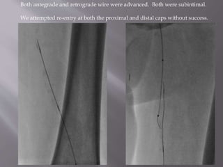
- 4. Both antegrade and retrograde wire were advanced. Both were subintimal. We attempted re-entry at both the proximal and distal caps without success.
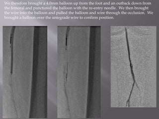
- 5. We therefore brought a 4.0mm balloon up from the foot and an outback down from the femoral and punctured the balloon with the re-entry needle. We then brought the wire into the balloon and pulled the balloon and wire through the occlusion. We brought a balloon over the antegrade wire to confirm position.
- 6. We used a 4.0 and 6.0 balloon to predilate. We stented with iDev Supera stents.
- 7. FINAL RESULT IN THE SFA
- 8. FINAL RUNOFF